

Abstract
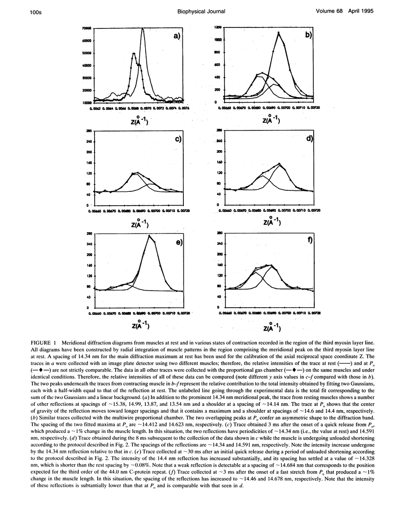
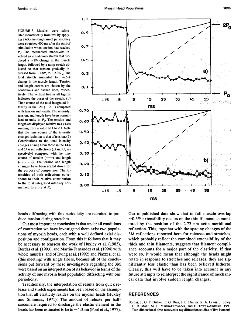
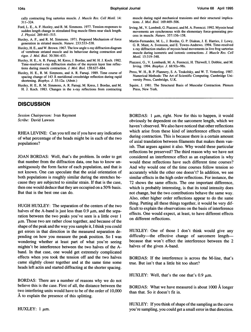
Using synchrotron radiation and whole muscles, 2 ms time-resolved x-ray diffraction patterns were recorded at 8 degrees C. The results show that in both isotonic and isometric contractions, as well as in length changes imposed at maximum tension [Po], the meridional third myosin layer line consists of two distinct reflections with different intensities and spacings that measure approximately 14,623 and 14,412 nm at Po. Although the intensity behavior of the two reflections is strikingly different during quick releases, it is very similar during stretches. Study of the time courses indicates that myosin heads diffracting at Po with the approximately 14.623 nm periodicity are actively involved in tension production. Those diffracting at Po with the periodicity of approximately 14.412 nm appear not be associated with tension production during isometric contraction and releases, but the results suggest that they are recruited during stretches and here contribute to tension production. Our most important conclusion is that under all conditions of contraction we have investigated there exist two populations of myosin heads, each with a well defined axial disposition and configuration.
Full text
PDF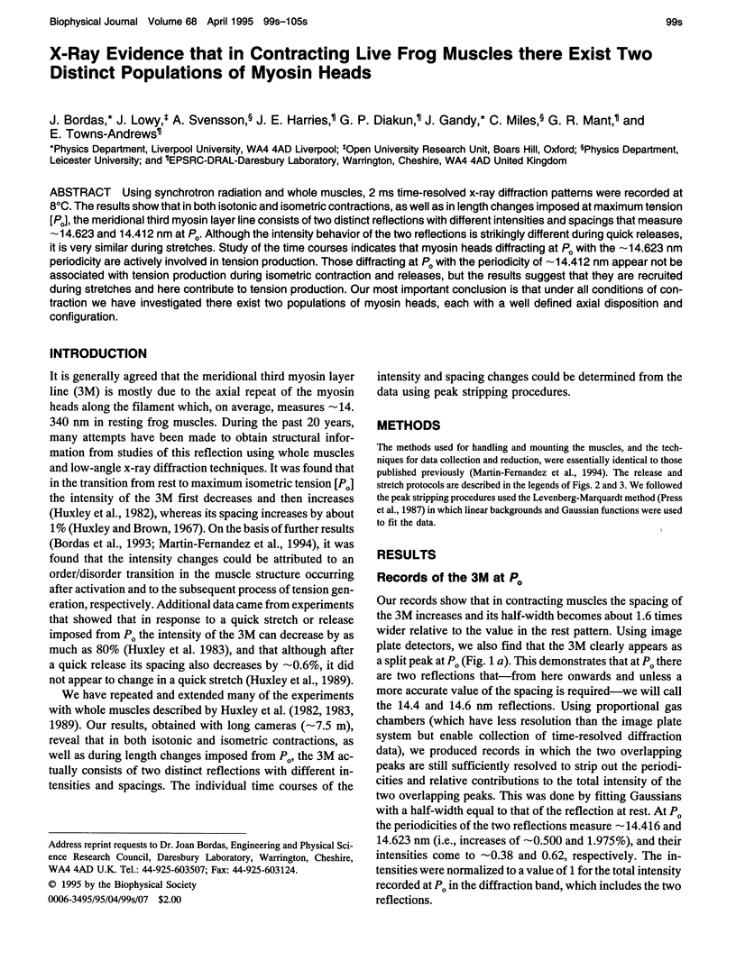
Selected References
These references are in PubMed. This may not be the complete list of references from this article.
- Bordas J., Diakun G. P., Diaz F. G., Harries J. E., Lewis R. A., Lowy J., Mant G. R., Martin-Fernandez M. L., Towns-Andrews E. Two-dimensional time-resolved X-ray diffraction studies of live isometrically contracting frog sartorius muscle. J Muscle Res Cell Motil. 1993 Jun;14(3):311–324. doi: 10.1007/BF00123096. [DOI] [PubMed] [Google Scholar]
- Ford L. E., Huxley A. F., Simmons R. M. Tension responses to sudden length change in stimulated frog muscle fibres near slack length. J Physiol. 1977 Jul;269(2):441–515. doi: 10.1113/jphysiol.1977.sp011911. [DOI] [PMC free article] [PubMed] [Google Scholar]
- Huxley A. F., Simmons R. M. Proposed mechanism of force generation in striated muscle. Nature. 1971 Oct 22;233(5321):533–538. doi: 10.1038/233533a0. [DOI] [PubMed] [Google Scholar]
- Huxley H. E., Faruqi A. R., Kress M., Bordas J., Koch M. H. Time-resolved X-ray diffraction studies of the myosin layer-line reflections during muscle contraction. J Mol Biol. 1982 Jul 15;158(4):637–684. doi: 10.1016/0022-2836(82)90253-4. [DOI] [PubMed] [Google Scholar]
- Huxley H. E., Simmons R. M., Faruqi A. R., Kress M., Bordas J., Koch M. H. Changes in the X-ray reflections from contracting muscle during rapid mechanical transients and their structural implications. J Mol Biol. 1983 Sep 15;169(2):469–506. doi: 10.1016/s0022-2836(83)80062-x. [DOI] [PubMed] [Google Scholar]
- Irving M., Lombardi V., Piazzesi G., Ferenczi M. A. Myosin head movements are synchronous with the elementary force-generating process in muscle. Nature. 1992 May 14;357(6374):156–158. doi: 10.1038/357156a0. [DOI] [PubMed] [Google Scholar]
- Martin-Fernandez M. L., Bordas J., Diakun G., Harries J., Lowy J., Mant G. R., Svensson A., Towns-Andrews E. Time-resolved X-ray diffraction studies of myosin head movements in live frog sartorius muscle during isometric and isotonic contractions. J Muscle Res Cell Motil. 1994 Jun;15(3):319–348. doi: 10.1007/BF00123484. [DOI] [PubMed] [Google Scholar]
- Piazzesi G., Lombardi V., Ferenczi M. A., Thirlwell H., Dobbie I., Irving M. Changes in the x-ray diffraction pattern from single, intact muscle fibers produced by rapid shortening and stretch. Biophys J. 1995 Apr;68(4 Suppl):92S–98S. [PMC free article] [PubMed] [Google Scholar]