Abstract
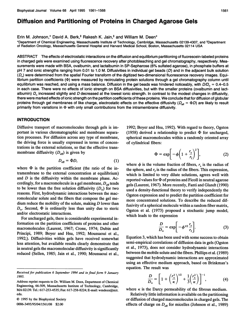
The effects of electrostatic interactions on the diffusion and equilibrium partitioning of fluorescein-labeled proteins in charged gels were examined using fluorescence recovery after photobleaching and gel chromatography, respectively. Measurements were made with BSA, ovalbumin, and lactalbumin in SP-Sepharose (6% sulfated agarose), in phosphate buffers at pH 7 and ionic strengths ranging from 0.01 to 1.0 M. Diffusivities in individual gel beads (D) and in the adjacent bulk solution (D infinity) were determined from the spatial Fourier transform of the digitized two-dimensional fluorescence recovery images. Equilibrium partition coefficients (phi) were measured by recirculating protein solutions through a gel chromatography column until equilibrium was reached, and using a mass balance. Diffusion in the gel beads was hindered noticeably, with D/D infinity = 0.4-0.5 in each case. There were no effects of ionic strength on BSA diffusivities, but with the smaller proteins (ovalbumin and lactalbumin) D infinity increased slightly and D decreased at the lowest ionic strength. In contrast to the modest changes in diffusivity, there were marked effects of ionic strength on the partition coefficients of these proteins. We conclude that for diffusion of globular proteins through gel membranes of like charge, electrostatic effects on the effective diffusivity (Deff = phi D) are likely to result primarily from variations in phi with only small contributions from the intramembrane diffusivity.
Full text
PDF


Selected References
These references are in PubMed. This may not be the complete list of references from this article.
- Amsterdam A., Er-el Z., Shaltiel S. Ultrastructure of beaded agarose. Arch Biochem Biophys. 1975 Dec;171(2):673–677. doi: 10.1016/0003-9861(75)90079-x. [DOI] [PubMed] [Google Scholar]
- Arnott S., Fulmer A., Scott W. E., Dea I. C., Moorhouse R., Rees D. A. The agarose double helix and its function in agarose gel structure. J Mol Biol. 1974 Dec 5;90(2):269–284. doi: 10.1016/0022-2836(74)90372-6. [DOI] [PubMed] [Google Scholar]
- Berk D. A., Yuan F., Leunig M., Jain R. K. Fluorescence photobleaching with spatial Fourier analysis: measurement of diffusion in light-scattering media. Biophys J. 1993 Dec;65(6):2428–2436. doi: 10.1016/S0006-3495(93)81326-2. [DOI] [PMC free article] [PubMed] [Google Scholar]
- Bor Fuh C., Levin S., Giddings J. C. Rapid diffusion coefficient measurements using analytical SPLITT fractionation: application to proteins. Anal Biochem. 1993 Jan;208(1):80–87. doi: 10.1006/abio.1993.1011. [DOI] [PubMed] [Google Scholar]
- Dubin P. L., Principi J. M. Optimization of size-exclusion separation of proteins on a Superose column. J Chromatogr. 1989 Sep 22;479(1):159–164. doi: 10.1016/s0021-9673(01)83327-6. [DOI] [PubMed] [Google Scholar]
- Giddings J. C., Yang F. J., Myers M. N. Flow-field-flow fractionation: a versatile new separation method. Science. 1976 Sep 24;193(4259):1244–1245. doi: 10.1126/science.959835. [DOI] [PubMed] [Google Scholar]
- Jain R. K., Stock R. J., Chary S. R., Rueter M. Convection and diffusion measurements using fluorescence recovery after photobleaching and video image analysis: in vitro calibration and assessment. Microvasc Res. 1990 Jan;39(1):77–93. doi: 10.1016/0026-2862(90)90060-5. [DOI] [PubMed] [Google Scholar]
- Laurent T. C. Determination of the structure of agarose gels by gel chromatography. Biochim Biophys Acta. 1967 Mar 22;136(2):199–205. doi: 10.1016/0304-4165(67)90064-5. [DOI] [PubMed] [Google Scholar]
- Moussaoui M., Benlyas M., Wahl P. Diffusion of proteins in Sepharose Cl-B gels. J Chromatogr. 1992 Feb 7;591(1-2):115–120. doi: 10.1016/0021-9673(92)80228-m. [DOI] [PubMed] [Google Scholar]
- Obrink B. Characterization of fiber parameters in agarose gels by light scattering. J Chromatogr. 1968 Oct 8;37(2):329–330. doi: 10.1016/s0021-9673(01)99117-4. [DOI] [PubMed] [Google Scholar]
- Raj T., Flygare W. H. Diffusion studies of bovine serum albumin by quasielastic light scattering. Biochemistry. 1974 Jul 30;13(16):3336–3340. doi: 10.1021/bi00713a024. [DOI] [PubMed] [Google Scholar]
- Righetti P. G., Caravaggio T. Isoelectric points and molecular weights of proteins. J Chromatogr. 1976 Apr 21;127(11):1–28. doi: 10.1016/s0021-9673(00)98537-6. [DOI] [PubMed] [Google Scholar]
- Spencer M. Reverse salt gradient chromatography of tRNA on unsubstituted agarose. III. Physical and chemical properties of different batches of Sepharose 4B. J Chromatogr. 1982 Apr 23;238(2):317–325. doi: 10.1016/s0021-9673(00)81317-5. [DOI] [PubMed] [Google Scholar]
- Tsay T. T., Jacobson K. A. Spatial Fourier analysis of video photobleaching measurements. Principles and optimization. Biophys J. 1991 Aug;60(2):360–368. doi: 10.1016/S0006-3495(91)82061-6. [DOI] [PMC free article] [PubMed] [Google Scholar]
- Waki S., Harvey J. D., Bellamy A. R. Study of agarose gels by electron microscopy of freeze-fractured surfaces. Biopolymers. 1982 Sep;21(9):1909–1926. doi: 10.1002/bip.360210917. [DOI] [PubMed] [Google Scholar]
- Walters R. R., Graham J. F., Moore R. M., Anderson D. J. Protein diffusion coefficient measurements by laminar flow analysis: method and applications. Anal Biochem. 1984 Jul;140(1):190–195. doi: 10.1016/0003-2697(84)90152-0. [DOI] [PubMed] [Google Scholar]


