

Abstract
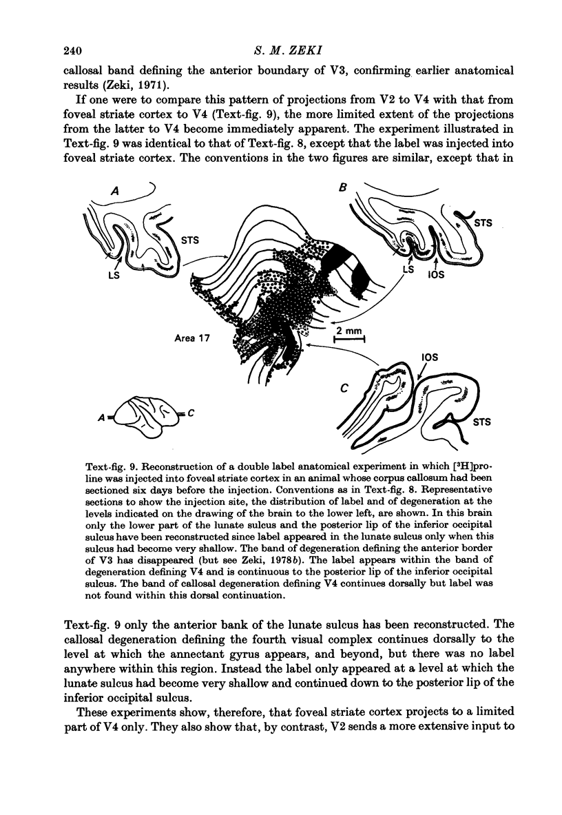
1. The cortical projections of the foveal and extrafoveal parts of the striate cortex have been compared, using conventional degeneration techniques, as well as combinations of anatomical methods. While both foveal and extrafoveal striate cortex share a common pattern of projections (to areas V2, V3 and the visual area in the medial part of the posterior bank of the superior temporal sulcus), foveal striate cortex was found to have an additional projection (to part of the cortex of the fourth visual areas, V4). The latter projection includes the posterior lip of the inferior occipital sulcus which, on anatomical grounds, is regarded as the ventral extension of V4. 2. Anatomical studies using double tracers were employed to clarify the nature of the projections from the striate cortex and from V2 to V4. In one such experiment, tritiated proline was injected into extra-foveal striate cortex and a small lesion was made in that part of V2 receiving a direct projection from the region of the striate cortex into which the radioactive tracer was injected. Only degenerating fibres (due to the lesion), and no radioactive label, was found in V4. Such an experiment showed that, unlike foveal striate cortex, the projections from extrafoveal striate cortex to V4 are not direct, but through V2. 3. In another type of anatomical experiment using double tracers, the corpus callosum was sectioned and tritiated proline was injected into foveal striate cortex. Such an experiment allowed a more accurate determination of the extent of V4, as judged from its callosal connexions, to which foveal striate cortex projects. 4. Considering the projections of V1 to areas V2, V3 and the visual area in the medial part of the posterior bank of the superior temporal sulcus, and considering the differences in the projections of foveal and extrafoveal striate cortex, it is suggested that, among other functions, the striate cortex acts as a distribution centre for the information coming over the retino-geniculo-cortical pathways, parcelling this information out to different visual areas of the prestriate cortex for further analysis.
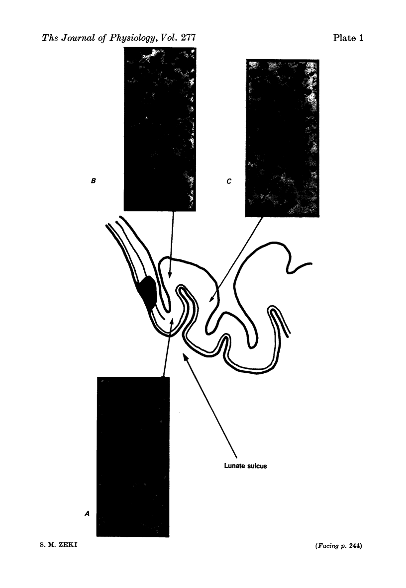
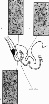
Full text
PDF
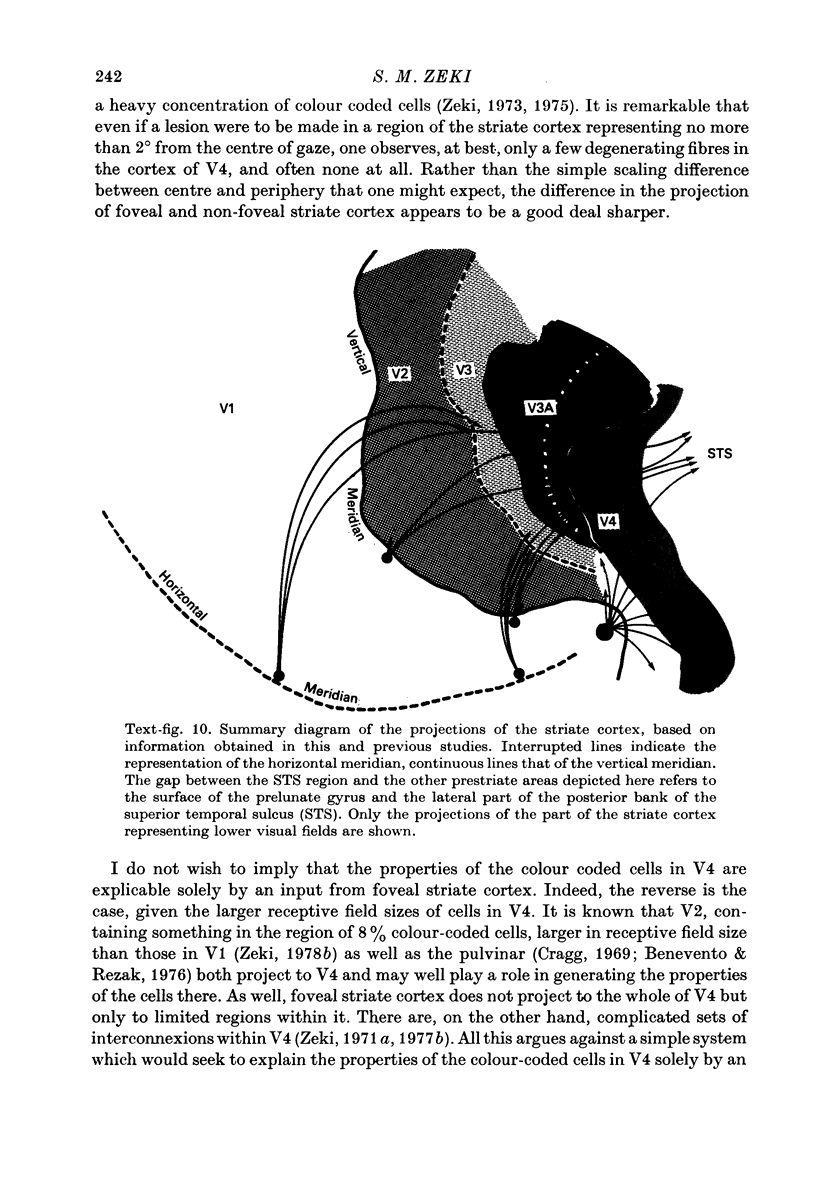
Images in this article
Selected References
These references are in PubMed. This may not be the complete list of references from this article.
- Benevento L. A., Rezak M. The cortical projections of the inferior pulvinar and adjacent lateral pulvinar in the rhesus monkey (Macaca mulatta): an autoradiographic study. Brain Res. 1976 May 21;108(1):1–24. doi: 10.1016/0006-8993(76)90160-8. [DOI] [PubMed] [Google Scholar]
- Cowan W. M., Gottlieb D. I., Hendrickson A. E., Price J. L., Woolsey T. A. The autoradiographic demonstration of axonal connections in the central nervous system. Brain Res. 1972 Feb 11;37(1):21–51. doi: 10.1016/0006-8993(72)90344-7. [DOI] [PubMed] [Google Scholar]
- Cragg B. G. The topography of the afferent projections in the circumstriate visual cortex of the monkey studied by the Nauta method. Vision Res. 1969 Jul;9(7):733–747. doi: 10.1016/0042-6989(69)90011-x. [DOI] [PubMed] [Google Scholar]
- DANIEL P. M., WHITTERIDGE D. The representation of the visual field on the cerebral cortex in monkeys. J Physiol. 1961 Dec;159:203–221. doi: 10.1113/jphysiol.1961.sp006803. [DOI] [PMC free article] [PubMed] [Google Scholar]
- Dow B. M., Gouras P. Color and spatial specificity of single units in Rhesus monkey foveal striate cortex. J Neurophysiol. 1973 Jan;36(1):79–100. doi: 10.1152/jn.1973.36.1.79. [DOI] [PubMed] [Google Scholar]
- Essen D. C., Zeki S. M. The topographic organization of rhesus monkey prestriate cortex. J Physiol. 1978 Apr;277:193–226. doi: 10.1113/jphysiol.1978.sp012269. [DOI] [PMC free article] [PubMed] [Google Scholar]
- Gouras P. Opponent-colour cells in different layers of foveal striate cortex. J Physiol. 1974 May;238(3):583–602. doi: 10.1113/jphysiol.1974.sp010545. [DOI] [PMC free article] [PubMed] [Google Scholar]
- Guld C., Bertulis A. Representation of fovea in the striate cortex of vervet monkey, Cercopithecus aethiops pygerythrus. Vision Res. 1976;16(6):629–631. doi: 10.1016/0042-6989(76)90010-9. [DOI] [PubMed] [Google Scholar]
- Hubel D. H., Wiesel T. N. Receptive fields and functional architecture of monkey striate cortex. J Physiol. 1968 Mar;195(1):215–243. doi: 10.1113/jphysiol.1968.sp008455. [DOI] [PMC free article] [PubMed] [Google Scholar]
- Hubel D. H., Wiesel T. N. Uniformity of monkey striate cortex: a parallel relationship between field size, scatter, and magnification factor. J Comp Neurol. 1974 Dec 1;158(3):295–305. doi: 10.1002/cne.901580305. [DOI] [PubMed] [Google Scholar]
- Wiitanen J. T. Selective silver impregnation of degenerating axons and axon terminals in the central nervous system of the monkey (Macaca mulatta). Brain Res. 1969 Jul;14(2):546–548. doi: 10.1016/0006-8993(69)90136-x. [DOI] [PubMed] [Google Scholar]
- Wilson M. E., Cragg B. G. Projections from the lateral geniculate nucleus in the cat and monkey. J Anat. 1967 Sep;101(Pt 4):677–692. [PMC free article] [PubMed] [Google Scholar]
- Zeki S. M. Colour coding in rhesus monkey prestriate cortex. Brain Res. 1973 Apr 27;53(2):422–427. doi: 10.1016/0006-8993(73)90227-8. [DOI] [PubMed] [Google Scholar]
- Zeki S. M. Colour coding in the superior temporal sulcus of rhesus monkey visual cortex. Proc R Soc Lond B Biol Sci. 1977 May 4;197(1127):195–223. doi: 10.1098/rspb.1977.0065. [DOI] [PubMed] [Google Scholar]
- Zeki S. M. Cortical projections from two prestriate areas in the monkey. Brain Res. 1971 Nov;34(1):19–35. doi: 10.1016/0006-8993(71)90348-9. [DOI] [PubMed] [Google Scholar]
- Zeki S. M. Representation of central visual fields in prestriate cortex of monkey. Brain Res. 1969 Jul;14(2):271–291. doi: 10.1016/0006-8993(69)90110-3. [DOI] [PubMed] [Google Scholar]
- Zeki S. M., Sandeman D. R. Combined anatomical and electrophysiological studies on the boundary between the second and third visual areas of rhesus monkey cortex. Proc R Soc Lond B Biol Sci. 1976 Nov 12;194(1117):555–562. doi: 10.1098/rspb.1976.0094. [DOI] [PubMed] [Google Scholar]
- Zeki S. M. Simultaneous anatomical demonstration of the representation of the vertical and horizontal meridians in areas V2 and V3 of rhesus monkey visual cortex. Proc R Soc Lond B Biol Sci. 1977 Feb 11;195(1121):517–523. doi: 10.1098/rspb.1977.0024. [DOI] [PubMed] [Google Scholar]
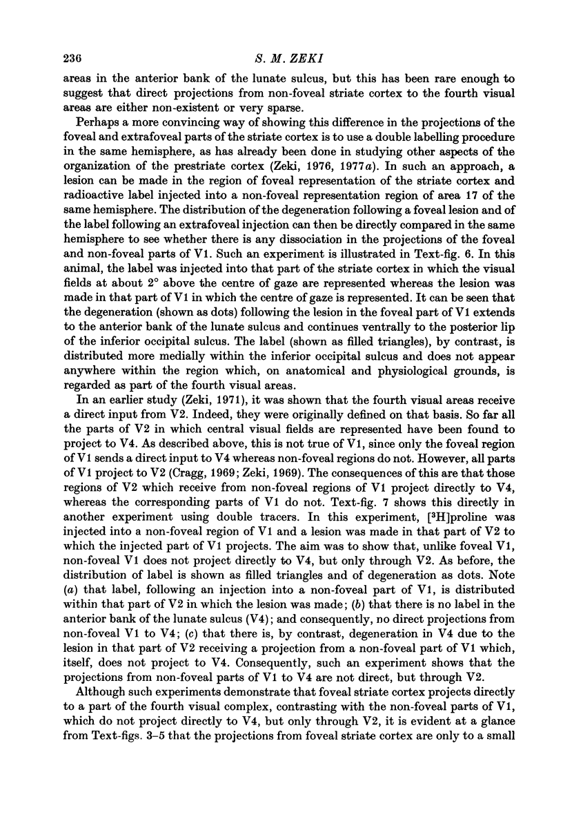
- Zeki S. M. The third visual complex of rhesus monkey prestriate cortex. J Physiol. 1978 Apr;277:245–272. doi: 10.1113/jphysiol.1978.sp012271. [DOI] [PMC free article] [PubMed] [Google Scholar]
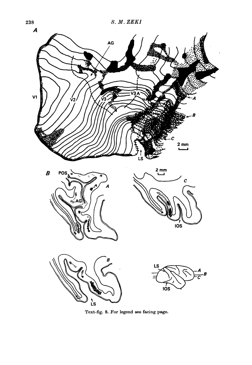
- Zeki S. M. Uniformity and diversity of structure and function in rhesus monkey prestriate visual cortex. J Physiol. 1978 Apr;277:273–290. doi: 10.1113/jphysiol.1978.sp012272. [DOI] [PMC free article] [PubMed] [Google Scholar]