

Abstract
1. Monosynaptic excitatory post-synaptic potentials (e.p.s.p.s) were recorded from medial (m.g.) and lateral gastrocnemius (l.g.) motoneurones in the cat 2-30 weeks after crushing the m.g. nerve.
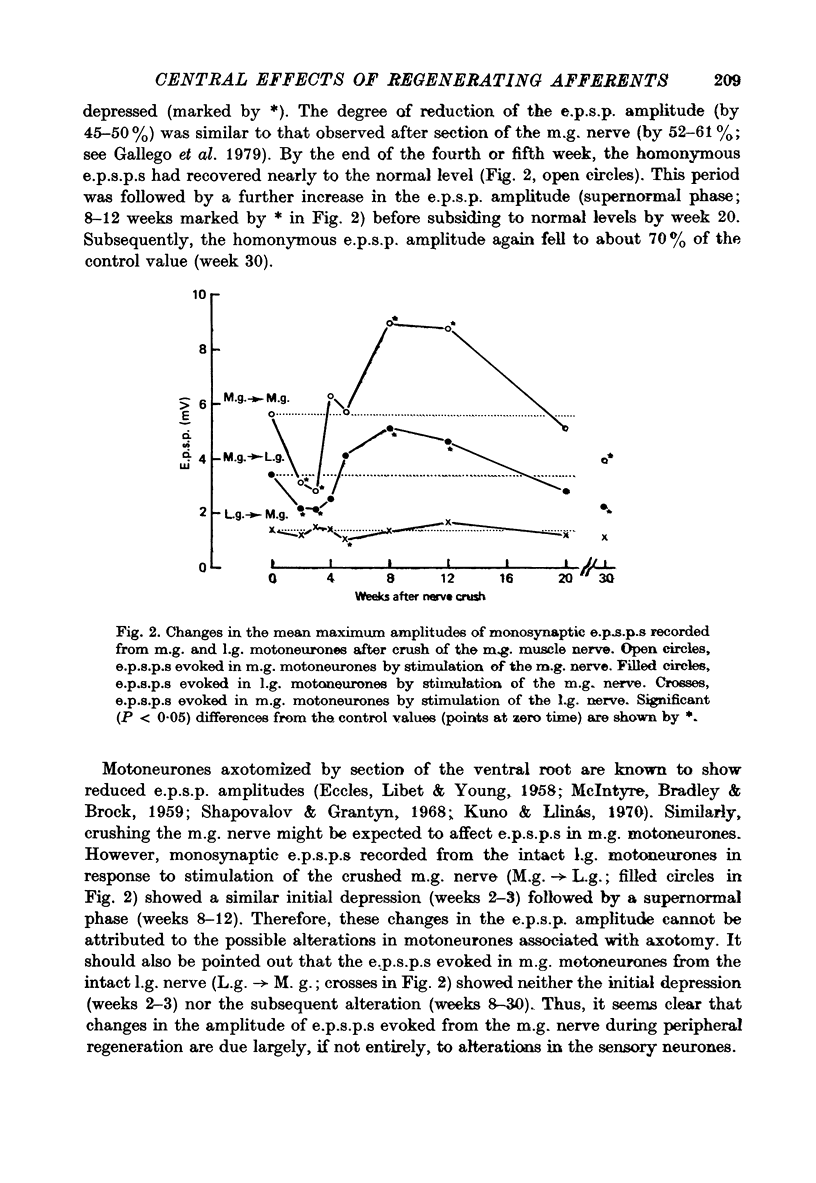
2. The mean amplitudes of homonymous and heteronymous e.p.s.p.s evoked from the m.g. nerve were initially depressed (2-3 weeks after injury) and subsequently reached levels greater than normal for a period (8-12 weeks) before slowly declining to about 70% of the normal values (by week 30).
3. Monosynaptic e.p.s.p.s evoked in m.g. motoneurones from the intact l.g. nerve showed neither initial depression nor subsequent alterations following crush of the m.g. nerve.
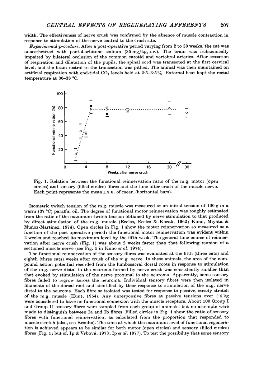
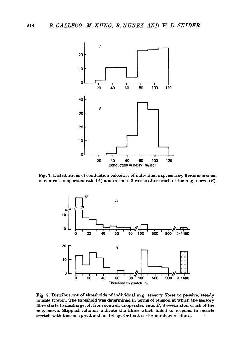
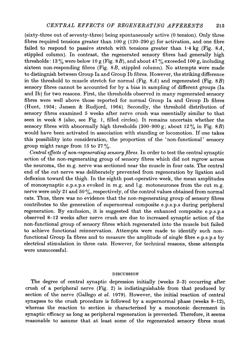
4. By the eighth week after nerve crush, about 70% of Group I and Group II sensory fibres in the m.g. nerve responded to muscle stretch, about 15% had regenerated into the muscle but did not respond to muscle stretch, and the remainder failed to regenerate across the neuroma formed by the nerve crush.
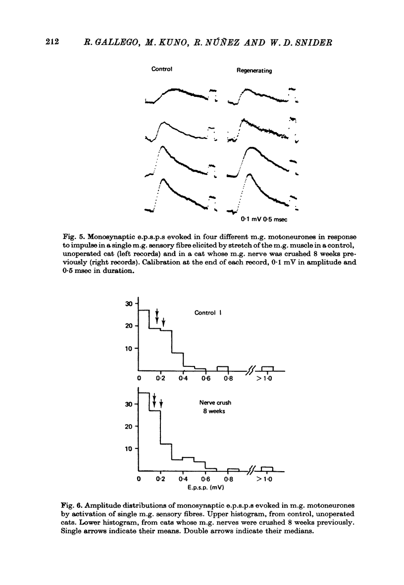
5. Homonymous, monosynaptic e.p.s.p.s produced by impulses in single sensory fibres responding to stretch of the m.g. muscle were recorded 8 weeks after crush of the m.g. nerve. Their amplitude distribution was indistinguishable from that obtained in normal, unoperated cats. Thus, there was no evidence that functionally reinnervated sensory fibres are responsible for the enhanced phase of composite e.p.s.p.s observed during peripheral regeneration.
6. When the m.g. nerve had been sectioned and prevented from regenerating into the muscle for 8 weeks, the amplitudes of homonymous and heteronymous e.p.s.p.s evoked from the m.g. nerve were significantly smaller than those observed in control animals. Thus, there was no evidence that non-regenerating sensory fibres are responsible for the enhanced phase of composite e.p.s.p.s after nerve crush.
7. It is suggested that the sensory fibres responsible for abnormally large composite e.p.s.p.s following nerve crush are those that regenerate into the muscle but do not achieve functional reinnervation. This possibility is discussed in relation to the increase in central synaptic efficacy observed after prolonged disuse of the sensory pathway.
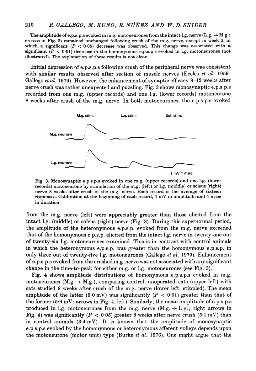
Full text
PDF
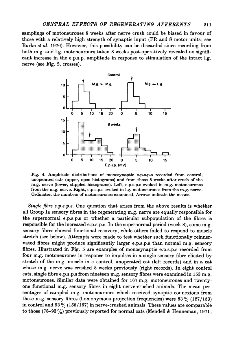
Selected References
These references are in PubMed. This may not be the complete list of references from this article.
- Brown M. C., Butler R. G. Regeneration of afferent and efferent fibres to muscle spindles after nerve injury in adults cats. J Physiol. 1976 Sep;260(2):253–266. doi: 10.1113/jphysiol.1976.sp011514. [DOI] [PMC free article] [PubMed] [Google Scholar]
- Burke R. E. Group Ia synaptic input to fast and slow twitch motor units of cat triceps surae. J Physiol. 1968 Jun;196(3):605–630. doi: 10.1113/jphysiol.1968.sp008526. [DOI] [PMC free article] [PubMed] [Google Scholar]
- ECCLES J. C., ECCLES R. M., SHEALY C. N. An investigation into the effect of degenerating primary afferent fibers on the monosynpatic innervation of motoneurons. J Neurophysiol. 1962 Jul;25:544–558. doi: 10.1152/jn.1962.25.4.544. [DOI] [PubMed] [Google Scholar]
- ECCLES J. C., KRNJEVIC K., MILEDI R. Delayed effects of peripheral severance of afferent nerve fibres on the efficacy of their central synapses. J Physiol. 1959 Jan 28;145(1):204–220. doi: 10.1113/jphysiol.1959.sp006136. [DOI] [PMC free article] [PubMed] [Google Scholar]
- ECCLES J. C., LIBET B., YOUNG R. R. The behaviour of chromatolysed motoneurones studied by intracellular recording. J Physiol. 1958 Aug 29;143(1):11–40. doi: 10.1113/jphysiol.1958.sp006041. [DOI] [PMC free article] [PubMed] [Google Scholar]
- ECCLES J. C., McINTYRE A. K. The effects of disuse and of activity on mammalian spinal reflexes. J Physiol. 1953 Sep;121(3):492–516. doi: 10.1113/jphysiol.1953.sp004961. [DOI] [PMC free article] [PubMed] [Google Scholar]
- Eccles J. C., Eccles R. M., Kozak W. Further investigations on the influence of motoneurones on the speed of muscle contraction. J Physiol. 1962 Sep;163(2):324–339. doi: 10.1113/jphysiol.1962.sp006978. [DOI] [PMC free article] [PubMed] [Google Scholar]
- Gallego R., Kuno M., Núez R., Snider W. D. Disuse enhances synaptic efficacy in spinal mononeurones. J Physiol. 1979 Jun;291:191–205. doi: 10.1113/jphysiol.1979.sp012807. [DOI] [PMC free article] [PubMed] [Google Scholar]
- HUNT C. C. Relation of function to diameter in afferent fibers of muscle nerves. J Gen Physiol. 1954 Sep 20;38(1):117–131. doi: 10.1085/jgp.38.1.117. [DOI] [PMC free article] [PubMed] [Google Scholar]
- Ip M. C., Vrbová G. Motor and sensory reinnervation of fast and slow mammalian muscles. Z Zellforsch Mikrosk Anat. 1973 Dec 31;146(2):261–279. doi: 10.1007/BF00307351. [DOI] [PubMed] [Google Scholar]
- Ip M. C., Vrbová G., Westbury D. R. The sensory reinnervation of hind limb muscles of the cat following denervation and de-efferentation. Neuroscience. 1977;2(3):423–434. doi: 10.1016/0306-4522(77)90007-0. [DOI] [PubMed] [Google Scholar]
- JANSEN J. K., RUDJORD T. ON THE SILENT PERIOD AND GOLGI TENDON ORGANS OF THE SOLEUS MUSCLE OF THE CAT. Acta Physiol Scand. 1964 Dec;62:364–379. doi: 10.1111/j.1748-1716.1964.tb10435.x. [DOI] [PubMed] [Google Scholar]
- Jack J. J., Miller S., Porter R., Redman S. J. The time course of minimal excitory post-synaptic potentials evoked in spinal motoneurones by group Ia afferent fibres. J Physiol. 1971 Jun;215(2):353–380. doi: 10.1113/jphysiol.1971.sp009474. [DOI] [PMC free article] [PubMed] [Google Scholar]
- Kuno M., Llinás R. Alterations of synaptic action in chromatolysed motoneurones of the cat. J Physiol. 1970 Nov;210(4):823–838. doi: 10.1113/jphysiol.1970.sp009244. [DOI] [PMC free article] [PubMed] [Google Scholar]
- Kuno M., Miyahara J. T. Non-linear summation of unit synaptic potentials in spinal motoneurones of the cat. J Physiol. 1969 Apr;201(2):465–477. doi: 10.1113/jphysiol.1969.sp008767. [DOI] [PMC free article] [PubMed] [Google Scholar]
- Kuno M., Miyata Y., Muñoz-Martinez E. J. Properties of fast and slow alpha motoneurones following motor reinnervation. J Physiol. 1974 Oct;242(1):273–288. doi: 10.1113/jphysiol.1974.sp010706. [DOI] [PMC free article] [PubMed] [Google Scholar]
- McINTYRE A. K., BRADLEY K., BROCK L. G. Responses of motoneurons undergoing chromatolysis. J Gen Physiol. 1959 May 20;42(5):931–958. doi: 10.1085/jgp.42.5.931. [DOI] [PMC free article] [PubMed] [Google Scholar]
- Mendell L. M., Henneman E. Terminals of single Ia fibers: distribution within a pool of 300 homonymous motor neurons. Science. 1968 Apr 5;160(3823):96–98. doi: 10.1126/science.160.3823.96. [DOI] [PubMed] [Google Scholar]
- Mendell L. M., Henneman E. Terminals of single Ia fibers: location, density, and distribution within a pool of 300 homonymous motoneurons. J Neurophysiol. 1971 Jan;34(1):171–187. doi: 10.1152/jn.1971.34.1.171. [DOI] [PubMed] [Google Scholar]
- Rall W., Burke R. E., Smith T. G., Nelson P. G., Frank K. Dendritic location of synapses and possible mechanisms for the monosynaptic EPSP in motoneurons. J Neurophysiol. 1967 Sep;30(5):1169–1193. doi: 10.1152/jn.1967.30.5.1169. [DOI] [PubMed] [Google Scholar]
- Scott J. G., Mendell L. M. Individual EPSPs produced by single triceps surae Ia afferent fibers in homonymous and heteronymous motoneurons. J Neurophysiol. 1976 Jul;39(4):679–692. doi: 10.1152/jn.1976.39.4.679. [DOI] [PubMed] [Google Scholar]
- Shapovalov A. I., Grantyn' A. A. Nadsegmentarnye sinapticheskie vliianiia na khromatolizirovannye motoneirony. Biofizika. 1968 Mar-Apr;13(2):260–269. [PubMed] [Google Scholar]
- Watt D. G., Stauffer E. K., Taylor A., Reinking R. M., Stuart D. G. Analysis of muscle receptor connections by spike-triggered averaging. 1. Spindle primary and tendon organ afferents. J Neurophysiol. 1976 Nov;39(6):1375–1392. doi: 10.1152/jn.1976.39.6.1375. [DOI] [PubMed] [Google Scholar]