Abstract
Cryptosporidium parvum TU502, a genotype 1 isolate of human origin, was passaged through three different mammalian hosts, including humans, pigs, and calves. It was confirmed to be genotype 1 by PCR-restriction fragment length polymorphism analysis of the Cryptosporidium oocyst wall protein gene, direct sequencing of PCR fragments of the small subunit rRNA and β-tubulin genes, and microsatellite analysis. This isolate was shown to be genetically stable when passaged through the three mammalian species, with no evidence of the emergence of new subpopulations as observed by a genotype-specific PCR assay. TU502 oocysts from different sources failed to infect gamma interferon knockout mice, a characteristic of genotype 1 isolates. The genotypic and phenotypic characterization of TU502 is significant since it is the isolate selected to sequence the genome of C. parvum genotype 1 and is currently used in several research projects including human volunteer studies.
The apicomplexan enteric parasite Cryptosporidium sp. infects a broad range of mammals, birds, fish, and reptiles (7, 12, 13, 15, 30). Cryptosporidium parvum, a major cause of diarrheal illness in humans and calves, has emerged as a serious contributor to waterborne outbreaks of cryptosporidiosis. Using a variety of genetic methods, C. parvum isolates are separated into two genetically distinct subgroups, designated genotype 1 and 2. Genotype 1 is anthroponotic and has so far been associated only with human and primate infections (21, 34). Genotype 2 is zoonotic and is found to infect a wide range of mammals, including humans (2, 10, 11, 16, 23, 31). The majority of sporadic cases of human cryptosporidiosis, including recent waterborne outbreaks, generally have one predominant genotype, but cryptosporidiosis is not restricted to one specific genotype (9, 10, 16, 18, 37; Tumwine et al., submitted for publication). Evidence, from animal and human studies in this laboratory and in others, indicates that genotype 1 and 2 display several distinct genotypic and phenotypic traits. The most common genotypic analyses are based on PCR-restriction fragment length polymorphism (PCR-RFLP) analysis and/or sequencing of the small subunit (SSU) rRNA (11, 17, 19, 21, 35), 70-kDa heat shock protein (25), β-tubulin (4, 24, 32), Cryptosporidium oocyst wall protein (COWP) (17-20, 36), or thrombospondin-related adhesive protein Cryptosporidium-1 (TRAP C1) or TRAP C2 (6, 19, 22, 23) genes. More recently, the introduction of multilocus microsatellite analysis to differentiate C. parvum isolates was reported (5, 8; A. E. Aiello et al., abstract from the 52nd Annual Meeting of the Society of Protozoologists 1999, J. Eukaryot. Microbiol. 46:46S-47S, 1999). Phenotypic differences between genotype 1 and 2 isolates, including host specificity and severity of clinical symptoms, have also been observed by our laboratory (34; Tumwine et al., submitted; Akiyoshi and Tzipori, unpublished data) and by others (13, 37). No evidence of recombination between the two genotypes has been reported, suggesting the possibility that these are two separate species (unpublished data).
In this study, we report the genotypic and phenotypic characterization of TU502, a genotype 1 isolate, and its passage through animal hosts, including humans, piglets, and calves. Genotyping methods including PCR-RFLP analysis of the COWP gene, sequencing of the SSU rRNA and β-tubulin genes, genotype-specific PCR assay, and microsatellite analyses were used to characterize the oocysts excreted from the different animal passages. Because of its genetic stability, TU502 has been designated our reference genotype 1 isolate, and its genome is currently being sequenced. This isolate is also used in several research projects, including challenge studies in human volunteers.
MATERIALS AND METHODS
Origin of the C. parvum TU502 genotype 1 isolate.
UG502 was originally isolated from a child with cryptosporidiosis as part of a recent survey conducted in hospitalized children in Uganda (Tumwine et al., submitted). This genotype 1 isolate was propagated three consecutive times in gnotobiotic piglets (see below) and was consistently shown to be genetically stable. Consequently, it was selected for continuous propagation in piglets. From time to time “caught” calves infected experimentally with oocysts purified from infected piglets were used to produce larger quantities of oocysts for laboratory investigations (see below). During the propagations in calves, three laboratory personnel caring for them became accidentally infected with the calf-propagated UG502 isolate. Oocysts purified from the last accidentally infected human, designated TU502, were selected for a comprehensive phenotypic and genotypic characterization study. Isolates UHP5 and TPH1 are the other two human-derived isolates.
Passage of genotype 1 isolates in gnotobiotic pigs.
Dichromate-treated oocysts of the original human UG502 isolate were used to infect gnotobiotic piglets (28, 29). Oocyst shedding was monitored by microscopic examination of fecal smears stained with modified acid-fast stain. The large intestines were sterilely harvested in a biological safety hood within a laboratory dedicated to the preparation and handling of fecal samples and intestinal contents of genotype 1-infected piglets. The pig intestinal contents were removed and used as inoculum for subsequent passages. All four genotype 1 human isolates (UG502, UHP5, TPH1, and TU502) were similarly propagated in piglets.
Propagation of genotype 1 isolates in calves.
To reduce the possibility of contamination with exogenous genotype 2, only calves caught during birth were used to propagate genotype 1. Briefly, the calves were caught by a worker gowned in sterile apparel as they were delivered and laid on sterile surgical drapes in a clean stall. The calves were dried and cleaned, and their navels were dipped in Betadine. The calves were transported from the farm to the university in a clean van within a few hours after birth. To further reduce the possibility of contamination, only gnotobiotic-piglet-propagated genotype 1 oocysts were used to infect these calves.
Mouse infectivity.
Gamma interferon knockout mice (Jackson Laboratories, Bar Harbor, Maine) were orally challenged with 1,000 purified pig-derived oocysts of isolate UG502, UHP5, or TU502 (26, 33). Oocyst shedding in feces was monitored by microscopic examination between days 5 and 20 postinoculation.
Oocyst purification and extraction of DNA from stool samples.
Oocysts were either purified from fecal samples by immunomagnetic separation (Crypto-Scan IMS; ImmuCell Corp., Portland, Maine) or by a previously described multistep purification protocol (33). If purified oocysts were used as the source of template for amplification, they were first heated at 95°C for 7 min. For some samples, DNA was extracted from stool samples using the protocol described by Buckholt et al. (3) and the DNA eluted in 50 μl of TE.
PCR-RFLP analysis.
COWP PCR amplifications were performed in 25-μl reaction mixtures containing 1× PCR buffer (10 mM Tris-HCl, pH 8.3; 50 mM KCl; 1.1 mM MgCl2; and 0.01% gelatin), 0.2 mM deoxynucleoside triphosphates, 0.4 μM forward primer (COWP-F1, 5′-GTAGATAATGGAAGAGATTGTGTTGC-3′), 0.4 μM reverse primer (cry9) (21), 1.25 U of RedTaq (Sigma-Aldrich, St. Louis, Mo.), and 1 μl of template DNA. The reaction mixtures were denatured at 94°C for 2 min and then cycled 35 times at 94°C for 30 s, 56°C for 30 s, and 72°C for 30 s. A final 72°C extension for 5 min completed the PCR program. For each set of PCRs, two C. parvum-positive controls (genotype 1 and 2) plus a negative PCR control were included. The resulting 553-bp PCR products were digested with RsaI and electrophoretically separated on a Tris-borate-EDTA-15% polyacrylamide gel (Criterion Precast gel; Bio-Rad, Inc., Richmond, Calif.).
5B12 microsatellite analysis.
The 5B12 marker is an AT repeat of variable length within noncoding sequence. PCR amplification of the 5B12 microsatellite was carried out as previously described (8).
PCR and DNA sequencing of the SSU rRNA and β-tubulin fragments.
The SSU rRNA and β-tubulin fragments from the four human genotype 1 isolates, UG502, UHP5, TPH1, and TU502, and their calf passages (all except TPH1) were amplified using the same PCR conditions as that for COWP, except a mixture of Taq DNA polymerase and proofreading polymerase (Expand High Fidelity PCR system; Roche Molecular Biochemicals, Indianapolis, Ind.) was used instead of RedTaq. A 490-bp SSU rRNA fragment was amplified using a forward primer, cry4a (5′-TCCTGACACAGGGAGGTAG-3′), and a reverse primer, cry2a (5′-TCCTTGGCAAATGCTTTCG-3′). A 536-bp β-tubulin PCR fragment was amplified using the forward primer btub5 and the reverse primer btub2 (32). The SSU rRNA and β-tubulin PCR fragments were cloned into pCR4-TOPO (Invitrogen Inc., San Diego, Calif.), and a minimum of four clones from each isolate and passage were double-strand sequenced. Multiple alignments of the DNA sequences were performed using the ClustalW program (27). GenBank searches against the nonredundant database for SSU rRNA and β-tubulin sequence similarities were performed using the BLAST algorithm (1).
Genotype-specific PCR assay.
To assay for the presence of genotype 2 DNA, a genotype-specific PCR assay was developed using TRAP C1. PCR was performed using a genotype 2-specific forward primer (TRAPC1-F2, 5′-TAAGGGTGGTGATAATGGCTGTA-3′) and a reverse primer (TRAPC1-Rc, 5′-CCTTCTGATAAAGTTGCATTATACGACC-3′) located within a conserved region. Each sample was also amplified with a genotype 1-specific forward primer (TRAPC1-F1, 5′-TAAAAGTGGTGATAACAGATGCG-3′) and TRAPC1-Rc to ensure there was no inhibition of amplification. PCRs and thermal cycler conditions were similar to those for COWP, except an annealing temperature of 54°C for 38 cycles was used.
RESULTS
C. parvum genotype 1 isolates.
C. parvum genotype 1 isolate TU502, originally isolated from a child with cryptosporidiosis, was passaged in piglets and calves. Experimental transmission of C. parvum in calves carries some risk of accidental infection of laboratory personnel. Infections with genotype 2 have been well-contained over the past 10 years, as the Division of Infectious Diseases at Tufts University School of Veterinary Medicine has instituted protective procedures to minimize accidental exposure of personnel. However, the propagation of genotype 1 in calves has increased considerably the risk due to, we believe, the fact that a smaller infectious dose is required for genotype 1 to cause infection. While caring for the calves, three laboratory personnel became infected with derivatives of the UG502 isolate (UHP5, TPH1, and TU502). TU502, one of the human-derived isolates, has now been passaged in both piglets and calves and is our laboratory genotype 1 standard isolate. The UHP5 and TPH1 isolates have also been passaged in gnotobiotic piglets, and the UHP5 isolate has been passaged in calves. We have serially passaged the UG502, UHP5, TPH1, and TU502 isolates in piglets 12, 1, 2, and 20 times, respectively.
Mouse infectivity.
Oocysts from UG502 pig passages 3 and 6, UHP5 human passage, and TU502 pig passage 12 were tested for infectivity in gamma interferon knockout mice, which appear to be susceptible to C. parvum genotype 2 isolates but not to genotype 1 isolates (33). No infection was detected for any of these isolates, confirming the absence of a genotype 2 subpopulation or introduction of genotype 2 oocysts during the propagation process. Newborn Mongolian gerbils and rats (Sprague Dawley) were also tested for susceptibility to infection with the UG502 isolate. No oocyst shedding by either rodent species was detected when the rodents were infected with the UG502 isolate, but shedding was detected when these rodents were infected with the genotype 2 isolate, GCH1 (data not shown).
COWP PCR-RFLP analysis.
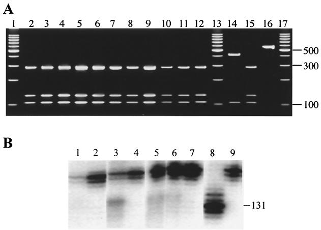
Oocysts excreted by the different animals infected with the four genotype 1 human isolates, displayed the typical COWP PCR-RFLP profile of genotype 1 isolates (Fig. 1A), in which RsaI restriction fragments of 34, 106, 129, and 284 bp were observed (21). The three RsaI sites were confirmed by sequence analysis of this 553-bp COWP PCR fragment from the UG502 human isolate. The COWP sequence from the UG502 isolate was identical to two other genotype 1 COWP sequences (AF266272 and AF266265) (34) in the GenBank database. PCR-RFLP analysis of the COWP gene fragment from the UG502-derived isolates passaged through the different host species showed no change in the restriction digest profile, with the genotype 1 profile maintained and no evidence of the presence of a genotype 2 subpopulation.
FIG. 1.
COWP PCR-RFLP (A) and 5B12 microsatellite (B) analyses of oocysts from the four C. parvum genotype 1 isolates passaged through different host species. (A) UG502 human (lane 2), pig (lane 3), and calf (lane 4) passages; UHP5 human (lane 5), pig (lane 6), and calf (lane 7) passages; TPH1 human (lane 8) and pig (lane 9) passages; and TU502 human (lane 10), pig (lane 11), and calf (lane 12) passages. A genotype 2 control (lane 14), genotype 1 control (lane 15), uncut PCR product (lane 16), and 100-bp DNA ladder (lanes 1, 13, and 17; Promega Corp., Madison, Wis.) are also included. (B) UG502 human passage (lane 1), UG502 calf passage (lane 2), UHP5 human passage (lane 3), UHP5 pig passage (lane 4), TPH1 human passage (lane 5), TU502 human passage (lane 6), and TU502 calf passage (lane 7). GCH1 (genotype 2) and NEMC1 (genotype 1) controls are shown in lanes 8 and 9, respectively.
Microsatellite analysis.
Microsatellite analysis using the 5B12 marker also confirmed that the UG502 isolate and its derivatives, when passaged through humans and calves, maintained the genotype 1 profile of the UG502 human isolate (Fig. 1B). No additional bands were seen in any of the UG502-derived isolates (UHP5, TPH1, and TU502) to indicate the introduction of a genotype 2 population. The microsatellite profiles for UG502, UHP5, TPH1, and TU502 were identical to two other genotype 1 isolates from Uganda and also to the NEMC1 isolate (8).
Sequence of the SSU rRNA and β-tubulin PCR fragments.
The sequences of the 490-bp SSU rRNA and 536-bp β-tubulin PCR fragments from the human, pig, and calf passages of the four isolates were identical to that of the original UG502 isolate from the Ugandan child. The calf passages represent the furthest animal passage from its human passage, and if genetic changes were to have taken place, these would be the most likely passages for their occurrence. Since the TPH1 isolate was not passaged in calves, DNA from a pig passage was sequenced instead. Sequences from a minimum of four clones for each sample were compared and found to be identical. The UG502 SSU rRNA sequence was compared to other sequences in the nonredundant GenBank database using the BLAST algorithm (1). Three C. parvum genotype 1 sequences (AF093489, AF108865, and L16997) were identical to the UG502 sequence. Similarly, the β-tubulin sequence from the UG502 isolate was compared to other sequences in the nonredundant GenBank database. The sequence of this 536-bp PCR fragment was identical to three other genotype 1 β-tubulin sequences (AF323578, AF323579, and AF323580) from human patients in the United Kingdom. This same fragment has been cloned and sequenced from 21 genotype 1 isolates and 13 genotype 2 isolates from other Ugandan children (Akiyoshi and Tzipori, unpublished data). The UG502 sequence of the coding region within this fragment was identical to those 21 genotype 1 isolates, although minor differences were observed within the intron sequences.
Genotype-specific PCR assay.
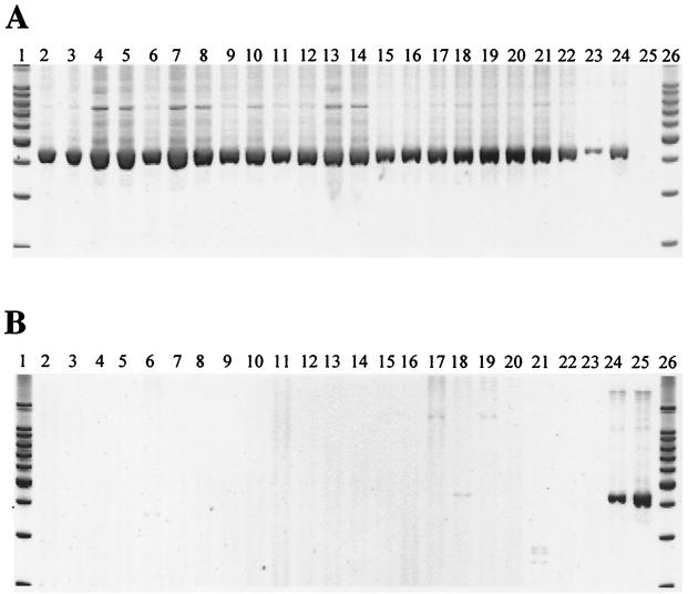
The PCR primers TRAPC1-F1 and TRAPC1-F2 differ by six bases, which is sufficient to confer genotype specificity. DNAs from each of the four isolates and from different animal passages were separately amplified with each primer pair (Fig. 2). No genotype 2-specific amplification was observed from DNAs isolated from oocysts excreted by humans and pigs. However, genotype 2-specific amplifications from three nonsequential calf propagations (UG502, calf 2; TU502, calves 2 and 14) were observed, despite our best efforts to maintain a clean environment for caught calves.
FIG. 2.
Genotype-specific PCR analysis of genotype 1 isolates passaged through different host species. (A) Each sample was assayed using the genotype 1-specific primers to demonstrate the absence of PCR inhibitors. (B) The presence of a genotype 2 subpopulation was assayed using the TRAP C1 genotype 2-specific primers. Samples are as follows: human isolate UG502 (lane 2), UG502 pig passage 1 (lane 3), UG502 pig passage 9 (lane 4), UG502 isolate from calf 3 (lane 5), UHP5 human (lane 6), UHP5 pig passage 1 (lane 7), UHP5 isolate from calf 1 (lane 8), UHP5 isolate from calf 2 (lane 9), human isolate TPH1 (lane 10), TPH1 pig passage 1 (lane 11), human isolate TU502 (lane 12), TU502 pig passage 1 (lane 13), TU502 pig passage 6 (lane 14), TU502 pig passage 11 (lane 15), TU502 pig passage 18 (lane 16), TU502 isolate from calf 1 (lane 17), TU502 isolate from calf 2 (lane 18), TU502 isolate from calf 3 (lane 19), TU502 isolate from calf 4 (lane 20), TU502 isolate from calf 9 (lane 21), and TU502 isolates calves 11 and 12 (lane 22). Genotype standards include genotype 1 (lane 23), mixed genotype 1 and 2 (lane 24), and genotype 2 (lane 25). DNA ladder (100 bp; Promega Corp.) is shown in lanes 1 and 26. A faint band is seen in TU502 isolate from calf 2 (lane 18) using the genotype 2-specific primers, indicating the presence of genotype 2 parasites.
Nucleotide sequence accession numbers.
Nucleotide sequences of the UG502 isolate COWP, β-tubulin and SSU rRNA sequences were submitted to GenBank and have accession numbers AF481960, AF481961 and AF481962, respectively.
DISCUSSION
Using molecular genetic tools, including COWP PCR-RFLP analysis and a TRAP C1 genotype-specific PCR assay, we have shown that homogeneous genotype 1 isolates of human origin can be passaged and maintained in gnotobiotic piglets. Extreme care was taken during the propagation of genotype 1 isolates in gnotobiotic piglets to avoid potential contamination with genotype 2 isolates that are routinely propagated in our laboratory. Our data clearly show that the UG502 isolate and its derivatives maintained genotypic profiles consistent with genotype 1 with the absence of other emerging subpopulations. DNA extracted from oocysts collected from each infected piglet, which was analyzed by COWP PCR-RFLP analysis, showed no changes in the genetic profile from the original UG502 (data not shown).
We have also successfully propagated four genotype 1 isolates, NEMC1 (34), UG502, UHP5, and TU502 in calves. Since caught calves have normal microflora, they may also contain C. parvum genotype 2 oocysts picked up on the farm during delivery. Consequently, calf-to-calf transmission of genotype 1 was avoided, and animals were therefore challenged only with oocysts derived from gnotobiotic piglets. To-date, TU502 from piglets has been propagated in 14 caught calves, of which two became contaminated with a genotype 2 population. The COWP PCR-RFLP and TRAP C1 genotype-specific PCR assays detected mixed populations in one of these calves, but only the TRAP C1 assay detected small amounts of genotype 2 oocysts in the second calf. This assay has the sensitivity of detecting 10 genotype 2 oocysts in a background population of 105 genotype 1 oocysts (data not shown).
While it has been shown that genotype 1 isolates can infect other mammals in addition to humans, there have been no reports of natural genotype 1 infections of other mammals, except captive monkeys (21; unpublished data). In a recent survey of Ugandan children with diarrhea, 72.8% of the Cryptosporidium-positive samples were identified as genotype 1, 18.4% were genotype 2, 4.1% were mixed genotype 1 and 2, and 4.7% were neither genotype 1 nor 2 or were unclassified (Tumwine et al., submitted). The isolation of a genetically homogeneous genotype 1 isolate, UG502, from this Ugandan study and reports from other human outbreaks of cryptosporidiosis suggest a unique mechanism of transmission from human to human. If genotype 1 isolates are truly anthroponotic, it is unclear how they are transmitted from human to human without introduction of genotype 2 isolates, which are ubiquitous in the environment and, from limited observations, appear to predominate when they occur concurrently with genotype 1 (Akiyoshi and Tzipori, unpublished). The lack of apparent genetic recombination between the two genotypes, even when they occur concurrently in the same host, also suggests that they maintain independent reproductive cycles and may be regarded as two distinct Cryptosporidium species (9, 13, 19, 21). Future studies focused on understanding the transmission cycles of the two genotypes and the basic biology of Cryptosporidium will provide these answers.
The identification of a genetically stable genotype 1 isolate is significant because of the absence of well-characterized Cryptosporidium isolates. There are several genotype 2 isolates including, IOWA, Moredun, and GCH1 (28), which are routinely used in laboratory investigations. However, these isolates are passaged in calves obtained from local farms where they may have been possibly exposed to farm-derived C. parvum oocysts. We have propagated UG502 and TU502 in gnotobiotic piglets for more than 2 years, from one animal to another, without exposing the oocysts assigned for further propagation to potential contamination. This eliminated the risk of cross-contamination of TU502 with oocysts from other sources and ensured the purity of the inoculum. Consequently, the TU502 isolate shows no trace of genotypic or phenotypic changes, making TU502 an excellent reference genotype 1 isolate. TU502 can be said to be the only currently existing isolate which is known to be free of potential contamination with other Cryptosporidium isolates since its initial isolation from a human patient. Therefore, TU502 has been selected for the genome sequencing project, challenge studies with human volunteers (Chappell et al., unpublished data), water treatment studies, and genetic recombination studies. A number of genes (partial and complete) from TU502 have been sequenced, including those coding for gp40/15 (14), β-tubulin, SSU rRNA, COWP, TRAP C1, and P23, and several microsatellite loci have been analyzed by our laboratory.
In conclusion, the piglet-propagated TU502 is a well-characterized genotype 1 C. parvum isolate and is the only currently available C. parvum isolate that is known to be free from contamination with other isolates or genotypes. While oocysts of genotype 1 can also infect calves, unless gnotobiotic calves are used, the risk of cross-contamination with genotype 2 eliminates calves as a potential animal species for propagation.
Acknowledgments
We thank Julia Dilo, Tammy Easter, Jianzhong Zhang, Kindra King, Christy Coulson, Katherine Hanson, Kim Deary, Jennifer Martineau, Jessica Brisben, and Paige Fox for technical or animal care support.
This study was supported by NIH grants NO1-AI-75321, PO1-DK-55510, and U01-AI-46418.
Editor: W. A. Petri, Jr.
REFERENCES
- 1.Altschul, S. F., W. Gish, W. Miller, E. W. Myers, and D. J. Lipman.1990. Basic local alignment search tool. J. Mol. Biol. 215:403-410. [DOI] [PubMed] [Google Scholar]
- 2.Awad-El-Kariem, F. M., H. A. Robinson, F. Petry, V. McDonald, D. Evans, and D. Casemore.1998. Differentiation between human and animal isolates of Cryptosporidium parvum using molecular and biological markers. Parasitol. Res. 84:97-101. [DOI] [PubMed] [Google Scholar]
- 3.Buckholt, M. A., J. H. Lee, and S. Tzipori. 2002. Prevalence of Enterocytozoon bieneusi in swine: an 18 month survey at a slaughterhouse in Massachusetts. Appl. Environ. Microbiol. 68:2595-2599. [DOI] [PMC free article] [PubMed] [Google Scholar]
- 4.Caccio, S., W. Homan, K. van Dijk, and E. Pozio. 1999. Genetic polymorphism at the β-tubulin locus among human and animal isolates of Cryptosporidium parvum. FEMS Microbiol. Lett. 170:173-179. [DOI] [PubMed] [Google Scholar]
- 5.Caccio, S., W. Homan, R. Camilli, G. Traldi, T. Koterbeek, and E. Pozio.2000. A microsatellite marker reveals population heterogeneity within human and animal genotypes of Cryptosporidium parvum. Parasitology 120:237-244. [DOI] [PubMed] [Google Scholar]
- 6.Elwin, K., R. M. Chalmers, R. Roberts, E. C. Guy, and D. P. Casemore. 2001. Modification of a rapid method for the identification of gene-specific polymorphisms in Cryptosporidium parvum and its application to clinical and epidemiological investigations. Appl. Environ. Microbiol. 67:5581-5584. [DOI] [PMC free article] [PubMed] [Google Scholar]
- 7.Fayer, R., C. A. Speer, and J. P. Dubey. 1997. The general biology of Cryptosporidium, p. 1-42. In R. Fayer (ed.), Cryptosporidium and cryptosporidiosis. CRC Press, Inc., Boca Raton, Fla.
- 8.Feng, X., S. M. Rich, D. Akiyoshi, J. K. Tumwine, A. Kekitiinwa, N. Nabukeera, S. Tzipori, and G. Widmer. 2000. Extensive polymorphism in Cryptosporidium parvum identified by multilocus microsatellite analysis. Appl. Environ. Microbiol. 66:3344-3349. [DOI] [PMC free article] [PubMed] [Google Scholar]
- 9.McLauchlin, J., S. Pedraza-Diaz, C. Amar-Hoetzeneder, and G. L. Nichols. 1999. Genetic characterization of Cryptosporidium strains from 218 patients with diarrhea diagnosed as having sporadic cryptosporidiosis. J. Clin. Microbiol. 37:3153-3158. [DOI] [PMC free article] [PubMed] [Google Scholar]
- 10.McLauchlin, J., C. Amar, S. Pedraza-Diaz, and G. L. Nichols. 2000. Molecular epidemiological analysis of Cryptosporidium spp. in the United Kingdom: results of genotyping Cryptosporidium spp. in 1,705 fecal samples from humans and 105 fecal samples from livestock animals. J. Clin. Microbiol. 38:3984-3990. [DOI] [PMC free article] [PubMed] [Google Scholar]
- 11.Morgan, U. M., C. C. Constantine, D. A. Forbes, and R. C. A. Thompson. 1997. Differentiation between human and animal isolates of Cryptosporidium parvum using rDNA sequencing and direct PCR analysis. J. Parasitol. 83:825-830. [PubMed] [Google Scholar]
- 12.Morgan, U. M., K. D. Sargent, P. Deplazes, D. A. Forbes, F. Spano, H. Hertzberg, A. Elliot, and R. C. A. Thompson. 1998. Molecular characterization of Cryptosporidium from various hosts. Parasitology 117:31-37. [DOI] [PubMed] [Google Scholar]
- 13.Morgan, U. M., L. Xiao, R. Fayer, A. A. Lal, and R. C. A. Thompson. 1999. Variation in Cryptosporidium: towards a taxonomic revision of the genus. Int. J. Parasitol. 29:1733-1751. [DOI] [PubMed] [Google Scholar]
- 14.O'Conner, R. M., C. M. Thorpe, A-M Cevallos, and H. D. Ward. 2002. Expression of the highly polymorphic Cryptosporidium parvum Cpgp40/15 gene in genotype I and II isolates. Mol. Biochem. Parasitol. 119:203-215. [DOI] [PubMed] [Google Scholar]
- 15.O'Donoghue, P. J. 1995. Cryptosporidium and cryptosporidiosis in man and animals. Int. J. Parasitol. 25:139-195. [DOI] [PubMed] [Google Scholar]
- 16.Patel, S., S. Pedraza-Diaz, J. McLauchlin, and D. P. Casemore. 1998. Molecular characterization of Cryptosporidium parvum from two large suspected waterborne outbreaks. Commun. Dis. Public Health 1:231-233. [PubMed] [Google Scholar]
- 17.Patel, S., S. Pedraza-Diaz, and J. McLauchlin. 1999. The identification of Cryptosporidium species and Cryptosporidium parvum directly from whole faeces by analysis of a multiplex PCR of the 18S rRNA gene and by PCR/RFLP of the Cryptosporidium outer wall protein (COWP) gene. Int. J. Parasitol. 29:1241-1247. [DOI] [PubMed] [Google Scholar]
- 18.Pedraza-Diaz, S., C. Amar, G. L. Nichols, and J. McLauchlin. 2001. Nested polymerase chain reaction for amplification of the Cryptosporidium oocyst wall protein gene. Emerg. Infect. Dis. 7:49-56. [DOI] [PMC free article] [PubMed] [Google Scholar]
- 19.Peng, M. M., L. Xiao, A. R. Freeman, J. Arrowood, A. A. Escalante, A. C. Weltman, C. S. L. Ong, W. R. MacKenzie, A. A. Lal, and C. B. Beard. 1997. Genetic polymorphism among Cryptosporidium parvum isolates: evidence of two distinct human transmission cycles. Emerg. Infect. Dis. 3:567-573. [DOI] [PMC free article] [PubMed] [Google Scholar]
- 20.Spano, F., L. Putignani, J. McLauchlin, D. P. Casemore, and A. Cristanti. 1997. PCR-RFLP analysis of the Cryptosporidium oocyst wall protein (COWP) gene discriminates between C. wrairi and C. parvum, and between C. parvum isolates of human and animal origin. FEMS Microbiol. Lett. 150:209-217. [DOI] [PubMed] [Google Scholar]
- 21.Spano, F., L. Putignani, A. Cristanti, P. Sallicandro, U. M. Morgan, S. M. LeBlancq, L. Tchack, S. Tzipori, and G. Widmer. 1998. Multilocus genotypic analysis of Cryptosporidium parvum isolates from different hosts and geographical origins. J. Clin. Microbiol. 36:3255-3259. [DOI] [PMC free article] [PubMed] [Google Scholar]
- 22.Spano, F., L. Putignani, S. Guida, and A. Cristanti. 1998. Cryptosporidium parvum: PCR-RFLP analysis of the TRAP-C1 (thrombospondin-related adhesive protein of Cryptosporidium-1) gene discriminates between two alleles differentially associated with parasite isolates of animal and human origin. Exp. Parasitol. 90:195-198. [DOI] [PubMed] [Google Scholar]
- 23.Sulaiman, I. M., L. Xiao, C. Yang, L. Escalante, A. Moore, C. B. Beard, M. J. Arrowood, and A. A. Lal. 1998. Differentiating human from animal isolates of Cryptosporidium parvum. Emerg. Infect. Dis. 4:681-685. [DOI] [PMC free article] [PubMed] [Google Scholar]
- 24.Sulaiman, I. M., A. A. Lal, M. J. Arrowood, and L. Xiao. 1999. Biallelic polymorphism in the intron region of β-tubulin gene of Cryptosporidium parasites. J. Parasitol. 85:154-157. [PubMed] [Google Scholar]
- 25.Sulaiman, I. M., U. M. Morgan, R. C. A. Thompson, A. A. Lal, and L. Xiao. 2000. Phylogenetic relationships of Cryptosporidium parasites based on the 70-kilodalton heat shock protein (HSP70) gene. Appl. Environ. Microbiol. 66:2385-2391. [DOI] [PMC free article] [PubMed] [Google Scholar]
- 26.Theodos, C. M., K. L. Sullivan, J. K. Griffiths, and S. Tzipori. 1997. Profiles of healing and nonhealing Cryptosporidium parvum infection in C57BL/6 mice with functional B and T lymphocytes: the extent of gamma interferon modulation determines the outcome of infection. Infect. Immun. 65:4761-4769. [DOI] [PMC free article] [PubMed] [Google Scholar]
- 27.Thompson, J. D., D. G. Higgins, and T. J. Gibson. 1994. CLUSTAL W: improving the sensitivity of progressive multiple sequence alignment through sequence weighting, position-specific gap penalties and weight matrix choice. Nucleic Acids Res. 22:4673-4680. [DOI] [PMC free article] [PubMed] [Google Scholar]
- 28.Tzipori, S., W. Rand, J. Griffiths, G. Widmer, and J. Crabb. 1994. Evaluation of an animal model system for cryptosporidiosis: therapeutic efficacy of paromomycin and hyperimmune bovine colostrum-immunoglobulin. Clin. Diagn. Lab. Immunol. 1:450-463. [DOI] [PMC free article] [PubMed] [Google Scholar]
- 29.Tzipori, S. 1998. Cryptosporidiosis: laboratory investigations and chemotherapy. Adv. Parasitol. 40:5-36. [DOI] [PubMed] [Google Scholar]
- 30.Tzipori, S., and G. Widmer. 2000. The biology of Cryptosporidium. Contrib. Microbiol. 6:1-32. [DOI] [PubMed] [Google Scholar]
- 31.Widmer, G. 1998. Genetic heterogeneity and PCR detection of Cryptosporidium parvum. Adv. Parasitol. 40:223-239. [DOI] [PubMed] [Google Scholar]
- 32.Widmer, G., L. Tchack, C. L. Chappell, and S. Tzipori. 1998. Sequence polymorphism in the β-tubulin gene reveals heterogeneous and variable population structures in Cryptosporidium parvum. Appl. Environ. Microbiol. 64:4477-4481. [DOI] [PMC free article] [PubMed] [Google Scholar]
- 33.Widmer, G., S. Tzipori, C. J. Fichtenbaum, and J. K. Griffiths. 1998. Genotypic and phenotypic characterization of Cryptosporidium parvum isolates from people with AIDS. J. Infect. Dis. 178:834-840. [DOI] [PubMed] [Google Scholar]
- 34.Widmer, G., D. Akiyoshi, M. A. Buckholt, X. Feng, S. M. Rich, K. M. Deary, C. A. Bowman, P. Xu, Y. Wang, G. A. Buck, and S. Tzipori. 2000. Animal propagation and genomic survey of a genotype 1 isolate of Cryptosporidium parvum. Mol. Biochem. Parasitol. 108:187-197. [DOI] [PubMed] [Google Scholar]
- 35.Xiao, L., L. E. Escalante, C. Yang, I. M. Sulaiman, A. A. Escalante, R. Montali, R. Fayer, and A. A. Lal. 1999. Phylogenetic analysis of Cryptosporidium parasites on the small-subunit ribosomal RNA gene locus. Appl. Environ. Microbiol. 65:1578-1583. [DOI] [PMC free article] [PubMed] [Google Scholar]
- 36.Xiao, L., J. Limor, U. M. Morgan, I. M. Sulaiman, R. C. A. Thompson, and A. A. Lal. 2000. Sequence differences in the diagnostic target region of the oocyst wall protein gene of Cryptosporidium parasites. Appl. Environ. Microbiol. 66:5499-5502. [DOI] [PMC free article] [PubMed] [Google Scholar]
- 37.Xiao, L., C. Bern, J. Limor, I. Sulaiman, J. Roberts, W. Checkley, L. Cabrera, R. H. Gillman, and A. A. Lal. 2001. Identification of 5 types of Cryptosporidium parasites in children in Lima, Peru. J. Infect. Dis. 183:492-497. [DOI] [PubMed] [Google Scholar]


