Abstract
Infection with high-risk human papillomavirus (HPV) types, particularly types 16 and 18, contributes to 90% of cervical cancer cases. HPV infects cutaneous or mucosal epithelium, tissue that is monitored for microbial infection or damage by Langerhans cells. In lesions produced by HPV type 16, there is a reduction in numbers of immune cells, especially Langerhans cells. Langerhans precursor cells selectively express CCR6, the receptor for macrophage inflammatory protein 3α (MIP-3α), and function as potent immune responders to inflamed epithelium and initiators of the innate immune response. It has been reported that E6 and E7 of high-risk HPVs interfere with immune mediators in order to suppress the recruitment of immune cells and antiviral activities of infected cells. Here we show that, following proinflammatory stimulus, HPV-16 E6 and E7 inhibit MIP-3α transcription, resulting in suppression of the migration of immature Langerhans precursor-like cells. Interestingly, the E6 and E7 proteins from the low-risk HPV types also inhibited MIP-3α transcription. These results suggest that one mechanism by which HPV-infected cells suppress the immune response may be through the inhibition of a vital alert signal, thus contributing to the persistence of HPV infection.
Human papillomaviruses (HPVs) are small DNA viruses which exhibit tropism for cutaneous or mucosal epithelium. Commonly categorized as either low- or high-risk HPV types on the basis of their oncogenic potential, infections with the high-risk HPV types such as HPV-16 and HPV-18 have been implicated in cervical-anogenital cancer and oral squamous cell carcinomas (41). The mucosal lesions caused by HPVs often resolve over time, and a high percentage of young women exposed to HPV clear the infection within a short period following initial contact with the virus (62). Empirical evidence for the critical role the immune response plays in controlling an HPV infection comes from the high prevalence and persistence of HPV infections in immunosuppressed individuals (62). In general, the mucosal immune response to an infection begins with the cells of the epidermis secreting alarm signals which act on immune cells as well as the infected cells. Chemokines, cytokines, adhesion molecules, and proteases direct the migration of leukocytes, monocytes, lymphocytes, neutrophils, eosinophils, basophils, natural killer cells, dendritic cells, and endothelial cells (56), while interferons inhibit viral replication within the infected cells (64).
Evasion of the immune response would contribute to the survival and propagation of HPV-infected cells. Two genes in the high-risk HPVs, E6 and E7, are associated with the virus's oncogenic potential to transform infected cells. In addition to their well-documented effects on cell cycle and differentiation, E6 and E7 also negatively impact the immune response by inhibiting the production of immune mediators. Expression of E6 is inversely correlated with the expression of interleukin-18 (IL-18), a proinflammatory cytokine similar to IL-1 that induces gamma interferon (IFN-γ) production (10). E6 and, to a lesser extent, E7 down-regulate the expression of IL-8, a chemoattractant for T cells (28). E6 and E7 also suppress the expression of a chemokine, monocyte chemoattractant protein 1 (MCP-1), in female genital epithelial cells (35). Additionally, E6 and E7 contribute to the escape from the antiviral and antiproliferative properties of tumor necrosis factor alpha (TNF-α) and IFN-α through growth stimulatory effects, loss of TNF sensitivity, and interaction with components of the IFN signaling pathway (62).
Following microbial infection, chemokines secreted by the epidermis initiate the adaptive immune response by recruiting dendritic cells. Dendritic cells are antigen-presenting cells which play an important role in the capture of antigen and stimulation of T lymphocytes (5). Many subsets of dendritic cells exist, characterized by differences in surface marker expression and developmental pathways. Dendritic cells differentiate from bone marrow progenitors and circulate in peripheral blood as immature precursors. Langerhans cells (LCs) are the dendritic cells that reside in the epidermis, and immature LCs, efficient in the uptake of antigens, continually monitor the epidermal microenvironment for damage and infection. Following antigen processing, LCs mature through differential regulation of surface markers and migrate to T-cell areas of the body, such as dermal lymphatic or peripheral lymphoid organs (5, 54). Although LCs have been shown to be maintained in the skin for long periods of time, LC precursors are recruited under conditions of local LC loss such as during inflammation (25, 43). Among the chemokines and cytokines secreted by epidermal cells, macrophage inflammatory protein-3α (MIP-3α) is the most potent chemokine for LC precursors and dermal dendritic cells (12). Originally found to be expressed in lung and liver tissue (57), liver and activation-induced chemokine/CCL20/MIP-3α is also produced in intestinal epithelium and epidermal keratinocytes (14, 26) but primarily in areas of inflamed epithelium (9, 47). MIP-3α expression can be increased by TNF-α, IL-1α, or IL-1β in oral squamous cell carcinomas, intestinal epithelial cells, and keratinocytes (1, 12, 26, 32, 36, 47, 67). Immature dendritic cells, memory T cells, and epidermal LC precursors migrate in response to MIP-3α due to their selective expression of CC chemokine receptor 6 (CCR6) (19, 37, 40, 55).
Investigations of cervical biopsies revealed that the number of LCs was significantly reduced in intraepithelial neoplasia and low-grade and high-grade lesions compared to normal tissue results (2, 11, 29, 70). Thus, areas of HPV persistence exhibiting decreases in LC density suggest decreased immune surveillance. Our studies show that, following a proinflammatory stimulus, human keratinocytes expressing HPV-16 E6 and E7 produce less MIP-3α than control cells. The result of this lack of secreted MIP-3α is a reduction in the migration of Langerhans precursor-like cells (LPLCs). Our results suggest that the down-regulation of MIP-3α is one mechanism by which HPV infections could evade the immune response and persist undetected in the epithelium.
MATERIALS AND METHODS
Keratinocyte culture and preparation of cell lines.
Primary human foreskin keratinocytes (HFKs) were isolated from neonatal foreskins and cultured in EpiLife medium supplemented with human keratinocyte growth supplement (bovine pituitary extract, bovine insulin, hydrocortisone, bovine transferrin, human epidermal growth factor) and penicillin-streptomycin-amphotericin B solution (Cascade Biologics). Cells were cultured in EpiLife medium supplemented without bovine pituitary extract and hydrocortisone (hereafter called minimal EpiLife) when culture supernatants were harvested for use in enzyme-linked immunosorbent assays (ELISAs) and migration assays.
Generation of stable cell lines expressing HPV-16 E6/E7, HPV-6 E6/E7, and HPV-11 E6/E7 was accomplished using the pBabe retroviral system (44). Retrovirus encoding either vector alone or E6/E7 from HPV-16, HPV-6, and HPV-11 was produced by transfecting ΦNX packaging cells (American Type Culture Collection) with vesicular stomatitis virus envelope gene and pBabe retroviral constructs by the calcium phosphate method (BD Biosciences), according to the manufacturer's protocol. At 48 h after transfection, retrovirus was concentrated by centrifugation for 90 min at 15,000 rpm and 4°C, and virus pellets were resuspended in 2 ml EpiLife overnight at 4°C. Resuspended virus was either used immediately to infect HFKs or frozen at −80°C for future use. Resuspended virus was used to infect HFKs in 8 μg Polybrene/ml of EpiLife for 5 h. Cells were allowed to recover for 24 h and then selected with 1.25 μg/ml puromycin for 2 to 4 days. Stable cell lines were expanded for treatment with polyinosine-poly(C) [Poly(I) · Poly(C)] (Amersham Biosciences).
Generation of E6 and E7 mutations.
Mutagenesis of E6/E7 was performed on HPV-16 E6/E7 in pGEM7Zf by use of a QuikChange kit (Stratagene). The following primers were used to generate a stop codon at the 16th amino acid in E7 gene of HPV-16 for E6/E7stop (forward, GATTTGTAACCAGAGACAACTG; reverse, CAGTTGTCTCTGGTTACAAATC) and a stop codon at the 15th amino acid in E6 gene of HPV-16 for E6stop/E7 (forward, CCACAGTAATGCACAGAGCTGC; reverse, GCAGCTCTGTGCATTACTGTGG). HPV-16 E6(C66G/C136G) and HPV-16 E7.2, previously characterized as unable to bind CBP/p300 and pCAF (28, 50), were generated in combination with E7stop and E6stop, respectively. The primers listed previously were used to mutate HPV-16 E6(C66G/C136G)/E7 in pGEM7ZF to generate E6(C66G/C136G)/E7stop. Mutagenesis of E7 was performed on HPV-16 E6stop/E7 in pGEM7Zf by use of a QuikChange kit (Stratagene). The following primers were used to generate an amino acid replacement of histidine by proline at position 2: forward, GCTGTAATCATGCCTGGAGATACACCTACA; reverse, TGTAGGTGTATCTCCAGGCATGATTACAGC. The mutants were sequenced and subcloned into the pBabe retroviral vector.
Measurement of MIP-3α by real-time quantitative PCR (qPCR) and ELISA.
A total of 1 × 10⁶ cells were seeded on 10-cm dishes 24 h prior to treatment with Poly(I) · Poly(C). Cells were treated with 100 μg/ml Poly(I) · Poly(C) in antibiotic-free EpiLife for 16 h, washed five times with phosphate-buffered saline (PBS), and refed with minimal EpiLife for 4 h. Cells were harvested for RNA isolation, and culture supernatants were collected for use in ELISAs and migration assays.
Total RNA was extracted using an RNeasy Mini kit (QIAGEN) according to the manufacturer's instructions. cDNA amplification was carried out in 25-μl reverse transcriptase (RT) reaction mixtures containing 3 μg RNA, 400 μM deoxyribonucleoside triphosphates, RNase out RNase inhibitor (Invitrogen), 10 ng random hexamer primer, 5 U avian myeloblastosis virus RT (Promega), and 5× RT buffer (Promega). The cDNA obtained was used in real-time qPCR reaction mixtures containing 2.5 μl each of (1 pM) forward and reverse primers, 12.5 μl 2× iQ SYBR green Supermix (Bio-Rad), and 1 μl cDNA template. The protocol used included a denaturation step (95°C for 30 s) followed by amplification repeated 45 times (95°C for 30 s, 60°C for 1 min, and 68°C for 1 min). A melt curve analysis performed following every run confirmed the amplification of a single product and no primer dimer. In each experiment, reactions were carried out in quadruplicate for each sample and glyceraldehyde phosphate dehydrogenase (GAPDH) was used for normalization. Primers used for real-time qPCR were obtained from Primer Bank (71) (MIP-3α forward, TGCTGTACCAAGAGTTTGCTC; MIP-3α reverse, CGCACACAGACAACTTTTTCTTT; GAPDH forward, TGTTGCCATCAATGACCCCTT; GAPDH reverse, CTCCACGACGTACTCAGCG). Paired t tests were used to generate P values.
MIP-3α was measured in culture supernatants collected from Poly(I) · Poly(C)-treated cells by use of a human MIP-3α DuoSet ELISA development kit (R & D Systems) according to the manufacturer's instructions. The lower limit of detection was 32 pg/ml. Each sample was run in triplicate in each experiment, and paired t tests were used to generate P values.
Culture of LPLCs.
Mononuclear cells were isolated from peripheral blood of healthy donors by centrifugation on lymphocyte separation medium (Cellgro). The mononuclear cell layer was collected from the interface, and monocytes were purified by positive magnetic selection using CD14 microbeads and an MS separation column (Miltenyi Biotec). Following magnetic bead selection on day 0 of culture, the purity of CD14+ cells was routinely >95%, as assessed by flow cytometry.
Purified CD14+ monocytes were cultured in 24-well tissue culture plates (Costar) in complete medium (RPMI 1640 supplemented with l-glutamine [Cellgro], 1% penicillin-streptomycin, and 10% heat-inactivated fetal calf serum [Fetalclone I; HyClone]) supplemented with 200 ng/ml granulocyte-macrophage colony stimulating factor (GM-CSF) and 10 ng/ml transforming growth factor β1 (TGF-β1) (Peprotech). At days 2 and 4, cells were fed with fresh medium plus cytokines. At day 6, LPLCs were harvested, analyzed by flow cytometry, and used in migration assays.
Analysis of LPLC surface marker expression and phagocytic capacity.
Cells (1 × 10⁵) were incubated for 30 min at 4°C in PBS-2% fetal calf serum (Fetalclone I; HyClone)-1% sodium azide with allophycocyanin-conjugated CD14 (eBioscience), phycoerythrin-conjugated CD83 (eBioscience), phycoerythrin-conjugated CCR6 (BD PharMingen), or fluorescein isothiocyanate-conjugated HLA-DR (BD PharMingen) monoclonal antibodies at the appropriate concentration or with control isotype-matched irrelevant monoclonal antibodies at the same concentration. Cells were washed twice and analyzed with a FACSCaliber system (Becton-Dickinson) using CellQuest software.
A total of 1 × 10⁵ cells were incubated with 10 μg/ml fluorescein isothiocyanate-conjugated ovalbumin (Molecular Probes) for 30 min at 37°C or 4°C (control cells). Cells were washed twice and analyzed with a FACSCaliber system. Fluorescein intensity provided a measurement of the phagocytic capacity of the cells.
Migration assays.
Migration assays were carried out using 24-well Transwell plates with 5-μm-pore-size polycarbonate filters (Costar 3421). Briefly, 2 × 10⁵ LPLCs in 100 μl of medium were added to the upper wells and 600 μl of conditioned medium was added to the lower wells. Cells were allowed to migrate for 4 h at 37°C under conditions of 5% CO2. Migrated cells and medium from the lower wells were collected, spun down for 5 min at 1,200 rpm and 4°C, resuspended in medium, and counted using a hemacytometer. The number of cells that migrated in response to unconditioned medium was subtracted from the number of cells migrating in response to conditioned medium to control for nonspecific migration. Recombinant human MIP-3α (Peprotech) and anti-human MIP-3α antibody (R & D Systems) were used in the compensation and neutralization migration assays. Each sample was run in duplicate in each experiment, and paired t tests were used to generate P values.
Reporter assay.
HFKs were seeded in six-well dishes at a density of 1 × 10⁵ cells per well. The luciferase reporter construct, kindly provided by Andrew Keates, consisted of an 849-bp fragment of the MIP-3α promoter cloned into the pGL3-Basic firefly luciferase expression vector (MIP-3αWT). A mutant reporter construct was also used which contained four nucleotide substitutions in the NF-κB binding element on the promoter (MIP-3αmNFκB). Cells were transfected 24 h later with 100 ng MIP-3αWT or MIP-3αmNFκB and 900 ng pSG5 by use of Fugene 6 (Roche). At 24 h after transfection, cells were washed with 1× PBS, refed with penicillin-streptomycin-amphotericin B-free EpiLife, and left untreated or treated with 10 μg/ml Poly(I) · Poly(C), 10 ng/ml TNF-α, or 10 ng/ml IL-1β for 6 h. Following treatment, cells were harvested, lysed, and measured for luciferase activity as described previously (42, 50). Assays were carried out in triplicate for each sample.
Microfluidic card analysis.
As described previously, 1 × 10⁶ cells were seeded on 10-cm dishes 24 h prior to treatment with Poly(I) · Poly(C). Following treatment and addition of minimal EpiLife, cells were harvested and total RNA was extracted using an RNeasy Mini kit (QIAGEN) according to the manufacturer's instructions. RNA was provided to the University of Rochester Functional Genomics Center for cDNA amplification and RT-PCR gene expression quantification using microfluidic cards (Applied Biosystems) containing primers specific for a panel of genes implicated in the immune response. Following sample loading, cards were processed with an ABI PRISM 7900HT Sequence Detection System (SDS; Applied Biosystems) and data was analyzed using SDS 2.2 software with GAPDH as the endogenous control and the control cell line (Babe) set as the calibrator sample (with expression equal to 1). Each sample was run in quadruplicate in each experiment, and paired t tests were used to generate P values. Results are expressed as the average severalfold reduction in gene expression for three experiments using three independent cell lines. Only those targets in which severalfold reduction was statistically significant (P < 0.05) are listed in Table 1.
TABLE 1.
Immune-related genes down-regulated in E6/E7-expressing keratinocytes
| Gene name | Accession no. | Avg fold decrease in E6/E7 cells |
|---|---|---|
| CD80a | NM_005191 | 20 |
| iNOS | NM_000625 | 11 |
| CYP7A1 | NM_000761 | 10 |
| LTα | NM_000595 | 6.7 |
| CD54 (ICAM) | NM_000201 | 5.9 |
| RANTES | NM_002985 | 5.6 |
| MIP-1α | NM_002983 | 4 |
| ITAC | NM_005409 | 3.6 |
| MIP-3αb | NM_004591 | 3 |
| IP10 | NM_001565 | 3 |
| CD40 | NM_001250 | 3 |
| C3 | NM_000064 | 2.8 |
| IL-8 | NM_000584 | 2.8 |
| FasL | NM_000639 | 2.6 |
| M-CSF | NM_005211 | 2.6 |
| IL-15 | NM_000585 | 2.4 |
| HO-1 | NM_002133 | 2.4 |
| ECE-1 | NM_001397 | 2.4 |
| Fas | NM_000043 | 2.4 |
| EDN1 | NM_001955 | 2.3 |
| IL-6 | NM_000600 | 2.1 |
| NFκB2 (p49) | NM_002502 | 2.1 |
| IL-1β | NM_000576 | 1.9 |
| MADH-7 | NM_005904 | 1.5 |
| BCL-XL | NM_001191 | 1.5 |
| Stat3 | NM_139276 | 1.5 |
| BAX | NM_004324 | 1.4 |
Expression was seen in two of three microfluidic cards.
Not a target gene included on microfluidic cards; value expressed is a result of separate real-time PCR validation.
RESULTS
MIP-3α mRNA and protein is decreased in Poly(I) · Poly(C)-treated HPV-16 E6/E7-expressing keratinocytes.
From microarray analysis performed in order to identify genes differentially regulated in E6/E7-expressing HFKs compared to control cells (51), several genes involved with immune regulation (IL-1α, IL-8, MIP-3α, TNF-α) were identified as being down-regulated in the E6/E7-expressing cells. To obtain a more accurate picture of immune response genes that are deregulated by E6/E7, microfluidic cards of 96 immune-related genes were used for real-time PCR quantitation. HFKs were stably infected with retrovirus carrying either vector alone (Babe) or a vector containing HPV-16 E6 and E7 in tandem (E6/E7). In order to stimulate significant cytokine-chemokine production, keratinocytes were activated with Poly(I) · Poly(C), which has been shown to efficiently activate keratinocytes (38). Poly(I) · Poly(C), a synthetic double-stranded ribonucleotide, mimics an intermediate of a viral infection and is recognized by Toll-like receptor 3. Activation of Toll-like receptor 3 results in induction of NF-κB activation and production of interferons and cytokines. In our hands, the use of Poly(I) · Poly(C) resulted in the strongest stimulation of MIP-3α production compared to lipopolysaccharide (LPS), TNF-α, and IL-1β results (data not shown). Control and E6/E7-expressing keratinocytes were activated with Poly(I) · Poly(C) for 16 h, washed, and refed with minimal EpiLife for 4 h. Each cell line of control and E6/E7-expressing keratinocytes was generated by the retroviral infection of pooled neonatal foreskin keratinocytes; thus, each cell line possesses a mixture of unique genetic backgrounds. To control for genetic background effects, three experiments were performed using cells from three different pooled foreskin samples that were infected with retrovirus and treated with Poly(I) · Poly(C). Following treatment, cells were harvested and RNA was purified for cDNA amplification. The microfluidic cards, containing 96 genes involved in the immune response, were processed with the ABI PRISM 7900HT SDS, and data were analyzed with SDS 2.2 software using GAPDH as the endogenous control. Results from the microfluidic cards validated the immune genes known to be down-regulated in E6/E7-expressing keratinocytes from the microarray and also identified additional cytokines, chemokines, receptors, and wound-healing factors that were down-regulated in Poly(I) · Poly(C)-stimulated E6/E7-expressing keratinocytes (Table 1).
Many of these genes may not be direct targets of E6/E7. For instance, IL-1β induces the production of TNF-α, and IL-15 stimulates inducible nitric oxide synthetase (iNOS) in keratinocytes (23, 73). The expression of Regulated on Activation, Normal T Expressed, and Secreted gene (RANTES), Enthothelin 1 (EDN1), and Complement Component 3 (C3) in keratinocytes is increased after stimulation with TNF-α (39, 49, 68). Thus, it is possible that down-regulation of RANTES, EDN1, C3, and iNOS may be an indirect result of repression of IL-1β and IL-15 by E6/E7. MIP-3α expression has been shown to up-regulated by TNF-α, IL-1α, and IL-1β (12, 26, 36, 47, 67). Initial experiments using a MIP-3α promoter assay and primary human keratinocytes demonstrated that E6 and E7, both in cooperation and individually, can repress MIP-3α promoter activity in the absence or presence of exogenously added IL-1β or TNF-α (data not shown). These results suggested that expression of E6/E7 was having a direct effect on MIP-3α transcription in the absence or presence of proinflammatory stimuli. Since MIP-3α was not provided as a target gene on the microfluidic card, it was necessary to perform separate real-time qPCR to validate the repression of MIP-3α observed in the microarray. Additionally, MIP-3α was selected for further investigation due to its selective and potent role in the recruitment of LCs during the innate immune response.
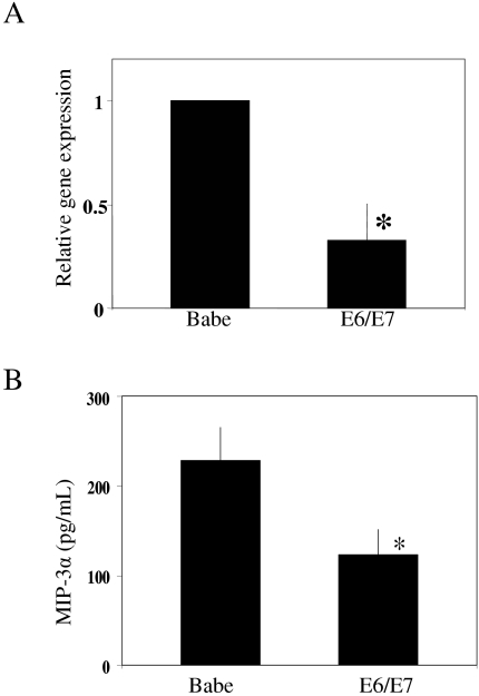
To examine the effect of E6/E7 on MIP-3α mRNA and protein levels, control and E6/E7-expressing cells were stimulated with Poly(I) · Poly(C) for 16 h. MIP-3α mRNA levels increase during the course of differentiation (data not shown); however, E6 and E7 differentially regulate numerous cell cycle, differentiation, and apoptotic genes during the differentiation process. In order to investigate the specific effect of E6/E7 on MIP-3α production, it was necessary to use cycling keratinocytes. However, in the absence of a proinflammatory stimuli, cycling keratinocytes express undetectable levels of MIP-3α mRNA or protein. Therefore, keratinocytes were treated with Poly(I) · Poly(C) to provide a proinflammatory stimulus and induce production of significant levels of MIP-3α. As stated previously, the use of Poly(I) · Poly(C) in our hands resulted in the strongest stimulation of MIP-3α production compared to LPS, TNF-α, and IL-1β results. Following treatment, cells were washed with PBS and refed with minimal EpiLife for 4 h. The cells were harvested for evaluation by real-time qPCR, and culture supernatants were collected for use in ELISA analysis and migration assays. Following Poly(I) · Poly(C) treatment of three independent cell lines, E6/E7-expressing keratinocytes showed an average threefold reduction in MIP-3α mRNA levels compared to control cells (Fig. 1A). We next examined the secretion of MIP-3α by Poly(I) · Poly(C)-treated keratinocytes. As shown in Fig. 1B, levels of secreted MIP-3α protein parallel the levels of mRNA expression, such that there was a reduced level of MIP-3α in cells expressing E6/E7 (123 pg/ml) compared to controls (228 pg/ml).
FIG. 1.
MIP-3α expression in Poly(I) · Poly(C)-treated control and E6/E7-expressing keratinocytes measured by real-time qPCR and ELISA. Primary human foreskin keratinocytes stably expressing vector alone (Babe) or HPV-16 E6/E7 (E6/E7) were treated with 100 μg/ml Poly(I) · Poly(C) for 16 h. Following treatment, cells were washed and refed with minimal EpiLife for 4 h. (A) RNA was harvested from the cells, and mRNA levels for MIP-3α were determined by real-time qPCR. Results are normalized to GAPDH and expressed as the means ± standard deviations (SD) of three experiments using three independent cell lines (* P < 0.001). (B) Culture supernatants were harvested, and levels of secreted MIP-3α were measured by ELISA. Results are expressed as the means ± SD of three experiments using three independent cell lines (* P < 0.001).
Culture supernatant from Poly(I) · Poly(C)-treated HPV-16 E6/E7-expressing keratinocytes reduces migration of LPLCs.
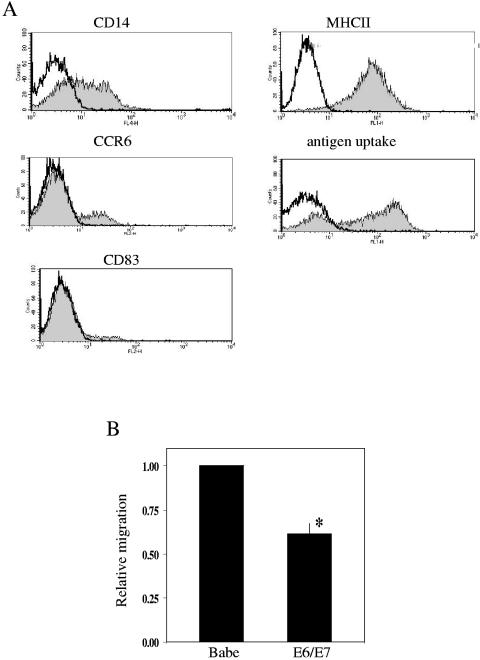
To evaluate the effect of factors secreted by Poly(I) · Poly(C)-treated keratinocytes on the migration of LPLCs, the cells needed for the migration assays were first generated from CD14+ monocytes. Dendritic cells can be generated in vitro by culturing either CD34+ hematopoietic progenitor cells from bone marrow or cord blood or peripheral blood CD14+ monocytes with combinations of GM-CSF, TNF-α, or IL-4 (66). LC precursor cultures, however, require the use of an additional cytokine, TGF-β1, which is critical for the development of LCs in vitro and in vivo (7, 33). The use of IL-4 suppresses the expression of CCR6 on LCs (13), so peripheral blood CD14+ monocytes cultured in the presence of GM-CSF, IL-4, and TGF-β1 show minimal expression of CCR6 (15), and monocytes cultured in GM-CSF and IL-4 do not express CCR6 (21). In order to generate LPLCs from CD14+ cells, the cells were cultured in GM-CSF and TGF-β1 for 6 days. On day 0 of culture, the cells expressed high levels of CD14 and class II molecules but low levels of CCR6 and CD83 and also failed to uptake antigen (data not shown). Following treatment with GM-CSF and TGF-β1 for 6 days, these culture conditions induced expression of CCR6 and ensured low levels of CD83 and high levels of class II molecules consistent with immature status and the ability of the LPLCs to uptake antigen. Results from fluorescence-activated cell sorting analysis of CD14+ monocytes cultured in the presence of GM-CSF and TGF-β1 revealed cells that expressed surface markers and antigen uptake characteristics of LPLCs for CD14 (53% ± 18%; range, 32 to 81), CCR6 (17% ± 9%; range, 8 to 33), CD83 (11% ± 3%; range, 7 to 15), class II molecules (88% ± 10%; range, 73 to 97), and antigen uptake (70% ± 2%; range, 68 to 72) (Fig. 2A). This profile is consistent with other studies either using CD14+ monocytes under similar culture conditions or LC precursors (21, 25). Additionally, double staining of the LPLCs for CD14 and CCR6 revealed that the majority of CCR6+ cells were also CD14+, which indicated the presence of a culture of precursor-like cells which have retained their precursor status but are acquiring the ability to respond to MIP-3α. Addition of 100 ng/ml LPS to the LPLC cultures on day 6 of culture for 24 h resulted in increased expression of CD14 and decreased antigen uptake and expression of class II molecules (data not shown) consistent with the ability of LPLCs to respond to LPS and begin maturation.
FIG. 2.
Characterization and migration of LPLCs. (A) LPLCs were generated from CD14+ peripheral blood monocytes as described in Materials and Methods. On day 6, LPLCs were stained with anti-CD14, CCR6, CD83, and major histocompatibility class II monoclonal antibodies or incubated with conjugated ovalbumin and were analyzed using a FACScan instrument and CellQuest software. Open histograms indicate cell staining with the isotype-matched negative-control antibody. (B) Primary human foreskin keratinocytes stably expressing vector alone (Babe) or HPV-16 E6/E7 (E6/E7) were treated with Poly(I) · Poly(C). Following treatment, cells were washed with PBS and refed with minimal media for 4 h. Culture supernatants were collected and used in multiwell chamber assays. Values are expressed as migration of LPLCs in response to E6/E7 supernatant relative to migration in response to supernatant from control cells (Babe). Results are expressed as the means ± SD of three experiments using three independent cell lines (* P < 0.001).
After 6 days in culture, LPLCs were used in Transwell cell culture chambers to determine their migratory response to culture supernatants from Poly(I) · Poly(C)-treated control and E6/E7-expressing keratinocytes. The migratory response of the LPLCs to MIP-3α was first tested to confirm that the level of CCR6 expression generated under the culture conditions described in this study would allow investigation of differences in migration between supernatants from control and E6/E7-expressing keratinocytes. In three independent experiments, the addition of 100 ng/ml MIP-3α to minimal media increased migration of LPLCs an average of 2.5-fold (data not shown). The migration of three independent LPLC cultures was then tested against matched supernatant from three independent cell lines of Poly(I) · Poly(C)-treated E6/E7-expressing keratinocytes. As shown in Fig. 2B, exposure to supernatant from E6/E7-expressing keratinocytes resulted in a 1.6-fold reduction in the number of migrating LPLCs (P < 0.0001).
Decreased migration of LPLCs is specific to the reduction of MIP-3α by Poly(I) · Poly(C)-treated HPV-16 E6/E7-expressing keratinocytes.
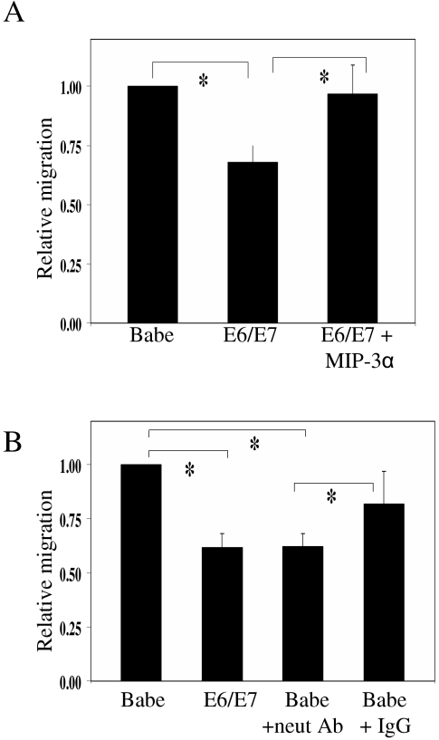
To determine the role of MIP-3α in the migratory response of LPLCs, MIP-3α was added to conditioned media from Poly(I) · Poly(C)-treated E6/E7-expressing keratinocytes. The amount of exogenous MIP-3α added to each migration assay was calculated from ELISAs carried out on the culture supernatants from Poly(I) · Poly(C)-treated control and E6/E7-expressing keratinocytes (Fig. 1B). The number of migrating LPLCs increased, following addition of exogenous MIP-3α to supernatant from E6/E7-expressing keratinocytes, to the level of LPLCs migrating in response to supernatant from control cells (Fig. 3A). To further examine the effect of MIP-3α on migrating LPLCs, neutralizing antibody to MIP-3α was added to supernatant from Poly(I) · Poly(C)-treated control cells. In similarity to the compensation assay, the amount of MIP-3α to be neutralized in the migration assay was calculated from ELISAs carried out on culture supernatants (Fig. 1B). The approximate amount of neutralizing antibody to be used was calculated using the Neutralization Dose50 provided on the technical data sheet. Use of the neutralizing antibody resulted in a reduction in the migration of LPLCs to the level of that seen with supernatant from E6/E7-expressing keratinocytes (Fig. 3B).
FIG. 3.
Effect of MIP-3α on migration of Langerhans precursor-like cells. Primary human foreskin keratinocytes stably expressing vector alone (Babe) or HPV-16 E6/E7 (E6/E7) were treated with Poly(I) · Poly(C). Following treatment, cells were washed with PBS and refed with minimal EpiLife for 4 h. Culture supernatants were collected and used in multiwell chamber assays. Values are expressed as migration of LPLCs in test samples relative to migration in response to supernatant from control cells (Babe). (A) Migration of LPLCs was increased upon addition of MIP-3α to E6/E7 supernatant. Results are expressed as the means ± SD of three experiments using three independent cell lines (* P < 0.003). (B) Migration of LPLCs was inhibited when MIP-3α-neutralizing antibody was added to control (Babe) supernatant. An equivalent concentration of goat immunoglobulin G (IgG) was added to duplicate wells as an internal control. Results are expressed as the means ± SD of three experiments using three independent cell lines (* P < 0.05).
Both E6 and E7 decrease MIP-3α mRNA and protein levels in Poly(I) · Poly(C)-treated keratinocytes.
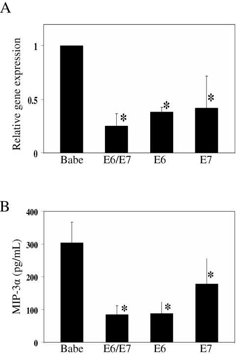
In order to investigate the individual contributions of E6 or E7 to the repression of MIP-3α, cell lines that expressed each oncoprotein were generated. Retroviral constructs containing stop codons in either E6 or E7 of HPV-16 were used to produce keratinocytes expressing E6 alone (E6/E7stop) and E7 alone (E6stop/E7). To determine the effects of E6 or E7 on the down-regulation of MIP-3α, control, E6/E7-, E6-, and E7-expressing cells were stimulated with Poly(I) · Poly(C) as described previously. Following treatment, cells were harvested for evaluation by real-time qPCR, and culture supernatants were collected for use in ELISA analysis. The results following treatment of three independent cell lines showed that Poly(I) · Poly(C)-treated E6/E7-expressing keratinocytes had the greatest repression (75%) in MIP-3α mRNA levels compared to control cells, followed by E6-expressing cells (62%) and E7-expressing cells (58%) (Fig. 4A). In similarity to the data shown in Fig. 1, the levels of secreted MIP-3α protein parallel the mRNA levels, with Poly(I) · Poly(C)-treated E6/E7-expressing keratinocytes secreting the least amount of MIP-3α (84 pg/ml) compared to control cells (304 pg/ml), followed by E6-expressing cells (88 pg/ml) and E7-expressing cells (178 pg/ml) (Fig. 4B).
FIG. 4.
Effect of E6 and E7 on MIP-3α mRNA and protein levels. Primary human foreskin keratinocytes stably expressing vector alone (Babe), HPV-16 E6/E7 (E6/E7), HPV-16 E6/E7stop (E6), or HPV-16 E6stop/E7 (E7) were treated with 100 μg/ml Poly(I) · Poly(C) for 16 h. Following treatment, cells were washed and refed with minimal EpiLife for 4 h. (A) RNA was harvested from the cells, and mRNA levels for MIP-3α were determined by real-time qPCR. Results are expressed as the means ± SD of three experiments using three independent cell lines (* P < 0.001). (B) Culture supernatants were harvested, and levels of secreted MIP-3α were measured by ELISA. Results are expressed as the means ± SD of three experiments using three independent cell lines (* P < 0.001).
Decreased MIP-3α expression also occurs in Poly(I) · Poly(C)-treated low-risk HPV types.
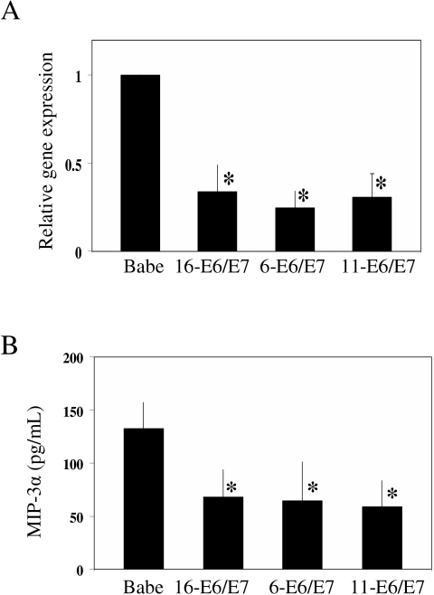
Although not prone to oncogenic transformation of the cells they infect, the capacity for low-risk HPVs to evade the host immune response would be beneficial to the persistence and propagation of the virus. To examine whether suppression of MIP-3α is unique to only the high-risk HPV types, cell lines were generated carrying HPV-16 E6/E7, HPV-6 E6/E7, or HPV-11 E6/E7. The cell lines and control keratinocytes were treated with Poly(I) · Poly(C) as before; then, cells and culture supernatants were harvested for qPCR and ELISA analysis. Real-time qPCR and ELISA results revealed similar reductions in MIP-3α mRNA levels among all three HPV types, as well as decreased production of MIP-3α protein, compared to control cells (Fig. 5A and 5B).
FIG. 5.
MIP-3α expression in Poly(I) · Poly(C)-treated control, HPV-16 E6/E7-, HPV-6 E6/E7-, and HPV-11 E6/E7-expressing keratinocytes measured by real-time qPCR and ELISA. Primary human foreskin keratinocytes stably expressing vector alone (Babe), HPV-16 E6/E7 (16-E6/E7), HPV-6 E6/E7 (6-E6/E7), or HPV-11 E6/E7 (11-E6/E7) were treated with 100 μg/ml Poly(I) · Poly(C) for 16 h. Following treatment, cells were washed and refed with minimal EpiLife for 4 h. (A) RNA was harvested from the cells, and mRNA levels for MIP-3α were determined by real-time qPCR. Results are expressed as the means ± SD of three experiments using two independent cell lines (* P < 0.001). (B) Culture supernatants were harvested and levels of secreted MIP-3α were measured by ELISA. Results are expressed as the mean ± SD of three experiments using two independent cell lines (* P < 0.001).
Results showing MIP-3α suppression in cells expressing E6 or E7 individually (Fig. 4A and 4B) suggest that both E6 and E7 contribute to the repression of MIP-3α; however, the mechanism underlying this inhibition is not known. For high-risk HPVs, their oncogenic potential has often been linked to the ability of E6 to bind and degrade p53 and of E7 to bind to retinoblastoma (Rb) and abrogate the functions of the Rb family members (27, 30, 52, 60). Therefore, one possible mechanism explaining the effect of HPV-16 E6 and E7 on MIP-3α expression would be through their interactions with p53 and Rb. However, E6 of HPV-6 and HPV-11 shows no binding to or degradation of p53 (59, 72), and E7 of HPV-6 and HPV-11, while binding to Rb, does so to a lesser extent than that seen with E7 of high-risk HPVs (24, 46). The ability of E6/E7 from high-risk and low-risk HPV types to repress MIP-3α transcription suggests that the inhibition of MIP-3α is through a mechanism independent of the effects of E6 and E7 on p53 and Rb.
NF-κB is required for MIP-3α expression in keratinocytes.
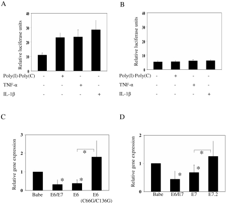
The proinflammatory cytokines TNF-α and IL-1β have been shown to induce MIP-3α through NF-κB activation (14, 22, 31, 36, 65). To determine whether MIP-3α expression in human keratinocytes requires NF-κB, reporter assays were performed using luciferase constructs containing either the full-length MIP-3α promoter or the full-length MIP-3α promoter containing substitution mutations in the NF-κB binding site. As shown in Fig. 6A and 6B, the mutated NF-κB binding site decreased basal MIP-3α activity as well as induction by Poly(I) · Poly(C), TNF-α, and IL-1β.
FIG. 6.
MIP-3α activity in keratinocytes requires NF-κB signaling. Keratinocytes (1 × 10⁵) were seeded in six-well dishes and transfected 24 h later with either 100 ng MIP-3αWT (A) or MIP-3αmNFκB (B) by use of Fugene 6. At 24 h after transfection, cells were washed with 1× PBS and treated with media alone or media containing 10 μg/ml Poly(I) · Poly(C), 10 ng/ml TNF-α, or 10 ng/ml IL-1β for 6 h. Following treatment, cells were harvested, lysed, and measured for luciferase activity. Results are expressed as the mean numbers of relative luciferase units ± SD of two independent experiments. (C and D) Primary human foreskin keratinocytes stably expressing vector alone (Babe), HPV-16 E6/E7 (E6/E7), HPV-16 E6/E7stop (E6), HPV-16 E6(C66G/C136G)/E7stop [E6(C66G/C136G)], HPV-16 E6stop/E7 (E7), or HPV-16 E6stop/E7.2 (E7.2) were treated with 100 μg/ml Poly(I) · Poly(C) for 16 h. Following treatment, cells were washed and refed with minimal EpiLife for 4 h. RNA was harvested from the cells, and mRNA levels for MIP-3α were determined by real-time qPCR. Results are expressed as the means ± SD of three experiments using two independent cell lines (* P < 0.01).
The central role of NF-κB in mediating the immune response has been clarified through the investigation of NF-κB activity in response to bacterial and viral infections as well as the regulation of numerous cytokines, chemokines, receptors, wound-healing factors, and cell adhesion molecules by NF-κB (17, 48, 58). Activation of transcription by NF-κB is regulated through the recruitment of coactivators such as CBP (CREB-binding protein), p300, and pCAF (p300/CBP-associated factor) (16, 53, 63). HPV-16 E6 has been shown to down-regulate NF-κB activation through its interaction with CBP/p300 (50). This effect of E6 is independent of its ability to degrade p53 (50), and this interaction between E6 and CBP/p300 results in down-regulation of promoters that required NF-κB, such as IL-8, another important chemoattractant of immune cells (28). Additionally, HPV-16 E7 has been shown to bind to pCAF as well as p300 (3, 6, 28) and inhibit its coactivator function.
To test whether the repressive effects of E6 and E7 on MIP-3α are occurring through similar mechanisms, we used a mutant of HPV-16 E6 (E6 C66G/C136G) that was shown previously not to bind to CBP/p300 (50) and a mutant of HPV-16 E7 (E7.2) that exhibits less binding to pCAF than wild-type HPV-16 E7 (28). In addition to control cells and E6/E7-expressing keratinocytes, cell lines were generated expressing E6 alone (E6/E7stop), E6 C66G/C136G (E6 C66G/C136G/E7stop), E7 alone (E6stop/E7), and E7.2 (E6stop/E7.2). Results following Poly(I) · Poly(C) treatment of two independent cells lines showed statistically significant repression of MIP-3α mRNA in the E6/E7- and E6-expressing keratinocytes compared to control cell results, but the MIP-3α mRNA level in the cell line expressing E6 C66G/C136G was close to the level of control cells (Fig. 6C). Similar results were observed using Poly(I) · Poly(C)-treated E6/E7-, E7-, and E7.2-expressing keratinocytes (Fig. 6D), suggesting that the interaction of HPV-16 E6 and E7 with CBP/p300 and pCAF contributes to the overall repression of MIP-3α production in keratinocytes. E6 and E7 of low-risk HPV types have also been found to bind p300 and pCAF (3, 6, 28, 50), thus suggesting that the interaction of E6 and E7 with these coactivators could be the mechanism underlying the repression of MIP-3α observed in HPV-16, HPV-6, and HPV-11 E6/E7-expressing cell lines.
DISCUSSION
Although highly correlated with the development of premalignant and malignant disease of the lower genital tract and oral cavity, infection with high-risk HPVs is not sufficient for the progression of disease. Many factors, including immune status, contribute to viral persistence and advancement to more-severe stages of disease. The role of the immune system in controlling HPV infection has been corroborated by studies showing increased prevalence and persistence of infections in individuals with suppressed immune function, such as transplant recipients and human immunodeficiency virus-positive individuals (62). The squamous epithelium that lines the reproductive tract, the oral cavity, and the skin provides the first line of defense against bacterial and viral pathogens. The initial innate immune response of the skin involves the release of cytokines, chemokines, antimicrobial molecules, and complement proteins by monocytes, macrophages, professional antigen-presenting cells, and keratinocytes (8, 34, 69). Keratinocytes play an active role in the innate immune response as cellular sources of cytokines in the epithelium (IL-1, IL-6, IL-7, IL-10, IL-12, IL-15, IL-18, IL-20) which not only influence the migration of immune cells but also stimulate the further release of cytokines by keratinocytes and immune cells (20). Also important in the initiation of the immune response are chemokines secreted by keratinocytes (IL-8, Gro-α/-β/-γ, IP-10, MCP-1) which are chemotactic for leukocytes, T lymphocytes, monocytes, eosinophils, and basophils (61).
Immune surveillance of the epithelium is maintained by immature Langerhans cells, which function as professional antigen-presenting cells and stimulate T lymphocytes following antigen uptake (5, 54). The expression of CCR6 by immature Langerhans cells, or Langerhans precursors, allows their selective migration in response to MIP-3α (19, 37, 40, 55), a potent chemokine released by inflamed epithelium (9, 47). However, in HPV lesions there is a reduction in the level of many of the immune cells needed to mount an effective immune response. Studies of cervical biopsies revealed that the number of Langerhans cells present in cervical tissue is decreased in low-grade and high-grade cervical intraepithelial neoplasias compared to normal tissue results (2, 11, 29, 71), which suggests that areas of HPV persistence also have decreased immune surveillance. The reduced levels of immune cells in lesions would allow persistence of the viruses and consequently the lesions for prolonged periods; this appears to be required for the eventual malignant conversion.
Results from this study demonstrate that expression of HPV-16 E6 and E7 in human keratinocytes down-regulates the level of MIP-3α mRNA and protein. The decreased MIP-3α production by E6/E7-expressing keratinocytes reduces the migration of immature Langerhans precursor-like cells. Interestingly, the E6/E7 proteins from low-risk HPV types 6 and 11 are also capable of modulating MIP-3α expression and this may account for the persistence of genital warts in infected individuals. This is the first instance where the E6 and E7 proteins from low-risk types exhibit a biological behavior that is similar in quality and quantity to that of E6/E7 from high-risk types.
However, E6 and E7 from HPV-16 appear to have an immune modulatory role that is more broad than the simple effects on MIP-3α, as indicated by the down-regulation of numerous other cytokines, chemokines, wound-healing factors, immune receptors, and adhesion molecules presented in this study (Table 1). Many of the immune factors identified in Table 1 bind to receptors found on a variety of immune cells and can, thus, direct the migration and influence the function of these cells. Immature dendritic cells migrate in response to RANTES, IL-8, and MIP-1α (4) during the innate immune response, and T-cell responses are regulated by IL-8 and I-CAM (62). The overall reduction in the expression of these and other proteins by E6/E7 may account for the low numbers of various immune cells in lesions caused by the virus and may further contribute to the ability of the virus to persist unobserved by the immune system. The correlation between HPV expression and a reduction in immune factors is substantiated by studies investigating clinical samples. Studies examining the immune environment of cervical intraepithelial neoplasia samples compared to normal tissue revealed a decrease in levels of HLA-DR, CD54, TNF-α, GM-CSF, and E-cadherin as well as an increase in IL-10, an immunosuppressive cytokine (18, 23, 29, 45). Additionally, the presence of HPV-16 E6 and E7 has been directly linked to the down-regulation of other immune mediators, such as IL-18, IL-8, and MCP-1 (10, 28, 35).
The activation of many immune response genes is mediated by NF-κB, a transcription factor that is a target of bacterial products, viral infections, and proinflammatory cytokines such as TNF-α and IL-1β and acts as a mediator in the expression of immune-related genes. Nearly two-thirds of the targets identified as being down-regulated in E6/E7-expressing keratinocytes (Table 1) are target genes of the NF-κB signaling cascade (17, 48). HPV-16 E6 and E7 have been shown to bind NF-κB coactivators, p300 and pCAF (3, 6, 28, 50). This interaction of E6 with p300 and E7 with pCAF resulted in decreased IL-8 promoter activity (28) and is implicated in the present study in the repression of MIP-3α transcription. Since E6 and E7 from both high- and low-risk viruses modulated MIP-3α production, the mechanisms may be shared between them. The ability of E6 and E7 of low-risk HPV types to also bind p300 and pCAF (3, 6, 28, 50) suggests that the interaction of E6 and E7 with these coactivators could be the mechanism underlying the repression of MIP-3α observed in HPV-16, HPV-6, and HPV-11 E6/E7-expressing cell lines.
In summary, the results reported here concentrated on MIP-3α, a potent chemoattractant for immature LC precursors, and showed that E6 and E7 from HPV-16 independently reduced transcription. The resulting decrease in MIP-3α production reduced the overall number of LPLCs migrating in response to this immune chemoattractant. Interestingly, the E6/E7 proteins from low-risk types also exhibited the same reduction in MIP-3α. The ability of E6 and E7 to lessen the production of immune mediators, such as MIP-3α, by infected cells would allow the virus to remain undetected and increase the probability of viral persistence and disease progression.
Acknowledgments
We thank H. McMurray, D. Patel, S. Dewhurst, and A. Livingstone for reviews of the manuscript. We also thank Helene McMurray for technical assistance and Andrew Keates for the gift of the MIP-3α promoter luciferase constructs.
This work was supported by grants NIAID RO1 AI30798 and NIDCR RO1 DE015935 (to D.J.M.) and NIAID PO1 AI48198. J.C.G. was supported by the United States Air Force.
The views expressed in this article are those of the authors and do not reflect the official policy or position of the United States Air Force, Department of Defense, or the U.S. government.
REFERENCES
- 1.Abiko, Y., M. Nishimura, K. Kusano, K. Nakashima, K. Okumura, T. Arakawa, T. Takuma, I. Mizoguchi, and T. Kaku. 2003. Expression of MIP-3α/CCL20, a macrophage inflammatory protein in oral squamous cell carcinoma. Arch. Oral Biol. 48:171-175. [DOI] [PubMed] [Google Scholar]
- 2.al-Saleh, W., P. Delvenne, J. E. Arrese, A. F. Nikkels, G. E. Pierard, and J. Boniver. 1995. Inverse modulation of intraepithelial Langerhans' cells and stromal macrophage/dendrocyte populations in human papillomavirus-associated squamous intraepithelial lesions of the cervix. Virchows Archiv. 427:41-48. [DOI] [PubMed] [Google Scholar]
- 3.Avvakumov, N., J. Torchia, and J. S. Mymryk. 2003. Interaction of the HPV E7 proteins with the pCAF acetyltransferase. Oncogene 22:3833-3841. [DOI] [PubMed] [Google Scholar]
- 4.Banchereau, J., F. Briere, C. Caux, J. Davoust, S. Lebecque, Y. J. Liu, B. Pulendran, and K. Palucka. 2000. Immunobiology of dendritic cells. Annu. Rev. Immunol. 18:767-811. [DOI] [PubMed] [Google Scholar]
- 5.Banchereau, J., and R. M. Steinman. 1998. Dendritic cells and the control of immunity. Nature 392:245-252. [DOI] [PubMed] [Google Scholar]
- 6.Bernat, A., N. Avvakumov, J. S. Mymryk, and L. Banks. 2003. Interaction between the HPV E7 oncoprotein and the transcriptional coactivator p300. Oncogene 22:7871-7881. [DOI] [PubMed] [Google Scholar]
- 7.Borkowski, T. A., J. J. Letterio, A. G. Farr, and M. C. Udey. 1996. A role for endogenous transforming growth factor beta 1 in Langerhans cell biology: the skin of transforming growth factor beta 1 null mice is devoid of epidermal Langerhans cells. J. Exp. Med. 184:2417-2422. [DOI] [PMC free article] [PubMed] [Google Scholar]
- 8.Bos, J. D. 1997. The skin as an organ of immunity. Clinical & Experimental Dermatology. 107:3-5. [PubMed] [Google Scholar]
- 9.Caux, C., S. Ait-Yahia, K. Chemin, O. de Bouteiller, M. C. Dieu-Nosjean, B. Homey, C. Massacrier, B. Vanbervliet, A. Zlotnik, and A. Vicari. 2000. Dendritic cell biology and regulation of dendritic cell trafficking by chemokines. Springer Seminars in Immunopathology. 22:345-369. [DOI] [PubMed] [Google Scholar]
- 10.Cho, Y. S., J. W. Kang, M. Cho, C. W. Cho, S. Lee, Y. K. Choe, Y. Kim, I. Choi, S. N. Park, S. Kim, C. A. Dinarello, and D. Y. Yoon. 2001. Down modulation of IL-18 expression by human papillomavirus type 16 E6 oncogene via binding to IL-18. FEBS Lett. 501:139-145. [DOI] [PubMed] [Google Scholar]
- 11.Connor, J. P., K. Ferrer, J. P. Kane, and J. M. Goldberg. 1999. Evaluation of Langerhans' cells in the cervical epithelium of women with cervical intraepithelial neoplasia. Gynecologic Oncology. 75:130-135. [DOI] [PubMed] [Google Scholar]
- 12.Dieu-Nosjean, M. C., C. Massacrier, B. Homey, B. Vanbervliet, J. J. Pin, A. Vicari, S. Lebecque, C. Dezutter-Dambuyant, D. Schmitt, A. Zlotnik, and C. Caux. 2000. Macrophage inflammatory protein 3alpha is expressed at inflamed epithelial surfaces and is the most potent chemokine known in attracting Langerhans cell precursors. J. Exp. Med. 192:705-718. [DOI] [PMC free article] [PubMed] [Google Scholar]
- 13.Dieu-Nosjean, M. C., C. Massacrier, B. Vanbervliet, W. H. Fridman, and C. Caux. 2001. IL-10 induces CCR6 expression during Langerhans cell development while IL-4 and IFN-gamma suppress it. J. Immunology. 167:5594-5602. [DOI] [PubMed] [Google Scholar]
- 14.Fujiie, S., K. Hieshima, D. Izawa, T. Nakayama, R. Fujisawa, H. Ohyanagi, and O. Yoshie. 2001. Proinflammatory cytokines induce liver and activation-regulated chemokine/macrophage inflammatory protein-3alpha/CCL20 in mucosal epithelial cells through NF-kappaB [correction of NK-kappaB]. Int. Immunol. 13:1255-1263. (Erratum, 13: 1443.) [DOI] [PubMed] [Google Scholar]
- 15.Geissmann, F., C. Prost, J. P. Monnet, M. Dy, N. Brousse, and O. Hermine. 1998. Transforming growth factor beta1, in the presence of granulocyte/macrophage colony-stimulating factor and interleukin 4, induces differentiation of human peripheral blood monocytes into dendritic Langerhans cells. J. Exp. Med. 187:961-966. [DOI] [PMC free article] [PubMed] [Google Scholar]
- 16.Gerritsen, M. E., A. J. Williams, A. S. Neish, S. Moore, Y. Shi, and T. Collins. 1997. CREB-binding protein/p300 are transcriptional coactivators of p65. Proc. Natl. Acad. Sci. USA 94:2927-2932. [DOI] [PMC free article] [PubMed] [Google Scholar]
- 17.Ghosh, S., M. J. May, and E. B. Kopp. 1998. NF-kappa B and Rel proteins: evolutionarily conserved mediators of immune responses. Annu. Rev. Immunol. 16:225-260. [DOI] [PubMed] [Google Scholar]
- 18.Giannini, S. L., W. Al-Saleh, H. Piron, N. Jacobs, J. Doyen, J. Boniver, and P. Delvenne. 1998. Cytokine expression in squamous intraepithelial lesions of the uterine cervix: implications for the generation of local immunosuppression. Clinical & Experimental Immunology. 113:183-189. [DOI] [PMC free article] [PubMed] [Google Scholar]
- 19.Greaves, D. R., W. Wang, D. J. Dairaghi, M. C. Dieu, B. Saint-Vis, K. Franz-Bacon, D. Rossi, C. Caux, T. McClanahan, S. Gordon, A. Zlotnik, and T. J. Schall. 1997. CCR6, a CC chemokine receptor that interacts with macrophage inflammatory protein 3alpha and is highly expressed in human dendritic cells. J. Exp. Med. 186:837-844. [DOI] [PMC free article] [PubMed] [Google Scholar]
- 20.Grone, A. 2002. Keratinocytes and cytokines. Vet. Immunol. Immunopathol. 88:1-12. [DOI] [PubMed] [Google Scholar]
- 21.Guironnet, G., C. Dezutter-Dambuyant, C. Vincent, N. Bechetoille, D. Schmitt, and J. Peguet-Navarro. 2002. Antagonistic effects of IL-4 and TGF-beta1 on Langerhans cell-related antigen expression by human monocytes. J. Leukoc. Biol. 71:845-853. [PubMed] [Google Scholar]
- 22.Harant, H., S. A. Eldershaw, and I. J. Lindley. 2001. Human macrophage inflammatory protein-3alpha/CCL20/LARC/Exodus/SCYA20 is transcriptionally upregulated by tumor necrosis factor-alpha via a non-standard NF-kappaB site. FEBS Lett. 509:439-445. [DOI] [PubMed] [Google Scholar]
- 23.Havard, L., P. Delvenne, P. Frare, J. Boniver, and S. L. Giannini. 2002. Differential production of cytokines and activation of NF-kappaB in HPV-transformed keratinocytes. Virology 298:271-285. [DOI] [PubMed] [Google Scholar]
- 24.Heck, D. V., C. L. Yee, P. M. Howley, and K. Munger. 1992. Efficiency of binding the retinoblastoma protein correlates with the transforming capacity of the E7 oncoproteins of the human papillomaviruses. Proc. Natl. Acad. Sci. USA 89:4442-4446. [DOI] [PMC free article] [PubMed] [Google Scholar]
- 25.Holzmann, S., C. H. Tripp, M. Schmuth, K. Janke, F. Koch, S. Saeland, P. Stoitzner, and N. Romani. 2004. A model system using tape stripping for characterization of Langerhans cell-precursors in vivo. J. Investig. Dermatol. 122:1165-1174. [DOI] [PubMed] [Google Scholar]
- 26.Homey, B., M. C. Dieu-Nosjean, A. Wiesenborn, C. Massacrier, J. J. Pin, E. Oldham, D. Catron, M. E. Buchanan, A. Muller, R. DeWaal Malefyt, G. Deng, R. Orozco, T. Ruzicka, P. Lehmann, S. Lebecque, C. Caux, and A. Zlotnik. 2000. Up-regulation of macrophage inflammatory protein-3 alpha/CCL20 and CC chemokine receptor 6 in psoriasis. J. Immunol. 164:6621-6632. [DOI] [PubMed] [Google Scholar]
- 27.Huang, P. S., D. R. Patrick, G. Edwards, P. J. Goodhart, H. E. Huber, L. Miles, V. M. Garsky, A. Oliff, and D. C. Heimbrook. 1993. Protein domains governing interactions between E2F, the retinoblastoma gene product, and human papillomavirus type 16 E7 protein. Mol. Cell. Biol. 13:953-960. [DOI] [PMC free article] [PubMed] [Google Scholar]
- 28.Huang, S.-M., and D. J. McCance. 2002. Down regulation of the interleukin-8 promoter by human papillomavirus type 16 E6 and E7 through effects on CREB binding protein/p300 and P/CAF. J. Virol. 76:8710-8721. [DOI] [PMC free article] [PubMed] [Google Scholar]
- 29.Hubert, P., J. H. Caberg, C. Gilles, L. Bousarghin, E. Franzen-Detrooz, J. Boniver, and P. Delvenne. 2005. E-cadherin-dependent adhesion of dendritic and Langerhans cells to keratinocytes is defective in cervical human papillomavirus-associated (pre)neoplastic lesions. J. Pathol. 206:346-355. [DOI] [PubMed] [Google Scholar]
- 30.Huibregtse, J. M., M. Scheffner, and P. M. Howley. 1991. A cellular protein mediates association of p53 with the E6 oncoprotein of human papillomavirus types 16 or 18. EMBO J. 10:4129-4135. [DOI] [PMC free article] [PubMed] [Google Scholar]
- 31.Imaizumi, Y., S. Sugita, K. Yamamoto, D. Imanishi, T. Kohno, M. Tomonaga, and T. Matsuyama. 2002. Human T cell leukemia virus type-I Tax activates human macrophage inflammatory protein-3 alpha/CCL20 gene transcription via the NF-kappa B pathway. Int. Immunol. 14:147-155. [DOI] [PubMed] [Google Scholar]
- 32.Izadpanah, A., M. B. Dwinell, L. Eckmann, N. M. Varki, and M. F. Kagnoff. 2001. Regulated MIP-3alpha/CCL20 production by human intestinal epithelium: mechanism for modulating mucosal immunity. Am. J. Physiol. Gastrointest. Liver Physiol. 280:G710-G719. [DOI] [PubMed] [Google Scholar]
- 33.Jaksits, S., E. Kriehuber, A. S. Charbonnier, K. Rappersberger, G. Stingl, and D. Maurer. 1999. CD34+ cell-derived CD14+ precursor cells develop into Langerhans cells in a TGF-beta 1-dependent manner. J. Immunol. 163:4869-4877. [PubMed] [Google Scholar]
- 34.Janeway, C. A., Jr., and R. Medzhitov. 2002. Innate immune recognition. Annu. Rev. Immunol. 20:197-216. [DOI] [PubMed] [Google Scholar]
- 35.Kleine-Lowinski, K., J. G. Rheinwald, R. N. Fichorova, D. J. Anderson, J. Basile, K. Munger, C. M. Daly, F. Rosl, and B. J. Rollins. 2003. Selective suppression of monocyte chemoattractant protein-1 expression by human papillomavirus E6 and E7 oncoproteins in human cervical epithelial and epidermal cells. Int. J. Cancer 107:407-415. [DOI] [PubMed] [Google Scholar]
- 36.Kwon, J. H., S. Keates, S. Simeonidis, F. Grall, T. A. Libermann, and A. C. Keates. 2003. ESE-1, an enterocyte-specific Ets transcription factor, regulates MIP-3alpha gene expression in Caco-2 human colonic epithelial cells. J. Biol. Chem. 278:875-884. [DOI] [PubMed] [Google Scholar]
- 37.Larregina, A. T., A. E. Morelli, L. A. Spencer, A. J. Logar, S. C. Watkins, A. W. Thomson, and L. D. J. Falo. 2001. Dermal-resident CD14+ cells differentiate into Langerhans cells. Nat. Immunol. 2:1151-1158. [DOI] [PubMed] [Google Scholar]
- 38.Lebre, M. C., J. C. Antons, P. Kalinski, J. H. N. Schuitemaker, T. M. M. van Capel, M. L. Kapsenberg, and E. C. de Jong. 2003. Double-stranded RNA-exposed human keratinocytes promote Th1 responses by inducing a type-1 polarized phenotype in dendritic cells: role of keratinocyte-derived tumor necrosis factor α, type I interferons, and interleukin-18. J. Investig. Dermatol. 120:990-997. [DOI] [PubMed] [Google Scholar]
- 39.Li, J., G. W. Ireland, P. M. Farthing, and M. H. Thornhill. 1996. Epidermal and oral keratinocytes are induced to produce RANTES and IL-8 by cytokine stimulation. J. Investig. Dermatol. 106:661-666. [DOI] [PubMed] [Google Scholar]
- 40.Liao, F., R. L. Rabin, C. S. Smith, G. Sharma, T. B. Nutman, and J. M. Farber. 1999. CC-chemokine receptor 6 is expressed on diverse memory subsets of T cells and determines responsiveness to macrophage inflammatory protein 3α. J. Immunol. 162:186-194. [PubMed] [Google Scholar]
- 41.McMurray, H. R., and D. J. McCance. 2004. Degradation of p53, not telomerase activation, by E6 is required for bypass of crisis and immortalization by human papillomavirus type 16 E6/E7. J. Virol. 78:5698-5706. [DOI] [PMC free article] [PubMed] [Google Scholar]
- 42.McMurray, H. R., and D. J. McCance. 2003. Human papillomavirus type 16 E6 activates TERT gene transcription through induction of c-Myc and release of USF-mediated repression. J. Virol. 77:9852-9861. [DOI] [PMC free article] [PubMed] [Google Scholar]
- 43.Merad, M., M. G. Manz, H. Karsunky, A. Wagers, W. Peters, I. Charo, I. L. Weissman, J. G. Cyster, and E. G. Engleman. 2002. Langerhans cells renew in the skin throughout life under steady-state conditions.[see comment][erratum appears in Nat Immunol. 2003 Jan;4(1):92]. Nat. Immunol. 3:1135-1141. [DOI] [PMC free article] [PubMed] [Google Scholar]
- 44.Morgenstern, J. P., and H. Land. 1990. Advanced mammalian gene transfer: high titre retroviral vectors with multiple drug selection markers and a complementary helper-free packaging cell line. Nucleic Acids Res. 18:3587-3596. [DOI] [PMC free article] [PubMed] [Google Scholar]
- 45.Mota, F., N. Rayment, S. Chong, A. Singer, and B. Chain. 1999. The antigen-presenting environment in normal and human papillomavirus (HPV)-related premalignant cervical epithelium. Clin. Exp. Immunol. 116:33-40. [DOI] [PMC free article] [PubMed] [Google Scholar]
- 46.Munger, K., B. A. Werness, N. Dyson, W. C. Phelps, E. Harlow, and P. M. Howley. 1989. Complex formation of human papillomavirus E7 proteins with the retinoblastoma tumor suppressor gene product. EMBO J. 8:4099-4105. [DOI] [PMC free article] [PubMed] [Google Scholar]
- 47.Nakayama, T., R. Fujisawa, H. Yamada, T. Horikawa, H. Kawasaki, K. Hieshima, D. Izawa, S. Fujiie, T. Tezuka, and O. Yoshie. 2001. Inducible expression of a CC chemokine liver- and activation-regulated chemokine (LARC)/macrophage inflammatory protein (MIP)-3 alpha/CCL20 by epidermal keratinocytes and its role in atopic dermatitis. Int. Immunol. 13:95-103. [DOI] [PubMed] [Google Scholar]
- 48.Pahl, H. L. 1999. Activators and target genes of Rel/NF-kappaB transcription factors. Oncogene 18:6853-6866. [DOI] [PubMed] [Google Scholar]
- 49.Pasch, M. C., N. H. Van Den Bosch, M. R. Daha, J. D. Bos, and S. S. Asghar. 2000. Synthesis of complement components C3 and factor B in human keratinocytes is differentially regulated by cytokines. J. Investig. Dermatol. 114:78-82. [DOI] [PubMed] [Google Scholar]
- 50.Patel, D., S. M. Huang, L. A. Baglia, and D. J. McCance. 1999. The E6 protein of human papillomavirus type 16 binds to and inhibits co-activation by CBP and p300. EMBO J. 18:5061-5072. [DOI] [PMC free article] [PubMed] [Google Scholar]
- 51.Patel, D., A. Incassati, W. Wang, and D. J. McCance. 2004. Human papillomavirus type 16 E6 and E7 cause polyploidy in human keratinocytes and up-regulation of G2-M-phase proteins. Cancer Res. 64:1299-1306. [DOI] [PubMed] [Google Scholar]
- 52.Patrick, D. R., A. Oliff, and D. C. Heimbrook. 1994. Identification of a novel retinoblastoma gene product binding site on human papillomavirus type 16 E7 protein. J. Biol. Chem. 269:6842-6850. [PubMed] [Google Scholar]
- 53.Perkins, N. D., L. K. Felzien, J. C. Betts, K. Leung, D. H. Beach, and G. J. Nabel. 1997. Regulation of NF-kappaB by cyclin-dependent kinases associated with the p300 coactivator. Science 275:523-527. [DOI] [PubMed] [Google Scholar]
- 54.Pieri, L., L. Domenici, and P. Romagnoli. 2001. Langerhans cells differentiation: a three-act play. Ital. J. Anat. Embryol. 106:47-69. (In Italian.) [PubMed] [Google Scholar]
- 55.Power, C. A., D. J. Church, A. Meyer, S. Alouani, A. E. Proudfoot, I. Clark-Lewis, S. Sozzani, A. Mantovani, and T. N. Wells. 1997. Cloning and characterization of a specific receptor for the novel CC chemokine MIP-3alpha from lung dendritic cells. J. Exp. Med. 186:825-835. [DOI] [PMC free article] [PubMed] [Google Scholar]
- 56.Proost, P., A. Wuyts, and J. van Damme. 1996. The role of chemokines in inflammation. Int. J. Clin. Lab. Res. 26:211-223. [DOI] [PubMed] [Google Scholar]
- 57.Rossi, D. L., A. P. Vicari, K. Franz-Bacon, T. K. McClanahan, and A. Zlotnik. 1997. Identification through bioinformatics of two new macrophage proinflammatory human chemokines: MIP-3alpha and MIP-3beta. J. Immunol. 158:1033-1036. (Erratum, 163: 1091, 1999.) [PubMed] [Google Scholar]
- 58.Santoro, M. G., A. Rossi, and C. Amici. 2003. NF-kappaB and virus infection: who controls whom. EMBO J. 22:2552-2560. [DOI] [PMC free article] [PubMed] [Google Scholar]
- 59.Scheffner, M., K. Munger, J. M. Huibregtse, and P. M. Howley. 1992. Targeted degradation of the retinoblastoma protein by human papillomavirus E7-E6 fusion proteins. EMBO J. 11:2425-2431. [DOI] [PMC free article] [PubMed] [Google Scholar]
- 60.Scheffner, M., B. A. Werness, J. M. Huibregtse, A. J. Levine, and P. M. Howley. 1990. The E6 oncoprotein encoded by human papillomavirus types 16 and 18 promotes the degradation of p53. Cell 63:1129-1136. [DOI] [PubMed] [Google Scholar]
- 61.Schroder, J. M.1995. Cytokine networks in the skin. J. Investig. Dermatol. 105:20S-24S. [PubMed] [Google Scholar]
- 62.Scott, M., M. Nakagawa, and A. Moscicki. 2001. Cell-mediated immune response to human papillomavirus infection. Clin. Diagn. Lab. Immunol. 8:209-220. [DOI] [PMC free article] [PubMed] [Google Scholar]
- 63.Sheppard, K.-A., D. W. Rose, Z. K. Haque, R. Kurokawa, E. McInerney, S. Westin, D. Thanos, M. G. Rosenfeld, C. K. Glass, and T. Collins. 1999. Transcriptional activation by NF-κB requires multiple coactivators. Mol. Cell. Biol. 19:6367-6378. [DOI] [PMC free article] [PubMed] [Google Scholar]
- 64.Stark, G. R., I. M. Kerr, B. R. Williams, R. H. Silverman, and R. D. Schreiber. 1998. How cells respond to interferons. Annu. Rev. Biochem. 67:227-264. [DOI] [PubMed] [Google Scholar]
- 65.Sugita, S., T. Kohno, K. Yamamoto, Y. Imaizumi, H. Nakajima, T. Ishimaru, and T. Matsuyama. 2002. Induction of macrophage-inflammatory protein-3alpha gene expression by TNF-dependent NF-kappaB activation. J. Immunol. 168:5621-5628. [DOI] [PubMed] [Google Scholar]
- 66.Syme, R., and S. Gluck. 2001. Generation of dendritic cells: role of cytokines and potential clinical applications. Transfus. Apher. Sci. 24:117-124. [DOI] [PubMed] [Google Scholar]
- 67.Tohyama, M., Y. Shirakara, K. Yamasaki, K. Sayama, and K. Hashimoto. 2001. Differentiated keratinocytes are responsible for TNF-α regulated production of macrophage inflammatory protein 3α/CCL20, a potent chemokine for Langerhans cells. J. Dermatol. Sci. 27:130-139. [DOI] [PubMed] [Google Scholar]
- 68.Tsuboi, R., C. Sato, C. M. Shi, T. Nakamura, T. Sakurai, and H. Ogawa. 1994. Endothelin-1 acts as an autocrine growth factor for normal human keratinocytes. J. Cell. Physiol. 159:213-220. [DOI] [PubMed] [Google Scholar]
- 69.Uthaisangsook, S., N. K. Day, S. L. Bahna, R. A. Good, and S. Haraguchi. 2002. Innate immunity and its role against infections. Ann. Allergy Asthma Immunol. 88:253-264. [DOI] [PubMed] [Google Scholar]
- 70.Viac, J., I. Guerin-Reverchon, Y. Chardonnet, and A. Bremond. 1990. Langerhans cells and epithelial cell modifications in cervical intraepithelial neoplasia: correlation with human papillomavirus infection. Immunobiology 180:328-338. [DOI] [PubMed] [Google Scholar]
- 71.Wang, X., and B. Seed. 15. December 2003, posting date. A PCR primer bank for quantitative gene expression analysis. Nucleic Acids Res. 31:1-8. [Online.] [DOI] [PMC free article] [PubMed] [Google Scholar]
- 72.Werness, B. A., A. J. Levine, and P. M. Howley. 1990. Association of human papillomavirus types 16 and 18 E6 proteins with p53. Science 248:76-79. [DOI] [PubMed] [Google Scholar]
- 73.Yanagita, M., Y. Shimabukuro, T. Nozaki, N. Yoshimura, J. Watanabe, H. Koide, M. Terakura, T. Saho, M. Takedachi, M. H. Jang, H. Kiyono, and S. Murakami. 2002. IL-15 up-regulates iNOS expression and NO production by gingival epithelial cells. Biochem. Biophys. Res. Commun. 297:329-334. [DOI] [PubMed] [Google Scholar]


