Abstract
Field-collected mosquitoes of the two main malaria vectors in Africa, Anopheles gambiae sensu lato and Anopheles funestus, were screened for their midgut bacterial contents. The midgut from each blood-fed mosquito was screened with two different detection pathways, one culture independent and one culture dependent. Bacterial species determination was achieved by sequence analysis of 16S rRNA genes. Altogether, 16 species from 14 genera were identified, 8 by each method. Interestingly, several of the bacteria identified are related to bacteria known to be symbionts in other insects. One isolate, Nocardia corynebacterioides, is a relative of the symbiont found in the vector for Chagas' disease that has been proven useful as a paratransgenic bacterium. Another isolate is a novel species within the γ-proteobacteria that could not be phylogenetically placed within any of the known orders in the class but is close to a group of insect symbionts. Bacteria representing three intracellular genera were identified, among them the first identifications of Anaplasma species from mosquitoes and a new mosquito-Spiroplasma association. The isolates will be further investigated for their suitability for a paratransgenic Anopheles mosquito.
Malaria remains the parasitic disease that kills the most people in the world. Anopheles gambiae sensu lato and Anopheles funestus mosquitoes are the main vectors in Africa, where 90% of malaria-related deaths occur. An approach to stop malaria transmission is paratransgenics. In this approach, suitable symbiotic bacteria are genetically modified to produce an antiparasitic factor and then reintroduced into the insect gut, where they kill or inhibit the development of the parasites (4).
A few studies have been performed to investigate bacterial species in field-collected Anopheles mosquitoes, all using culturing techniques. Jadin et al. (22) found Pseudomonas sp. in the midgut of mosquitoes from the Democratic Republic of the Congo. Straif et al. (31) identified 20 different genera of midgut bacteria from A. gambiae sensu lato and A. funestus mosquitoes caught in Kenya and Mali. They identified Pantoea agglomerans (synonym Enterobacter agglomerans) as the most frequently isolated bacterium, apart from Escherichia coli (31). Gonzalez-Ceron et al. (14) isolated Enterobacter amnigenus, Enterobacter cloacae, Enterobacter sp., Serratia marcescens, and Serratia sp. from Anopheles albimanus mosquitoes caught in southern Mexico.
To identify bacterial candidates for a paratransgenic mosquito, we conducted a screen for uncultured and cultured midgut bacteria from wild-caught A. gambiae and A. funestus mosquitoes.
MATERIALS AND METHODS
Field site, mosquitoes, and dissections.
Indoor-resting, blood-fed female A. gambiae sensu lato and A. funestus mosquitoes were caught in Lwanda, 12 km east of Mbita Point Research and Training Centre, ICIPE, Suba district, Western Kenya. In total, 116 Anopheles mosquitoes were caught on eight different occasions (A2 to H2). Living mosquitoes were anesthetized with chloroform, the species were determined by morphology and PCR (A. gambiae sensu lato) (30a). The mosquitoes were dissected in a sterile hood. Individual midguts were mashed in 50 μl of sterile saline (0.9% NaCl); this suspension was later used for isolation of bacteria and cloning of the 16S rRNA gene from bacteria. Controls for the efficiency of sterilization were treated like the other samples.
Bacterial isolation and phenotypic characterization.
The midgut suspension was streaked on Luria-Bertani agar (LA) plates and incubated for 48 h at room temperature. All bacteria were restreaked and preserved as deep-stick cultures during transport to Sweden. The morphology of the bacteria was examined using visual investigation and a light microscope. Motility tests were performed using the hanging-drop technique and motility medium plates (1% nutrient broth, 5.3% gelatin, 0.3% agar, 0.1% KNO3, pH 7.2) that were incubated overnight at 30°C. Anaerobic growth was determined by incubating LA plates overnight at 30°C in bioMerieux GENbox anaer generators. Optimum growth temperatures were determined in LB by shaking at 160 rpm and spectrophotometric reading. The isolates were sent to the Culture Collection, University of Gothenburg (CCUG), for classical phenotyping; different types of analyses were used depending on the bacterial genus.
Amplification, cloning, and sequencing of 16S rRNA genes.
Chromosomal DNA from the remaining midgut suspension was prepared using a guanidine-thiocyanate method (21). PCRs were performed to amplify 1.3 to 1.5 kb of the 16S rRNA gene from all the DNA samples by using PCR beads (0.5-ml Ready-To-Go PCR beads; Amersham Pharmacia Biotech). As the forward primer, 8f (5′-AGAGTTTGATIITGGCTCAG-3′; I = inosine) was used, and as the reverse primer, 1401r (5′-CGGTGTGTACAAGACCC-3′) was used for clones from sampling occasions G2 and H2 and 1501r (5′-CGGITACCTTGTTAC GAC-3′) was used for all other samples. The PCR program was as follows: 94°C for 3 min, followed by 30 cycles of 94°C for 30 s, 58 to 48°C for 30 s (the temperature was decreased by 1°C every cycle for 10 cycles and then held at 48°C for 20 cycles), 72°C for 1 min, followed by a final extension step at 72°C for 20 min. To construct a gene library with the 16S rRNA genes amplified from the DNA preparation, the PCR products of the expected size were cloned into TOPO 2.1 vectors utilizing TA cloning (Invitrogen).
For 16S rRNA gene cloning of cultured isolates, templates were prepared by boiling a bacterial colony for 10 min in a Tris-EDTA buffer (20 mM Tris, 2 mM EDTA, 1% Triton). PCR with primers 8f and 1501r and cloning were performed as described above. The 16S rRNA gene inserts in the plasmids were sequenced at Macrogen, Korea, using M13 primers.
Sequence analysis.
For preliminary identifications, the 16S rRNA gene sequences were analyzed in BLASTn (http://www.ncbi.nlm.nih.gov/BLAST/) and the Ribosomal Database Project II (RDP II) (http://rdp.cme.msu.edu). Chimeric sequences were searched for using the Ribosomal Database Project II Chimera Check program (http://rdp8.cme.msu.edu/cgis/chimera.cgi). The ARB system (26) was used for phylogenetic analysis with the ssujun02 database (http://www.arb-home.de). The 16S rRNA gene sequences were imported into the database and aligned using the ARB tool Fast Aligner, and the alignment was then checked manually. The aligned sequences were inserted into the main tree using the parsimony insertion tool of ARB to show their approximate positions; these positions were verified using distance (neighbor joining) and parsimony (100 bootstrap replicates) analyses with default settings in ARB and the ARB filter corresponding to the respective class or phylum of bacteria.
RESULTS AND DISCUSSION
In this study, two different detection pathways were used to screen for bacteria in Anopheles mosquito midguts, one culture independent and one culture dependent. A total of 116 mosquitoes (91 A. gambiae sensu lato and 25 A. funestus mosquitoes) and 19 sterility controls were examined. Four bacterial species that were identified in both mosquito samples and sterility control samples and two chimeric clones, found by the RDP II Chimera Check program, were removed from the data set.
Sixteen species of bacteria were identified as habitants of Anopheles mosquito midguts. They represent 14 genera, 7 genera obtained using the culture-independent pathway (Table 1) and 7 other genera obtained with the culture-dependent pathway (Table 2). Since streaks on LA plates and DNA isolation were performed on each midgut, it is surprising that the PCR-based method did not retrieve the genera found with the culture-dependent method. One explanation might be that remnants from the midgut cells or human blood interfere with the PCR. Another explanation could be competition between the DNAs from different bacteria favoring the ones with higher abundance. All previous studies of midgut flora of Anopheles mosquitoes exclusively utilized cultivation methods for screening. By including a culture-independent method, we obtained a broader picture of the mosquito midgut flora. The first study describing identification of uncultured and cultured microbiota in mosquitoes, investigating wild-caught Culex quinquefasciatus, was recently published by Pidiyar et al. (27). Similar to our study, different bacteria were found with the culture-dependent and the culture-independent methods.
TABLE 1.
Phylogenetic affiliations of the uncultured bacteria based on 16S rRNA gene analysisa
| Cloneb | GenBank accession no. | Anopheles sp. | Closest relative according to BLASTn (% identity) | Closest relative(s) according to phylogenetic analysis |
|---|---|---|---|---|
| B2.3.17 | AY837725 | arabiensis | Acidovorax temperans AF078766c(99) | A. temperans AF078766, bromate-reducing bacterium AF442523 |
| B2.5.31 | AY837724 | arabiensis | Mycoplasma wenyoniid AF016546 (96) | M. wenyoniid AF016546 |
| B2.3.14 B2.13.13 | AY837726AY837727 | arabiensis | Stenotrophomonas maltophilia AB180661 (99) | S. maltophilia AJ131913, Pseudomonas hibiscicolae AB021405 |
| B2.15.35 | AY837728 | gambiae sensu stricto | Stenotrophomonas maltophilia AB180661 (99) | |
| B2.8.27 B2.18.23 | AY837729AY837730 | gambiae | Stenotrophomonas sp. strain AJ002814c(99) | S. maltophilia AJ131910 |
| D2.2.2-3f | AY837745-46 | funestus | Spiroplasma sp. strain AB048263 (99) | See Fig. 1Bg |
| D2.2.12-14 | AY837731-33 | funestus | Spiroplasma sp. strain AJ245996 (98) | See Fig. 1Bg |
| G2.9.23 | AY837734 | arabiensis | Paenibacillus sp. strain AY382189 (93) | Paenibacillus sp. strain AF290916 |
| G2.12.2, -25, -46 | AY837735, -36, -37 | arabiensis | Anaplasma ovis AF414870 (99) | See Fig. 1A |
| G2.12.7B, -31, -35 | AY837738, -39, -40 | arabiensis | Ehrlichia sp. strain Bom Pastor AF318023h (99-100) | See Fig. 1A |
| H2.26.2 | AY837741 | gambiae sensu stricto | Aeromonas hydrophila X87271 (99) | A. hydrophila X87271 |
| H2.26.11 | AY837742 | gambiae sensu stricto | Aeromonas sp. strain U88656 (99) | Aeromonas sp. strain AF099027, Aeromonas caviaei X60409, Aeromonas sp. strain U88656 |
| H2.26.29 | AY837743 | gambiae sensu stricto | Aeromonas sp. strain AF099027 (99) | Aeromonas sp. strain AF099027, A. caviaei X60409, Aeromonas sp. strain U88656 |
Sequence analyses are based on 1.4 to 1.5 kb, where nothing else is stated, and were performed in November 2004.
The mosquito label consists of two parts: first, the sampling occasion (A2 to H2), and second, a number in order of dissection. Clones retrieved from a mosquito have the same label as the mosquito plus an additional number to separate them.
Best hit with a sequence having a species name.
Synonym Eperythrozoon wenyonii.
Synonyms Xanthomonas maltophilia, Stenotrophomonas maltophilia.
Sequence analysis based on 800-bp sequence.
The clones group together with D2.2.12 in the phylogenetic tree (not shown).
Belongs to the clade of Ehrlichia species renamed Anaplasma (Dumler et al. [9] and PAUP analysis [not shown]).
Synonym Aeromonas hydrophila subsp. anaerogenes.
TABLE 2.
Phenotypic dataa and phylogenetic affiliations (based on 16S rRNA gene analysisb) of the cultured bacteria
| Isolatec/Anopheles sp. | GenBank accession no./CCUG no. | Growth temp interval (°C) (growth temp optimum [°C]/GT [min])d | Phylogenetic affiliation | Closest relative according to BLASTn (% identity) | Closest relative(s) according to phylogenetic analysis |
|---|---|---|---|---|---|
| B2.1B/arabiensis | AY837746/49717 | 10-40, (35/32) | Bacillaceae | Bacillus simplex AJ628747 (99) | Bacillus sp. strains AJ315059 and AJ315062 |
| E2.5/arabiensis | AY837747/49718 | 10-40, (35/20) | Vibrionaceae | Vibrio metschnikovii X74712 (98) | V. metschnikovii X74711 and X74712 |
| H2.1/arabiensis | AY837748/49520 | 15-45, (30/100) | γ-Proteobacteria | Serratia odorifera AJ233432 (91) | See Fig. 2A |
| H2.3/arabiensis | AY837749/49711 | 20-40, (40/104) | Nocardiaceae | Nocardia corynebacterioidese AF430066 (99) | See Fig. 3 |
| H2.5/arabiensis | AY837750/49712 | 10-40, (35/92) | Bacillales | Bacillus silvestris AJ006086 (99) | B. silvestris AJ006086 |
| H2.14/arabiensis | AY837751/49713 | 10-40, (35/28) | Enterobacteriaceae | Escherichia senegalensisAY217654f (98) | See Fig. 2B |
| H2.16B/arabiensis | AY837752/49715 | 20-40, (35/44) | Intrasporangiaceae | Janibacter limosus Y08539 (98) | Janibacter sp. strain AF170746 |
| H2.26/gambiae sensu stricto | AY837753/49716 | 10-35, (30/32) | Pseudomonadaceae | Pseudomonas putida AF447394 (99) | P. monteilii AF181576, P. mosselii AF072688 |
Additional data are available at http://www.ccug.se.
Sequence analyses are based on 1.4 to 1.5 kb and were performed in November 2004.
The mosquito label consists of two parts: first, the sampling occasion (A2 to H2), and second, a number in order of dissection. Isolates retrieved from a mosquito have the same label as the mosquito.
10°C was the lowest temperature examined. GT, generation time.
Synonyms Rhodococcus corynebacterioides and Nocardia corynebacteroides.
Best hit with sequence having a species name.
In the present study, bacteria were found in 15% of the mosquitoes. Few of the mosquitoes harbored more than one bacterial species, possibly reflecting internal competition among the bacteria, and only one species (Stenotrophomonas sp.) was found in more than one mosquito. We consider the number of mosquitoes in our study too small to be representative of field-caught Anopheles mosquitoes from this area; however, another study performed in Kenya showed similar results (31). The low prevalence may reflect one of two things, either that mosquitoes in nature do not harbor more bacteria or that the methods available for bacterial screening are not sufficient to obtain the whole picture of the mosquito midgut flora.
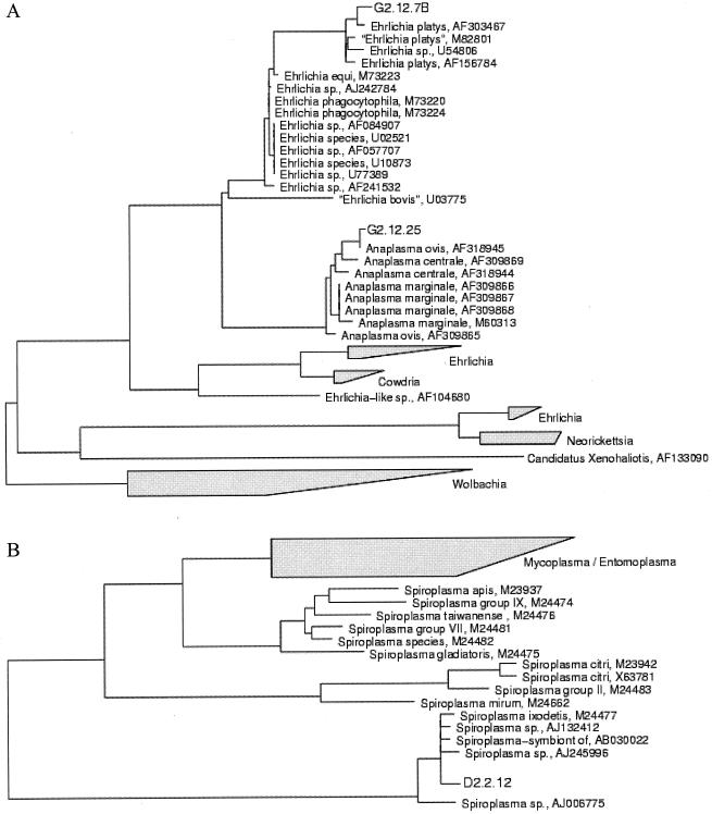
Interestingly, three genera of intracellular bacteria were identified by the culture-independent method in this study. The bacterial DNA clones from mosquito D2.2 represent the fifth Spiroplasma species found in mosquitoes. It does not group with any of the previously known mosquito spiroplasmas, Spiroplasma culicicola (19), Spiroplasma diminutum (34), Spiroplasma saubaudiense (1), or Spiroplasma taiwanense (2), in the phylogenetic analysis (Fig. 1B) and phylogenetic analysis using PAUP (not shown). S. taiwanense was previously demonstrated to be pathogenic to adult Aedes aegypti and Aedes stephensi mosquitoes (17) and A. aegypti larvae (16), and the potential role of mosquito spiroplasmas as vector control agents has been discussed (16-18). Several species of Spiroplasma are male-killing bacteria (3, 20, 23, 25); however, this has not been shown for mosquito-Spiroplasma associations. Although Spiroplasma spp. most often are considered pathogens (33), they have also been reported to be symbionts in some insects (12, 13). Two different Anaplasma species were identified (Fig. 1A), making this the first report of Anaplasma in mosquitoes. The genus Anaplasma, which is a sister taxon to Wolbachia (Fig. 1A), contains several tick-borne species that are pathogenic to ruminants, including Anaplasma ovis, a sheep and goat pathogen (5, 24). In 1994, Anaplasma phagocytophilum (synonym Ehrlichia phagocytophila) (Fig. 1A) was described as a human pathogen for the first time (15). By 2004, over 600 cases had been reported in the United States and 19 in Europe (26). We cannot exclude the possibility that the bacterial DNA recovered from Anaplasma in this study was present in the blood ingested by the mosquito, since these bacteria live intracellularly in blood cells. The vectorial capacity of mosquitoes for Anaplasma remains to be investigated. Clone B2.5.31, Mycoplasma wenyonii (synonym Eperythrozoon wenyonii) (Table 1), is a close relative of Mycoplasma suis (synonym Eperythrozoon suis) that can be mechanically transmitted between pigs by A. aegypti mosquitoes, according to Prullage et al. (28).
FIG. 1.
Phylogenetic dendrograms constructed in ARB based on 16S rRNA gene sequences (1,350 to 1,500 bp). (A) Bacterial DNA clones from mosquito G2.12. The clones belong to the α-proteobacteria class, and their positions within the genus Anaplasma are shown. The Ehrlichia spp. in the upper clade have recently been renamed Anaplasma spp. (10). (B) Position of the bacterial DNA clone D2.2.12 within the genus Spiroplasma belonging to the class Mollicutes.
All of the bacteria isolated showed rod-shaped forms of various lengths; however, H2.16B changes shape from rods to cocci as it grows. Isolates H2.14 and E2.5 are facultative anaerobes, and the rest are obligate aerobes. All isolates except H2.3 and H2.16B are motile. The results from the phenotypic characterization performed at CCUG (http://www.ccug.se) correspond well with the phylogenetic results for the isolates.
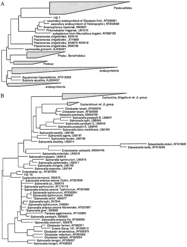
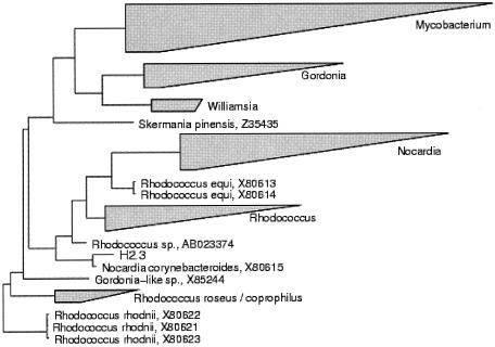
Our isolate designated H2.1 is a novel species within the γ-proteobacteria. Phylogenetically it is placed outside all families and orders within this class and close to a group of insect symbionts (Fig. 2A); among these are the Arsenophonus endosymbionts often found in whiteflies (Hemiptera: Aleyrodidae) (32). Further characterizations of isolate H2.1 is in progress. Isolate H2.14 could be identified only to the family level, Enterobacteriaceae (Fig. 2B) (http://www.ccug.se). The family Enterobacteriaceae contains species previously identified in mosquito midgut screens (6-8, 14, 27, 29-31) and, in addition, several species that have been described as insect symbionts (35). H2.5 is a close relative of Bacillus silvestris (AJ006066) (Table 2) according to the phylogenetic tree; however, RDP II analysis places H2.5 closest to Caryophanon sp. (AF385535). The phenotypic analysis identifies H2.5 as a Bacillus sp. (http://www.ccug.se). Several Bacillus spp. have previously been identified in mosquitoes. Straif et al. (31) found different Bacillus species in field-caught A. gambiae and A. funestus mosquitoes. Fouda et al. (11) concluded that Bacillus and Staphylococcus, isolated from the midguts of a laboratory colony of Culex pipiens mosquitoes and then reintroduced, were essential for high and normal fecundity. Our isolate H2.3, identified as Nocardia corynebacterioides (synonyms Rhodococcus corynebacterioides and Nocardia corynebacteroides), may be important as a candidate to test for paratransgenics, since it is a relative of Rhodococcus rhodnii (Fig. 3). The last is a true symbiont found in Rhodnius prolixus (Hemiptera: Reduviidae) and has been successfully used in a paratransgenic approach (4). This isolated bacterium was reintroduced into the Rhodnius gut and killed the trypanosome causing Chagas' disease after it had been transformed with a plasmid expressing a cecropin gene (10). Isolate H2.16B is the first reported bacterium of the family Intrasporangiaceae to be isolated from mosquitoes. Further analyses of this isolate revealed that it is a novel species within the genus Janibacter (P. Kämpfer, J. M. Lindh, O. Terenius, and I. Faye, unpublished data).
FIG. 2.
Phylogenetic dendrograms constructed in ARB based on 16S rRNA gene sequences (1,400 to 1,500 bp). (A) Position of bacterial isolate H2.1 within the γ-proteobacteria class. (B) Position of bacterial isolate H2.14 within Enterobacteriaceae. Groups marked “JL group” have been created to simplify the overview of the tree.
FIG. 3.
Phylogenetic dendrogram constructed in ARB based on 16S rRNA gene sequences (∼1,500 bp). Shown is the position of the bacterial isolate H2.3 within the genus Rhodococcus.
All of the bacterial isolates from this study will be further evaluated for their suitability as paratransgenic tools. A first step will be to study sustainability in mosquito midguts after reintroduction. A second step is to assess the immune response induced by the bacteria. The survival of a reintroduced bacterium will depend on its level of tolerance to the immune response mounted by the mosquito and putative antagonistic effects from other midgut bacteria. Several studies have shown that gram-negative midgut bacteria can suppress Plasmodium parasites (14, 29, 30), possibly by acting as elicitors of the mosquito immune response affecting Plasmodium development (30). Hence, an ideal bacterium for paratransgenics would be one that elicits a strong immune response that suppresses other bacteria and Plasmodium parasites but does not affect its own survival. In addition, genetic modification of this bacterium by introducing a gene expressing an antiparasitic molecule could achieve total clearance of Plasmodium parasites from the mosquito midgut. From this point of view, it is promising that several of the isolates are gram-negative γ-proteobacteria, for which there are means of genetic modification.
ADDENDUM IN PROOF
The cultured isolates H2.1 and H2.16B have now been further classified and named Thorsellia anophelis (P. Kämpfer, J. M. Lindh, O. Terenius, S. Hagdost, E. Falsen, H. J. Busse, and I. Faye, Int. J. Syst. Evol. Microbiol., in press) and Janibacter anophelis (P. Kämpfer, O. Terenius, J. M. Lindh, and I. Faye, Int. J. Syst. Evol. Microbiol., in press).
Acknowledgments
We thank Ulrike Fillinger, Hassan Akelo, Stephen Aluoch, Michael Kephers, and George Sonye for essential help with collecting mosquitoes. We also thank John Githure, Louis Gouagna, and Ahmed Hassanali for the opportunity to conduct the fieldwork at Mbita Point Research and Training Centre (ICIPE).
The research was supported by Sida/SAREC (I.F.), the Swedish Research Council (I.F.), the Royal Swedish Academy of Sciences (J.M.L.), and the Sven and Lilly Lawski foundation (O.T.).
REFERENCES
- 1.Abalain-Colloc, M. L., C. Chastel, J. G. Tully, J. M. Bove, R. F. Whitcomb, B. Gilot, and D. L. Williamson. 1987. Spiroplasma sabaudiense sp. nov. from mosquitos collected in France. Int. J. Syst. Bacteriol. 37:260-265. [Google Scholar]
- 2.Abalain-Colloc, M. L., L. Rosen, J. G. Tully, J. M. Bove, C. Chastel, and D. L. Williamson. 1988. Spiroplasma taiwanense sp. nov. from Culex tritaeniorhynchus mosquitos collected in Taiwan. Int. J. Syst. Bacteriol. 38:103-107. [DOI] [PubMed] [Google Scholar]
- 3.Anbutsu, H., and T. Fukatsu. 2003. Population dynamics of male-killing and non-male-killing spiroplasmas in Drosophila melanogaster. Appl. Environ Microbiol. 69:1428-1434. [DOI] [PMC free article] [PubMed] [Google Scholar]
- 4.Beard, C., C. Cordon-Rosales, and R. V. Durvasula. 2002. Bacterial symbionts of the triatominae and their potential use in control of Chagas disease transmission. Annu. Rev. Entomol. 47:123-141. [DOI] [PubMed] [Google Scholar]
- 5.Bekker, C. P., S. de Vos, A. Taoufik, O. A. Sparagano, and F. Jongejan. 2002. Simultaneous detection of Anaplasma and Ehrlichia species in ruminants and detection of Ehrlichia ruminantium in Amblyomma variegatum ticks by reverse line blot hybridization. Vet. Microbiol. 89:223-238. [DOI] [PubMed] [Google Scholar]
- 6.Chao, J., and G. Wistreich. 1959. Microbial isolation from the midgut of Culex tarsalis Coquillett. J. Insect Pathol. 1:311-318. [Google Scholar]
- 7.Chao, J., and G. Wistreich. 1960. Microorganisms from the midgut of larval and adult Culex quinquefasciatus Say. J. Insect Pathol. 2:220-224. [Google Scholar]
- 8.Demaio, J., C. B. Pumpuni, M. Kent, and J. C. Beier. 1996. The midgut bacterial flora of wild Aedes triseriatus, Culex pipiens and Psorophora columbiae mosquitoes. Am. J. Trop. Med. Hyg. 54:219-223. [DOI] [PubMed] [Google Scholar]
- 9.Dumler, J. S., A. F. Barbet, C. P. J. Bekker, G. A. Dasch, G. H. Palmer, S. C. Ray, Y. Rikihisa, and F. R. Rurangirwa. 2001. Reorganization of genera in the families Rickettsiaceae and Anaplasmataceae in the order Rickettsiales: unification of some species of Ehrlichia with Anaplasma, Cowdria with Ehrlichia and Ehrlichia with Neorickettsia, descriptions of six new species combinations and designation of Ehrlichia equi and ‘HGE agent’ as subjective synonyms of Ehrlichia phagocytophila. Int. J. Syst. Evol. Microbiol. 51:2145-2165. [DOI] [PubMed] [Google Scholar]
- 10.Durvasula, R. V., A. Gumbs, A. Panackal, O. Kruglov, S. Aksoy, R. B. Merrifield, F. F. Richards, and C. B. Beard. 1997. Prevention of insect-borne disease: an approach using transgenic symbiotic bacteria. Proc. Natl. Acad. Sci. USA 94:3274-3278. [DOI] [PMC free article] [PubMed] [Google Scholar]
- 11.Fouda, M. A., M. I. Hassan, A. G. Al-Daly, and K. M. Hammad. 2001. Effect of midgut bacteria of Culex pipiens L. on digestion and reproduction. J. Egypt Soc Parasitol. 31:767-780. [PubMed] [Google Scholar]
- 12.Fukatsu, T., and N. Nikoh. 2000. Endosymbiotic microbiota of the bamboo pseudococcid Antonina crawii (Insecta: Homoptera). Appl. Environ. Microbiol. 66:643-650. [DOI] [PMC free article] [PubMed] [Google Scholar]
- 13.Fukatsu, T., T. Tsuchida, N. Nikoh, and R. Koga. 2001. Spiroplasma symbiont of the pea aphid, Acyrthosiphon pisum (Insecta: Homoptera). Appl. Environ. Microbiol. 67:1284-1291. [DOI] [PMC free article] [PubMed] [Google Scholar]
- 14.Gonzalez-Ceron, L., F. Santillan, M. H. Rodriguez, D. Mendez, and J. E. Hernandez-Avila. 2003. Bacteria in midguts of field-collected Anopheles albimanus block Plasmodium vivax sporogonic development. J. Med. Entomol. 40:371-374. [DOI] [PubMed] [Google Scholar]
- 15.Grzeszczuk, A., B. Puzanowska, H. Miegoc, and D. Prokopowicz. 2004. Incidence and prevalence of infection with Anaplasma phagocytophilum. Prospective study in healthy individuals exposed to ticks. Ann. Agric. Environ. Med. 11:155-157. [PubMed] [Google Scholar]
- 16.Humphery-Smith, I., O. Grulet, and C. Chastel. 1991. Pathogenicity of Spiroplasma taiwanense for larval Aedes aegypti mosquitoes. Med. Vet. Entomol. 5:229-232. [DOI] [PubMed] [Google Scholar]
- 17.Humphery-Smith, I., O. Grulet, F. Le Goff, and C. Chastel. 1991. Spiroplasma (Mollicutes: Spiroplasmataceae) pathogenic for Aedes aegypti and Anopheles stephensi (Diptera: Culicidae). J. Med. Entomol. 28:219-222. [DOI] [PubMed] [Google Scholar]
- 18.Humphery-Smith, I., O. Grulet, F. Legoff, P. Robaux, and C. Chastel. 1991. Mosquito spiroplasmas and their role in the fight against the major tropical diseases transmitted by mosquitos. Bull. Soc. Pathol. Exot. 84:693-696. [PubMed] [Google Scholar]
- 19.Hung, S. H. Y., T. A. Chen, R. F. Whitcomb, J. G. Tully, and Y. X. Chen. 1987. Spiroplasma culicicola sp. nov. from the salt marsh mosquito Aedes sollicitans. Int. J. Syst. Bacteriol. 37:365-370. [Google Scholar]
- 20.Hurst, G. D. D., J. H. G. von der Schulenburg, T. M. O. Majerus, D. Bertrand, I. A. Zakharov, J. Baungaard, W. Volkl, R. Stouthamer, and M. E. N. Majerus. 1999. Invasion of one insect species, Adalia bipunctata, by two different male-killing bacteria. Insect Mol. Biol. 8:133-139. [DOI] [PubMed] [Google Scholar]
- 21.Ibrahim, A., L. Norlander, A. Macellaro, and A. Sjostedt. 1997. Specific detection of Coxiella burnetii through partial amplification of 23S rDNA. Eur. J. Epidemiol. 13:329-334. [DOI] [PubMed] [Google Scholar]
- 22.Jadin, J., I. H. Vincke, A. Dunjic, J. P. Delville, M. Wery, J. Bafort, and M. Scheepers-Biva. 1966. Role of Pseudomonas in the sporogenesis of the hematozoon of malaria in the mosquito. Bull. Soc. Pathol. Exot. Filiales 59:514-525. [PubMed] [Google Scholar]
- 23.Jiggins, F. M., G. D. D. Hurst, C. D. Jiggins, J. H. G. Von der Schulenburg, and M. E. N. Majerus. 2000. The butterfly Danaus chrysippus is infected by a male-killing Spiroplasma bacterium. Parasitology 120:439-446. [DOI] [PubMed] [Google Scholar]
- 24.Lew, A. E., K. R. Gale, C. M. Minchin, V. Shkap, and D. T. de Waal. 2003. Phylogenetic analysis of the erythrocytic Anaplasma species based on 16S rDNA and GroEL (HSP60) sequences of A. marginale, A. centrale, and A. ovis and the specific detection of A. centrale vaccine strain. Vet. Microbiol. 92:145-160. [DOI] [PubMed] [Google Scholar]
- 25.Majerus, T. M. O., J. H. G. von der Schulenburg, M. E. N. Majerus, and G. D. D. Hurst. 1999. Molecular identification of a male-killing agent in the ladybird Harmonia axyridis (Pallas) (Coleoptera: Coccinellidae). Insect Mol. Biol. 8:551-555. [DOI] [PubMed] [Google Scholar]
- 26.Parola, P. 2004. Tick-borne rickettsial diseases: emerging risks in Europe. Comp. Immunol. Microbiol. Infect. Dis. 27:297-304. [DOI] [PubMed] [Google Scholar]
- 27.Pidiyar, V. J., K. Jangid, M. S. Patole, and Y. S. Shouche. 2004. Studies on cultured and uncultured microbiota of wild Culex quinquefasciatus mosquito midgut based on 16S ribosomal RNA gene analysis. Am. J. Trop. Med. Hyg. 70:597-603. [PubMed] [Google Scholar]
- 28.Prullage, J. B., R. E. Williams, and S. M. Gaafar. 1993. On the transmissibility of Eperythrozoon suis by Stomoxys calcitrans and Aedes aegypti. Vet. Parasitol. 50:125-135. [DOI] [PubMed] [Google Scholar]
- 29.Pumpuni, C. B., M. S. Beier, J. P. Nataro, L. D. Guers, and J. R. Davis. 1993. Plasmodium falciparum—Inhibition of sporogonic development in Anopheles stephensi by Gram-negative bacteria. Exp. Parasitol. 77:195-199. [DOI] [PubMed] [Google Scholar]
- 30.Pumpuni, C. B., J. Demaio, M. Kent, J. R. Davis, and J. C. Beier. 1996. Bacterial population dynamics in three anopheline species: the impact on Plasmodium sporogonic development. Am. J. Trop. Med. Hyg. 54:214-218. [DOI] [PubMed] [Google Scholar]
- 30a.Scott, J. A., W. G. Brogdon, and F. H. Collins. 1993. Identification of single specimens of Anopheles gambiae complex by polymerase chain reaction. Am. J. Trop. Med. Hyg. 49:520-529. [DOI] [PubMed] [Google Scholar]
- 31.Straif, S. C., C. N. Mbogo, A. M. Toure, E. D. Walker, M. Kaufman, Y. T. Toure, and J. C. Beier. 1998. Midgut bacteria in Anopheles gambiae and An. funestus (Diptera: Culicidae) from Kenya and Mali. J. Med. Entomol. 35:222-226. [DOI] [PubMed] [Google Scholar]
- 32.Thao, M. L., and P. Baumann. 2004. Evidence for multiple acquisition of Arsenophonus by whitefly species (Sternorrhyncha: Aleyrodidae). Curr. Microbiol. 48:140-144. [DOI] [PubMed] [Google Scholar]
- 33.Whitcomb, R. 1981. The biology of spiroplasmas. Annu. Rev. Entomol. 26:397-425. [Google Scholar]
- 34.Williamson, D. L., J. G. Tully, L. Rosen, D. L. Rose, R. F. Whitcomb, M. L. AbalainColloc, P. Carle, J. M. Bove, and H. Smyth. 1996. Spiroplasma diminutum sp. nov., from Culex annulus mosquitoes collected in Taiwan. Int. J. Syst. Bacteriol. 46:229-233. [DOI] [PubMed] [Google Scholar]
- 35.Zientz, E., F. J. Silva, and R. Gross. 2001. Genome interdependence in insect-bacterium symbioses. Genome Biol. 2:1032.1-1032.6. [DOI] [PMC free article] [PubMed] [Google Scholar]


