Abstract
The beta and gamma genera of papillomaviruses consist of epidermodysplasia verruciformis-related human papillomaviruses (HPVs) and phylogenetically related cutaneous HPVs. Here, we have developed a consensus primer PCR assay and reverse line blot typing system coupled thereto (referred to as beta and gamma cutaneous HPV PCR [BGC-PCR]) for detection and typing of 24 beta and gamma HPVs (HPV types 4, 5, 8, 9, 12, 14, 15, 17, 19, 20, 21, 22, 23, 24, 25, 36, 37, 38, 47, 48, 49, 50, 60, and 65). Because the HPV-specific PCR products are only 72 bp in size, the system is suitable for formalin-fixed, paraffin-embedded specimens and other samples in which the DNA is of suboptimal quality. This system was able to detect and type as little as 100 ag to 1 fg HPV DNA per reaction (depending on the HPV type) in a background of 100 ng human DNA without any cross-reactivity between the tested types. Beta and gamma HPVs were detected in DNA extracted from plucked eyebrow hairs of 31 of 34 renal transplant recipients. In addition, formalin-fixed, paraffin-embedded specimens from nonmelanoma skin tumors of renal transplant recipients (n = 25) and immunocompetent individuals (n = 15) scored BGC-PCR positive in 21 and 6 cases, respectively, with HPV type 5 (HPV5) and HPV8 being the predominant types. The data indicate that this method can be a valuable, user-friendly tool for the detection and typing of cutaneous HPV in clinical specimens and may have implications for future monitoring of vaccines or alternative treatment modalities for diseases caused by these cutaneous HPVs.
Human papillomaviruses (HPVs) are small DNA viruses that infect cells of epithelial origin and can be grouped into either mucosal or cutaneous HPV types based on tissue tropism. The mucosal types include those HPVs that infect mucosa of the genital and respiratory tracts. They are divided into low-risk and high-risk types according to their ability to induce benign lesions and cancer, respectively. Low-risk types such as HPV type 6 (HPV6) and HPV11 can cause genital warts (condylomata acuminata). High-risk types such as HPV16 and HPV18 are causally involved in carcinogenesis of the uterine cervix and other mucosa of anogenital and oropharyngeal sites(13).
The cutaneous HPV types are phylogenetically distant from the mucosal types, and they infect the external skin. A subset of cutaneous HPV types is known to cause benign cutaneous warts. There is also evidence for an association of cutaneous HPV types with skin cancer. This was demonstrated first in patients with epidermodysplasia verruciformis (EV), a rare hereditary disease characterized by disseminated, persistent, flat warts and macular lesions that arise during childhood (reviewed in reference 14). In the macular lesions, the following phylogenetically related HPV types have been detected: HPV types 5, 8, 9, 12, 14, 15, 17, and 19 to 25. Therefore, these HPV types are classified as EV HPVs. EV patients have a high risk of developing cutaneous squamous cell carcinoma (SCC) later in life. HPV DNA, particularly that of types 5 and 8, is found in nearly all of these SCCs, and these HPV types can thus be considered high-risk types (14).
Formerly, the EV HPVs and HPV types related to them (e.g., HPV types 36, 37, 38, 47, and 49) as well as the type 4-related HPVs (including HPV types 4, 48, 50, 60, and 65) were classified collectively as the supergroup B HPV types. However, according to the most updated papillomavirus classification, the EV HPVs and phylogenetically related types are grouped together into the beta papillomavirus genus, whereas the type 4-related HPVs belong to the gamma papillomavirus genus (5). Altogether, these HPVs make up the largest group of cutaneous HPV types.
Recent data suggest that beta and gamma HPVs might also be involved in the pathogenesis of nonmelanoma skin cancer (NMSC) of non-EV patients, particularly of immunosuppressed individuals (e.g., renal transplant recipients) (1, 2, 6, 8, 10, 12, 17). In addition, cutaneous HPV DNA of types 5, 36, and 38 was detected in up to 90% of psoriasis lesions, suggesting an etiological relationship with this disease as well (7, 11, 21). Studies on cutaneous HPVs are, however, hampered by the fact that these comprise a large and highly heterogeneous virus group. As a result, with previously published DNA detection systems, several separate PCR assays had to be performed in order to cover a broad spectrum of these HPV types (12, 16). Moreover, these tests amplify relatively large DNA fragments and are difficult to use on archival formalin-fixed, paraffin-embedded material, which is the most widely available material (2, 8, 12, 16).
An additional practical limitation of the currently available cutaneous HPV consensus PCR systems is that they only allow typing via sequencing of PCR products (2, 8, 12, 16). This is a very laborious process, especially when infections with multiple HPV types are to be expected, which calls for cloning of the PCR products prior to sequencing.
In order to overcome the above-mentioned limitations, we developed a novel consensus beta and gamma cutaneous HPV PCR (BGC-PCR) for the detection of beta and gamma HPVs. The reverse primers are biotinylated, which allows subsequent typing of the PCR products by reverse line blotting (RLB), as we described previously for typing of mucosal HPVs (19). The RLB system contains specific probes for 24 HPV types, i.e., HPV types 4, 5, 8, 9, 12, 14, 15, 17, 19, 20, 21, 22, 23, 24, 25, 36, 37, 38, 47, 48, 49, 50, 60, and 65. Because the generated PCR products are only 72 bp in size, the system is suitable for use on formalin-fixed, paraffin-embedded specimens or other clinical samples in which the DNA is partially fragmented.
MATERIALS AND METHODS
Cloned HPV genomes.
Cloned genomes of HPV types 5, 8, and 38 were kindly provided by E. M. DeVilliers (Deutsches Krebsforschungszentrum, Heidelberg, Germany); HPV types 9, 12, 14, 15, 17, 19, 20, 21, and 24 were kindly provided by G. Orth and M. Favre (Institut Pasteur, Paris, France). Chemically competent Escherichia coli cells (One-Shot competent cells; Invitrogen, Breda, The Netherlands) were transformed with either of these plasmids. Single colonies were picked and grown overnight in 3 ml LB medium with ampicillin selection. Confirmation of the authenticity and purity of the clones was done by restriction enzyme analysis following miniprep. To ensure high purity of the plasmid DNA, the preparations were treated with 0.4 mg/ml RNase at 37°C for 1 h, followed by purification using the High Pure PCR Template Purification kit (Roche, Mannheim, Germany) according to the manufacturer's protocol. Serially diluted plasmid DNAs ranging from 1 pg to 10 ag (the lowest amount being equivalent to approximately one plasmid copy) in a background of 100 ng human placental DNA were used in reconstruction experiments.
Clinical specimens. (i) DNA from plucked eyebrow hairs.
Five eyebrow hairs were collected from each of 34 renal transplant recipients. Plucked hairs were obtained from patients of the Department of Dermatology, Charité, University Hospital of Berlin, Berlin, Germany, after informed consent was given. DNA was extracted from the hairs using the QIAmp DNA Mini kit (QIAGEN, Hilden, Germany) according to the manufacturer's instructions. Briefly, hairs were lysed in 180 μl ATL buffer (QIAGEN) with 20 μl proteinase K at 56°C for 3 h. After a short centrifugation step, 200 μl AL buffer (QIAGEN) was added and proteinase K was heat inactivated at 70°C for 10 min, and finally, 200 μl absolute ethanol was added. DNA was isolated using columns and eluted in 200 μl AE buffer (QIAGEN). For each PCR experiment, 5 μl of this DNA solution was used. This protocol was approved by the local ethics committee.
(ii) Formalin-fixed, paraffin-embedded skin biopsy samples.
For the evaluation of HPV detection and typing on clinical specimens, we used formalin-fixed, paraffin-embedded biopsy specimens of nonmelanoma skin tumors from immunocompetent individuals (three biopsies of actinic keratosis [AK], four biopsies of basal cell carcinoma [BCC], and eight biopsies of SCC) and renal transplant recipients (5, 8, and 12 biopsies of AK, BCC, and SCC, respectively). These samples were collected at the Department of Dermatology, Hospital Germans Trias i Pujol, Badalona, Spain. Signed informed consent was obtained from the participating patients.
From each biopsy, a series of 5-μm sections was cut using the so-called sandwich method (20). Briefly, outer sections were hematoxylin-eosin stained for histology, whereas inner sections (in total, approximately 1 cm2 of tissue) were placed into a tube and used for PCR purposes. A total of 250 μl lysis solution (10 mM Tris-HCl [pH 7.5], 0.45% Tween 20, and 500 μg proteinase K [Roche, Mannheim, Germany]) was added, and incubation was performed overnight at 37°C. Subsequently, the samples were boiled and centrifuged for 1 min in a standard table-top centrifuge (10,000 rpm). After cooling, 10 μl of these crude samples was used for PCR.
BGC-PCR. (i) Primers.
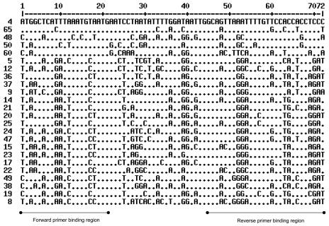
To ensure sensitive PCR amplification of a broad spectrum of, if not all, HPV genotypes from the beta and gamma genera, we used a mixture of six overlapping forward and eight overlapping reverse primers. The forward primer binding region comprises a conserved domain from nucleotide position 6539 to position 6559 of the HPV type 4 genome (EMBL accession number X70827) and the corresponding region within the genomes of other cutaneous beta and gamma HPV types. The reverse primer binding region is a conserved domain at nucleotide positions 6583 to 6610 of the HPV4 genome and the corresponding region of other cutaneous beta and gamma HPV types. Reverse primers are biotinylated to allow HPV typing of the PCR products by reverse line blotting (see below). An alignment of 24 common HPV types detected by this system and the positions of the primer binding regions are given in Fig. 1.
FIG. 1.
Alignment of the amplified region of the 24 supergroup B HPV types detected in the BGC-RLB assay. The conserved primer binding regions are indicated. The nonconserved region between the primer binding sites was used to select the RLB probes. Primer and probe sequences are available upon request.
(ii) BGC-PCR protocol.
The PCR mixtures of 50 μl contained 1.5 mM MgCl2, 200 μM of each deoxynucleoside triphosphate, 12.5 pmol of each primer, 1 unit AmpliTaq Gold DNA polymerase (Perkin-Elmer, Foster City, CA), and 10 μl of various concentrations of DNA or 10 μl of crude sample extract in Taq Gold buffer (Perkin-Elmer, Foster City, CA). Forty PCR cycles were performed using a PE 9700 thermocycler (Perkin-Elmer, Foster City, CA). The PCR was carried out under rather low-stringency annealing conditions at 38°C to allow a certain degree of mismatch acceptance. Each PCR was initiated by a preheating step for 6 min at 94°C followed by 40 cycles consisting of a denaturation step (20 seconds at 94°C), a primer-annealing step (30 seconds at 38°C), and an elongation step (80 seconds at 71°C). Ramping times were described previously (19). The final elongation step was extended for 4 min.
(iii) HPV RLB of BGC-PCR products.
HPV-specific 5′ amino-linked oligonucleotides (Isogen, Maarssen, The Netherlands) for RLB typing were selected for 24 beta and gamma HPV types (HPV types 4, 5, 8, 9, 12, 14, 15, 17, 19, 20, 21, 22, 23, 24, 25, 36, 37, 38, 47, 48, 49, 50, 60, and 65). Oligonucleotide binding regions are shown in Fig. 1. Selection was performed in such a way that the melting temperature was approximately 55°C and the length was approximately 20 bases. Moreover, selected oligonucleotide sequences were subjected to a database search (BLAST [http://www.ncbi.nlm.nih.gov/BLAST/]) and to multiple alignment with the sequences within the BGC-PCR region of cutaneous HPV types (http://prodes.toulouse.inra.fr/multalin/multalin.html) in order to verify their specificity. RLB analysis was performed essentially as described previously for mucosotropic HPV types (19). The system uses a miniblotter for spotting up to 42 different oligoprobes containing a 5′ amino group in parallel on a carboxyl-coated nylon membrane. Subsequently, up to 42 PCR products can be applied to the parallel channels of the miniblotter in such a way that the channels are perpendicular to the rows of oligoprobes deposited previously. In this format, hybridization was carried out, followed by incubation of the membrane with anti-biotin conjugate and enhanced chemiluminescence (ECL) detection (19).
Beta-globin and HPV TS-PCR.
Each sample was also subjected to a beta-globin PCR as described previously (4) to determine the quality of the DNA. All samples tested in this series were beta-globin PCR positive. A subset of the eyebrow hair DNA samples was subjected to HPV type-specific PCR (TS-PCR) for HPV types 5, 8, 12, 20, and/or 23 using a PCR mixture as described above but with 25 pmol/reaction of a given TS forward and reverse primer instead (sequences are available upon request). The TS-PCR protocol consisted of 6 min of preheating at 94°C followed by 40 cycles of 1 min at 94°C, 1 min at 60°C, and 1min at 72°C. TS-PCR products were analyzed on agarose gels, blotted, and hybridized with radioactive TS probes as described previously (18).
RESULTS
Development of the BGC-PCR. (i) Selection of PCR primers and RLB oligoprobes.
Based on two short regions encompassing a 72-bp DNA fragment in the L1 open reading frame that are highly conserved among beta and gamma HPVs, we selected a set of overlapping forward primers and a set of overlapping reverse primers of 21 and 28 nucleotides, respectively (Fig. 1). In combination, these would allow PCR amplification of DNA of multiple HPV types belonging to these genera under conditions of reduced stringency to allow some degree of primer/target mismatch acceptance. The primer regions were selected in such a way that the in-between region is sufficiently heterogeneous for HPV typing by RLB. The 24HPV types analyzed in this study had at maximum four mismatches with the best-matching forward primer and seven mismatches with the best-matching reverse primer. The numbers of mismatches with the best-matching primer within each forward and reverse primer panel, respectively, are as follows for the different HPV types: HPV4, 1 and 3; HPV5, 3 and 2; HPV8, 2 and 3; HPV9, 2 and 5; HPV12, 1 and 6; HPV14, 0 and 5; HPV15, 1 and 3; HPV17, 1 and 4; HPV19, 2 and 7; HPV20, 1 and 6; HPV21, 0 and 5; HPV22, 3 and 5; HPV23, 2 and 4; HPV24, 2 and 6; HPV25, 2 and 3; HPV36, 4 and 2; HPV37, 1 and 3; HPV38, 4 and 0; HPV47, 1 and 5; HPV48, 0 and 2; HPV49, 3 and 4; HPV50, 4 and 2; HPV60, 1 and 0; HPV65, 2 and 6.
All selected oligoprobes for RLB typing, except those for HPV14 and HPV15, had at least two mismatches with one or more other HPV genotypes for which the sequences in the primer binding regions are known. Thirteen of the oligoprobes had at least three mismatches with other HPV genotypes. The oligoprobe sequences of HPV14 and HPV15 showed one mismatch with the putative novel HPV types X34 and X26 (1), respectively.
(ii) Sensitivity and specificity of the assay.
Cloned genomes of various HPV types (i.e., HPV types 5, 8, 12, 14, 19, 20, 21, and 24 from the beta-1 group and HPV types 9, 15, 17, and 38 from the beta-2 group) that represent different degrees of mismatches with the PCR primers were selected for extensive reconstruction experiments. These included types having the maximum number of four mismatches with the best-matching forward primer (i.e., HPV38) or seven mismatches with the best-matching reverse primer (i.e., HPV19). The sensitivity of the BGC-PCR assay was determined by serial dilutions of the cloned HPV genomes in 100 ng human placental DNA per reaction.
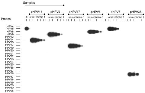
By RLB analysis of PCR products derived from these dilution series, the analytical sensitivity of the assay was found to be between 100 ag/reaction (equivalent to approximately 10HPV genome copies/reaction) and 10 fg/reaction (1,000 copies/ reaction) (Table 1). A representative experiment is shown in Fig. 2 for HPV types for which a sensitivity of 10 (HPV types 9, 14, and 17), 100 (HPV types 5 and 8), or 1,000 (HPV38) HPV genome copies/reaction was reached. Together, the tested cloned HPV genomes have mismatch distributions relative to the primers that are similar to, or less favorable than, those of the other HPV types that are detected by the assay. Therefore, we anticipate that the analytical sensitivity for the other types will also be in the range from 100 ag to 10 fg per reaction.
TABLE 1.
Analytical sensitivity of the BGC-PCR for different common cutaneous HPV types
| HPV type | Analytical sensitivity of BGC-PCRa |
|---|---|
| 5 | 100 |
| 8 | 100 |
| 9 | 10 |
| 12 | 10 |
| 14 | 10 |
| 15 | 100 |
| 17 | 10 |
| 19 | 10 |
| 20 | 10 |
| 21 | 100 |
| 24 | 10 |
| 38 | 1,000 |
Copies of cloned HPV genomes diluted in 100 ng human placental DNA.
FIG. 2.
Representative results of BGC-PCR on dilution series of genomes of cloned HPV types 5, 8, 9, 14, 17, and 38. The numbers (105, etc.) represent the approximate number of HPV copies used as input in the PCR. All reactions were done in a background of 100 ng human placental DNA. Abbreviation: p, positive control (1,000 copies of cloned HPV5 in 100 ng human placenta DNA).
RLB analysis of the PCR products obtained with the dilution series of these HPV types furthermore suggests that the assay is highly specific. None of the probes of the HPV types tested, including the HPV9 probe, which had only two mismatches with the HPV15 target, showed any cross-hybridization, not even when the input for the PCR was increased to 1ng of cloned DNA (108 copies) per reaction. Since the probes designed for most of the other HPV types have an equal number of or even more mismatches compared to other cutaneous types, cross-reactivity is very unlikely to occur.
Reproducibility of the assay.
Reproducibility was tested first by performing three additional tests with the dilution series of the cloned HPV genomes. The outcome of the tests with respect to sensitivity and specificity was the same every time.
Subsequently, reproducibility of the test was determined by analysis of DNA from plucked eyebrow hairs of kidney transplant recipients (n = 34). We chose this sample series because plucked eyebrow hairs were previously shown to commonly contain cutaneous beta and gamma HPVs (3).
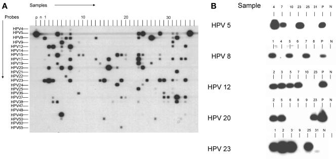
A representative result of the BGC-PCR on DNA of plucked eyebrow hairs is shown in Fig. 3A. The assay was done in triplicate on these samples. Results of the triplicate assay are summarized in Table 2. In 31 of these samples, 1 or more of 21 HPV types were detected in one or more of the assays. Only three samples remained negative throughout all three assays. DNAs of HPV types 4, 47, and 60 were not found in this series. Whereas infections with multiple types were seen frequently, many signals from samples with multiple infections were weak and did not appear in all three assays. However, all signals that appeared relatively strong at RLB were reproducible. The most frequently observed HPV types were HPV23 (detected in triplicate in 11 samples), HPV15 (detected in seven samples in triplicate), and HPV20 (detected in six samples in triplicate). No signals for any HPV type were seen in association with signals for a certain other type, not even among the HPV types for which the probes have only two mismatches with other types in the assay (e.g., HPV5 with HPV36, HPV9 with HPV15 and HPV23, HPV14 with HPV20, HPV20 with HPV14, HPV36 with HPV5 and HPV49, and HPV49 with HPV36). This suggests again that cross-hybridization does not occur.
FIG. 3.
BGC-PCR (A) and TS-PCR for HPV types 5, 8, 12, 20, and 23 (B) on DNA isolated from plucked eyebrow hairs. The sample numbers correspond with the sample numbers listed in Table 2. Abbreviations: p, positive control (1,000 copies of the respective cloned HPV genome in 100 ng human placenta DNA); n, negative control (no DNA input in PCR).
TABLE 2.
Results of triplicate BGC-PCR RLB and HPV types 5, 8, 12, 20, and 23 type-specific PCR on eyebrow hair DNA samples of renal transplant recipients
| Sample no. | HPV type(s) detected by BGC-PCR RLB
|
|
|---|---|---|
| Detected in triplicate | Detected once/twice | |
| 1 | 8, 23 | 12, 17, 36 |
| 2 | 12, 15, 20, 23 | None |
| 3 | 12, 23, 37, 38 | 15 |
| 4 | 5, 15, 19, 38 | 9, 20 |
| 5 | 12, 14, 20, 49 | 5, 8, 9, 17, 50 |
| 6 | None | 8, 22, 37 |
| 7 | 5, 8, 12, 14, 17, 20, 23 | 4, 15, 38 |
| 8 | None | None |
| 9 | 15, 36 | 9, 14 |
| 10 | None | 14, 15, 17 |
| 11 | None | 9, 23 |
| 12 | None | None |
| 13 | None | 8, 9, 17, 21, 23, 38 |
| 14 | None | 14, 19, 23 |
| 15 | 23 | 14, 24, 50 |
| 16 | 17, 20 | 14, 15, 23 |
| 17 | None | 15, 38 |
| 18 | 15, 24 | 14, 20, 23, 48 |
| 19 | 15, 23 | 8, 37, 65 |
| 20 | 37 | 15, 17 |
| 21 | None | None |
| 22 | None | 14, 23 |
| 23 | 5, 15, 17, 19, 20, 25, 38 | 8, 14, 23 |
| 24 | 5, 15, 20, 24 | 14, 17, 19, 23 |
| 25 | 23 | 5, 19 |
| 26 | 8, 19 | 5, 9 |
| 27 | None | 15, 25, 36, 50 |
| 28 | 5, 23 | 14, 15, 17 |
| 29 | 23, 36, 37 | 38, 50 |
| 30 | None | 37, 38 |
| 31 | 23, 36 | 5, 8, 9, 12, 17, 19, 25, 36 |
| 32 | 8, 21, 23, 37 | 5, 14, 38 |
| 33 | None | 5, 14, 15, 17, 21, 23, 50 |
| 34 | 17, 37 | 5, 12, 23, 38, 48 |
To confirm specificity of the BGC-PCR, we performed TS-PCR followed by Southern blotting of PCR products and radioactive hybridization with TS oligonucleotides on a random set of eyebrow hair samples (Fig. 3B). All HPV types that were detected in triplicate by BGC-PCR were confirmed with TS-PCR, with the exception of one type, type 23, that was missed in sample 31. Types that were not detected by BGC-PCR were also not seen with TS-PCR. Sometimes, a type that had been detected in only two of the triplicate BGC-PCRs was detected by TS-PCR.
Detection of beta and gamma HPVs in formalin-fixed, paraffin-embedded specimens.
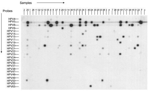
To evaluate the suitability of the BGC-PCR for use on formalin-fixed, paraffin-embedded specimens, 39 nonmelanoma skin tumors of renal transplant recipients and immunocompetent individuals were subjected to the test. Positivity for at least one HPV type was found in 27 of the samples (68%). Altogether, the HPV types detected in these skin lesions included the following 14 types: HPV types 4, 5, 8, 9, 12, 14, 15, 17, 19, 20, 21, 23, 50, and 65 (Fig. 4 and Table 3). Positivity was seen more frequently in biopsies from renal transplant recipients (21 of 25 [84%]) than in biopsies from immunocompetent individuals (6 of 15 [40%]). Moreover, 20 of the 25 (80%) samples from renal transplant recipients, compared to only 3 of the 15 (20%) samples from immunocompetent individuals, showed a relatively strong RLB signal, suggestive of relatively higher copy numbers. The most prevalent HPV types in the renal transplant recipients that gave relatively strong PCR signals were HPV5 and HPV8, which were both or individually identified in 8 of the 25 cases.
FIG. 4.
BGC-PCR RLB on DNA isolated from formalin-fixed tissue specimens of skin tumors of renal transplant recipients and immunocompetent individuals. The sample numbers correspond with the sample numbers listed in Table 3.
TABLE 3.
BGC-PCR on biopsy samples of skin lesions from immunocompetent individuals and renal transplant recipients
| Sample type and no. | Histology | HPV type(s)a |
|---|---|---|
| Immune competent | ||
| 1 | AK | (5) |
| 2 | AK | —b |
| 3 | AK | — |
| 4 | BCC | (4, 23) |
| 5 | BCC | 5 (8, 12, 20) |
| 6 | BCC | — |
| 7 | BCC | (65) |
| 8 | SCC | 8 (4) |
| 9 | SCC | (20) |
| 10 | SCC | 20 (5, 8) |
| 11 | SCC | — |
| 12 | SCC | — |
| 13 | SCC | — |
| 14 | SCC | — |
| 15 | SCC | — |
| Renal transplant recipients | ||
| 16 | AK | 8 |
| 17 | AK | 15 |
| 18 | AK | 17 |
| 19 | AK | 5, 8 (19, 23) |
| 20 | AK | 8 (15, 17) |
| 21 | BCC | 5 |
| 22 | BCC | 20 (8, 15) |
| 23 | BCC | 4, 5, 8 (19, 21) |
| 24 | BCC | 5 (65) |
| 25 | BCC | 5, 8 (17, 20, 23, 50) |
| 26 | BCC | — |
| 27 | BCC | — |
| 28 | BCC | — |
| 29 | SCC | 5, 9 |
| 30 | SCC | 8 |
| 31 | SCC | 21 |
| 32 | SCC | (8, 21) |
| 33 | SCC | 15 (23) |
| 34 | SCC | 20 (14) |
| 35 | SCC | 5 (12, 15, 20) |
| 36 | SCC | 5 (21, 50) |
| 37 | SCC | 5 (8) |
| 38 | SCC | 8 (15, 21) |
| 39 | SCC | 8 (20, 23) |
| 40 | SCC | — |
HPV types in parentheses revealed weak RLB signals.
—, HPV negative.
DISCUSSION
In this paper, we describe the BGC-PCR, a novel PCR-based detection and typing method for cutaneous beta and gamma HPV types. We found that the test can detect as little as 10 to 1,000 HPV DNA copies per reaction, depending on the HPV type. In addition, it appeared to be suitable for clinical samples in which the DNA is of a suboptimal quality, such as formalin-fixed, paraffin-embedded specimens. An additional major advantage is that this system allows high-throughput applications and greatly facilitates the detection of multiple infections.
Our finding that NMSC from renal transplant recipients more frequently contain HPV DNA than NMSC from immunocompetent individuals is in line with previous findings, as is the frequent detection of HPV5 and HPV8 in these samples (reviewed in reference 15). By contrast, plucked eyebrow hairs from renal transplant recipients showed a high rate of HPV type heterogeneity, which not only was detected by BGC-PCR but could be confirmed by type-specific PCR. This is also in agreement with results of a previous study (3). Therefore, the BGC-PCR seems sufficiently sensitive for the assessment of cutaneous HPV DNA in hairs and tissues for large epidemiological studies.
When the test was performed in triplicate, strong signals were always reproducible, but weak signals were sometimes present in one experiment and absent in the other. This suggests that the HPV types that give rise to these weak signals are present in copy numbers approaching the sensitivity limit of the test (i.e., between 10 and 1,000 copies per reaction). Viral load determinations have also shown that in human skin tumors, there may be as little as one cutaneous HPV DNA copy per 5,000 human cells, indicating that not every tumor cell contains HPV (14). Combined with the use of less-optimal, i.e., formalin-fixed, tissue, this may be an explanation for the weak RLB signals seen for some of the skin tumors analyzed in this study. Although this assumption needs to be tested further by type-specific quantitative PCR assays, our data suggest that duplicate or triplicate reactions are necessary to assess the predominant HPV type(s) in a specimen among multiple infections. Alternatively, the use of fewer PCR cycles may be an option to focus specifically on the predominant types. It should be noted that in the DNA isolated from eyebrow hairs, TS-PCR confirmed the presence of HPV types that were detected consistently by BGC-PCR, except for one sample. By contrast, samples that were consistently negative by BGC-PCR also did not give signals in type-specific PCR. These type-specific PCR experiments confirmed the specificity of the BGC-PCR test. The one HPV type that was missed by type-specific PCR probably reflects the high sensitivity of the short-fragment BGC-PCR compared to type-specific PCR.
As was our experience for the RLB assay developed for mucosotropic HPV types (19), the presence of two nucleotide mismatches of type-specific oligoprobes with corresponding sequences of other HPV types was already sufficient to prevent cross-reactivity. Given the fact that all except two probes (i.e., those of HPV14 and HPV15) had at least two mismatches with other HPV types, this ensures a high specificity of this typing assay. Whether the probes selected for HPV14 and HPV15, having only one mismatch with the sequences of the putative novel types X34 and X26, respectively, would show any cross-reactivity with these types remains to be determined. In a parallel study of patients with NMSC, type-specific PCR for HPV types 8, 14, 21, and 36 confirmed BGC-PCR typing data for these types in almost 90% of the cases (I. Nindl et al., unpublished data). This further supports the high specificity of the assay.
In recent studies, several other cutaneous HPV types, many of them putatively novel types, were identified by other consensus primer systems in skin lesions of immunocompromised and immunocompetent individuals (1, 2, 6, 8, 17). Therefore, future epidemiological studies may also include these types in a high-throughput detection and typing format. To a large extent, the assay we describe in the present paper would also be suitable for this purpose for two reasons. First, the region of the HPV genome that is amplified by our method maps within the amplified region of the previously published PCR systems that yielded, among others, many novel HPV sequences (1, 2, 6). Second, the alignment studies performed so far suggest that our primer combination would indeed allow amplification of most of these HPV types as well. We are currently expanding the number of probes of our RLB system for the detection of these additional types.
Other possibilities include the use of this PCR system as a tool for nested PCR strategies combined with other consensus PCR assays, such as the CP61/70 (1) and MY09/11 (9) systems, and the use of the RLB system as a typing system for other consensus PCR assays that overlap our target region in L1.
In conclusion, this new method (BGC-PCR) is applicable for large epidemiological studies that require high-throughput testing to analyze the role of these HPVs in human disease. Moreover, it may be valuable as a prescreen for subsequent type-specific viral load analysis, HPV expression, and/or in situ analysis. Whether it also has clinical applications, such as monitoring of the diversity of cutaneous HPV types in transplant recipients during immunosuppressive treatment or monitoring of antiviral therapy efficiency, needs to be investigated in future studies. Finally, this technique is the first technique to allow large-scale investigation of the association of cutaneous HPV types with NMSC within the same patient and between patients.
Acknowledgments
We are grateful to D. Boon, K. Benschop, and A. Schoenmaker for excellent technical assistance. We thank C. Ferrandiz for contributing to the sample collection.
REFERENCES
- 1.Berkhout, R. J., J. N. B. Bavinck, and J. ter Schegget. 2000. Persistence of human papillomavirus DNA in benign and (pre)malignant skin lesions from renal transplant recipients. J. Clin. Microbiol. 38:2087-2096. [DOI] [PMC free article] [PubMed] [Google Scholar]
- 2.Berkhout, R. J., L. M. Tieben, H. L. Smits, J. N. Bavinck, B. J. Vermeer, and J. ter Schegget. 1995. Nested PCR approach for detection and typing of epidermodysplasia verruciformis-associated human papillomavirus types in cutaneous cancers from renal transplant recipients. J. Clin. Microbiol. 33:690-695. [DOI] [PMC free article] [PubMed] [Google Scholar]
- 3.Boxman, I. L., R. J. Berkhout, L. H. Mulder, M. C. Wolkers, J. N. B. Bavinck, B. J. Vermeer, and J. ter Schegget. 1997. Detection of human papillomavirus DNA in plucked hairs from renal transplant recipients and healthy volunteers. J. Investig. Dermatol. 108:712-715. [DOI] [PubMed] [Google Scholar]
- 4.de Roda Husman, A. M., P. J. Snijders, H. V. Stel, A. J. van den Brule, C. J. Meijer, and J. M. Walboomers. 1995. Processing of long-stored archival cervical smears for human papillomavirus detection by the polymerase chain reaction. Br. J. Cancer 72:412-417. [DOI] [PMC free article] [PubMed] [Google Scholar]
- 5.de Villiers, E. M., C. Fauquet, T. R. Broker, H. U. Bernard, and H. zur Hausen. 2004. Classification of papillomaviruses. Virology 324:17-27. [DOI] [PubMed] [Google Scholar]
- 6.de Villiers, E. M., D. Lavergne, K. McLaren, and E. C. Benton. 1997. Prevailing papillomavirus types in non-melanoma carcinomas of the skin in renal allograft recipients. Int. J. Cancer 73:356-361. [DOI] [PubMed] [Google Scholar]
- 7.Favre, M., G. Orth, S. Majewski, S. Baloul, A. Pura, and S. Jablonska. 1998. Psoriasis: a possible reservoir for human papillomavirus type 5, the virus associated with skin carcinomas of epidermodysplasia verruciformis. J. Investig. Dermatol. 110:311-317. [DOI] [PubMed] [Google Scholar]
- 8.Forslund, O., A. Antonsson, P. Nordin, B. Stenquist, and B. G. Hansson. 1999. A broad range of human papillomavirus types detected with a general PCR method suitable for analysis of cutaneous tumours and normal skin. J. Gen. Virol. 80:2437-2443. [DOI] [PubMed] [Google Scholar]
- 9.Gravitt, P. E., C. L. Peyton, R. J. Apple, and C. M. Wheeler. 1998. Genotyping of 27 human papillomavirus types by using L1 consensus PCR products by a single-hybridization, reverse line blot detection method. J. Clin. Microbiol. 36:3020-3027. [DOI] [PMC free article] [PubMed] [Google Scholar]
- 10.Harwood, C. A., T. Surentheran, J. M. McGregor, P. J. Spink, I. M. Leigh, J. Breuer, and C. M. Proby. 2000. Human papillomavirus infection and non-melanoma skin cancer in immunosuppressed and immunocompetent individuals. J. Med. Virol. 61:289-297. [DOI] [PubMed] [Google Scholar]
- 11.Mahe, E., C. Bodemer, V. Descamps, I. Mahe, B. Crickx, Y. De Prost, and M. Favre. 2003. High frequency of detection of human papillomaviruses associated with epidermodysplasia verruciformis in children with psoriasis. Br. J. Dermatol. 149:819-825. [DOI] [PubMed] [Google Scholar]
- 12.Meyer, T., R. Arndt, E. Christophers, I. Nindl, and E. Stockfleth. 2001. Importance of human papillomaviruses for the development of skin cancer. Cancer Detect. Prev. 25:533-547. [PubMed] [Google Scholar]
- 13.Munoz, N., F. X. Bosch, S. de Sanjose, R. Herrero, X. Castellsague, K. V. Shah, P. J. Snijders, and C. J. Meijer. 2003. Epidemiologic classification of human papillomavirus types associated with cervical cancer. N. Engl. J. Med. 348:518-527. [DOI] [PubMed] [Google Scholar]
- 14.Pfister, H. 2003. Chapter 8: human papillomavirus and skin cancer. J. Natl. Cancer Inst. Monogr. 31:52-56. [DOI] [PubMed] [Google Scholar]
- 15.Pfister, H., and J. ter Schegget. 1997. Role of HPV in cutaneous premalignant and malignant tumors. Clin. Dermatol. 15:335-347. [DOI] [PubMed] [Google Scholar]
- 16.Shamanin, V., H. Delius, and E. M. de Villiers. 1994. Development of a broad spectrum PCR assay for papillomaviruses and its application in screening lung cancer biopsies. J. Gen. Virol. 75:1149-1156. [DOI] [PubMed] [Google Scholar]
- 17.Shamanin, V., M. Glover, C. Rausch, C. Proby, I. M. Leigh, H. zur Hausen, and E. M. de Villiers. 1994. Specific types of human papillomavirus found in benign proliferations and carcinomas of the skin in immunosuppressed patients. Cancer Res. 54:4610-4613. [PubMed] [Google Scholar]
- 18.van den Brule, A. J., C. J. Meijer, V. Bakels, P. Kenemans, and J. M. Walboomers. 1990. Rapid detection of human papillomavirus in cervical scrapes by combined general primer-mediated and type-specific polymerase chain reaction. J. Clin. Microbiol. 28:2739-2743. [DOI] [PMC free article] [PubMed] [Google Scholar]
- 19.van den Brule, A. J., R. Pol, N. Fransen-Daalmeijer, L. M. Schouls, C. J. Meijer, and P. J. Snijders. 2002. GP5+/6+ PCR followed by reverse line blot analysis enables rapid and high-throughput identification of human papillomavirus genotypes. J. Clin. Microbiol. 40:779-787. [DOI] [PMC free article] [PubMed] [Google Scholar]
- 20.Walboomers, J. M., M. V. Jacobs, M. M. Manos, F. X. Bosch, J. A. Kummer, K. V. Shah, P. J. Snijders, J. Peto, C. J. Meijer, and N. Munoz. 1999. Human papillomavirus is a necessary cause of invasive cervical cancer worldwide. J. Pathol. 189:12-19. [DOI] [PubMed] [Google Scholar]
- 21.Weissenborn, S. J., R. Hopfl, F. Weber, H. Smola, H. J. Pfister, and P. G. Fuchs. 1999. High prevalence of a variety of epidermodysplasia verruciformis-associated human papillomaviruses in psoriatic skin of patients treated or not treated with PUVA. J. Investig. Dermatol. 113:122-126. [DOI] [PubMed] [Google Scholar]


