

Abstract
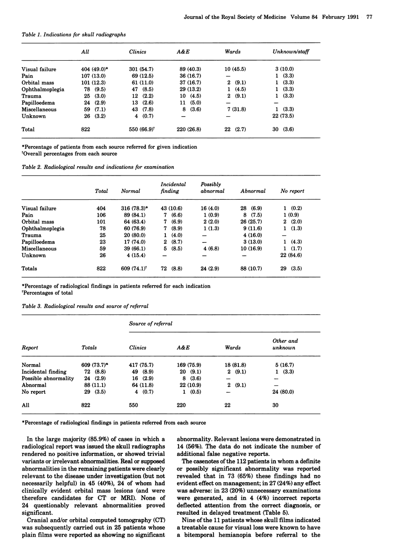
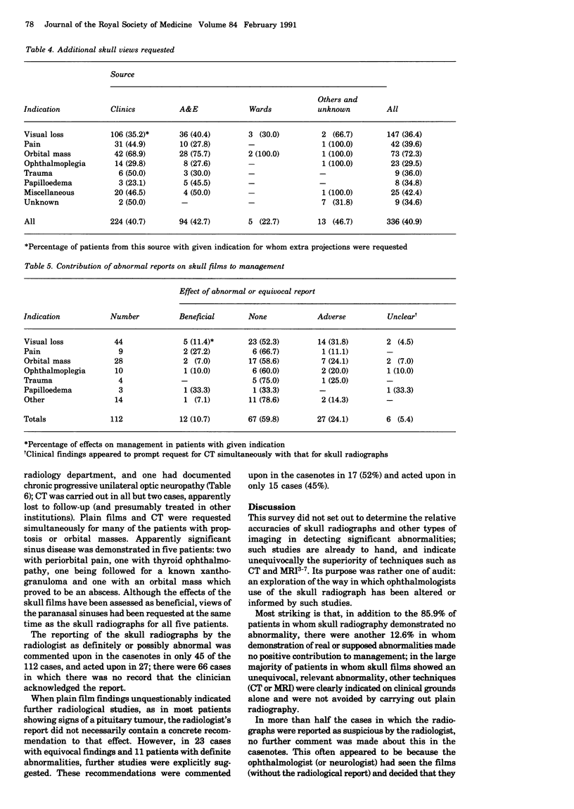
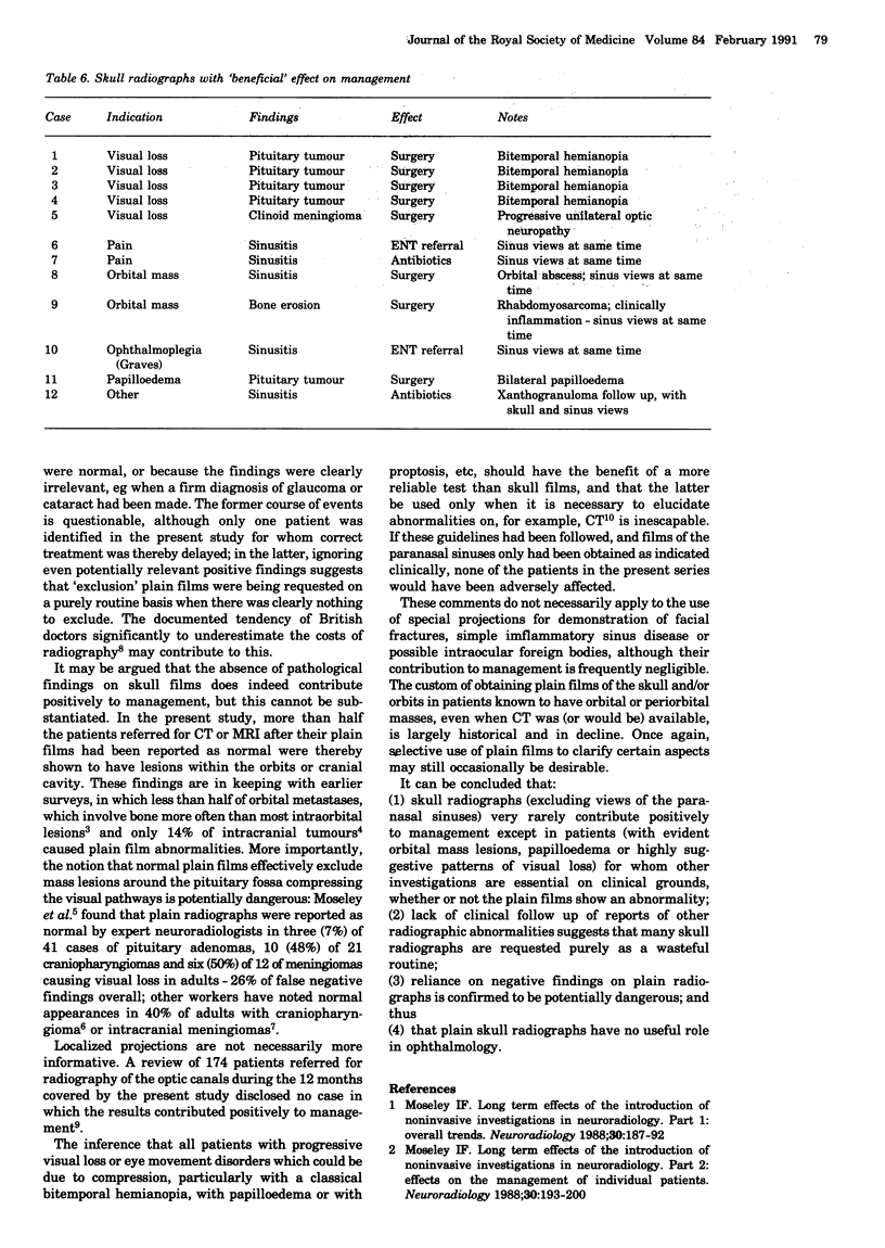
The indications for 822 consecutive referrals for skull radiography were prospectively studied in a large eye hospital over a one year period. In 85.9% of patients the results were normal, and in 89% of the remainder they had no positive effects on management; all patients in whom a 'beneficial' effect could be identified would have been more appropriately investigated by other means. Fourteen of 25 patients whose skull radiographs were normal were shown by computed tomography or magnetic resonance imaging to have orbital or intracranial lesions. Views of the optic canals, orbits or paranasal sinuses were also requested in 336 patients. With appropriate use of alternative imaging methods, no patient's treatment would have been adversely affected if none of the skull radiographs had been obtained.
Full text
PDF

Selected References
These references are in PubMed. This may not be the complete list of references from this article.
- Caillé J. M., Dautheribes M., Guibert-Tranier D., Piton J., Salamon R. The value of plain skull X-rays in the diagnosis of intracranial meningiomas. J Neuroradiol. 1981;8(1):13–20. [PubMed] [Google Scholar]
- Hesselink J. R., Davis K. R., Weber A. L., Davis J. M., Taveras J. M. Radiological evaluation of orbital metastases, with emphasis on computed tomography. Radiology. 1980 Nov;137(2):363–366. doi: 10.1148/radiology.137.2.7433669. [DOI] [PubMed] [Google Scholar]
- Hillemacher A. Der Wert anamnestischer und klinischer Daten sowie apparativer Untersuchungsbefunde bei der Diagnose von Hirntumoren. Fortschr Neurol Psychiatr. 1982 Apr;50(4):93–112. doi: 10.1055/s-2007-1002253. [DOI] [PubMed] [Google Scholar]
- Moseley I. F. Long term effects of the introduction of noninvasive investigations in neuroradiology. Part 1: Overall trends. Neuroradiology. 1988;30(3):187–192. doi: 10.1007/BF00341827. [DOI] [PubMed] [Google Scholar]
- Moseley I. Diagnostic value of 'optic foramen views': experience from an eye hospital. Br J Ophthalmol. 1990 Apr;74(4):235–237. doi: 10.1136/bjo.74.4.235. [DOI] [PMC free article] [PubMed] [Google Scholar]
- Moseley I. Long term effects of the introduction of noninvasive investigations in neuroradiology. Part 2: Effects on management of individual patients. Neuroradiology. 1988;30(3):193–200. doi: 10.1007/BF00341828. [DOI] [PubMed] [Google Scholar]
- Sorva R., Jäskinen J., Heiskanen O. Craniopharyngioma in children and adults. Correlations between radiological and clinical manifestations. Acta Neurochir (Wien) 1987;89(1-2):3–9. doi: 10.1007/BF01406660. [DOI] [PubMed] [Google Scholar]
- Tress B. M. The need for skull radiography in patients presenting for CT. Radiology. 1983 Jan;146(1):87–89. doi: 10.1148/radiology.146.1.6849072. [DOI] [PubMed] [Google Scholar]
- Wynick D., Jessop J. H. A survey of cost awareness among hospital medical staff. Health Trends. 1985 Feb;17(1):24–24. [PubMed] [Google Scholar]