

Abstract
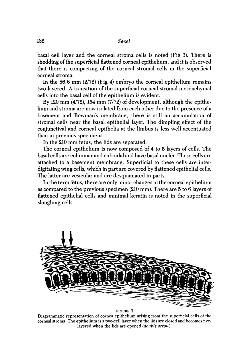
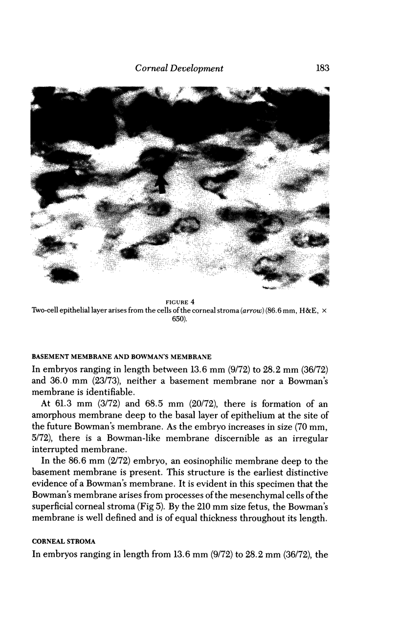
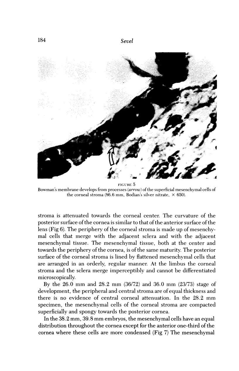
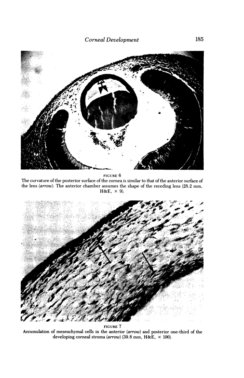
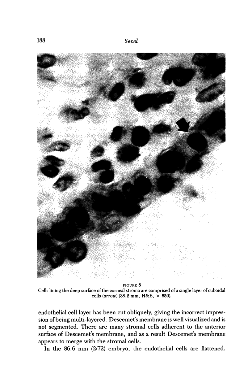
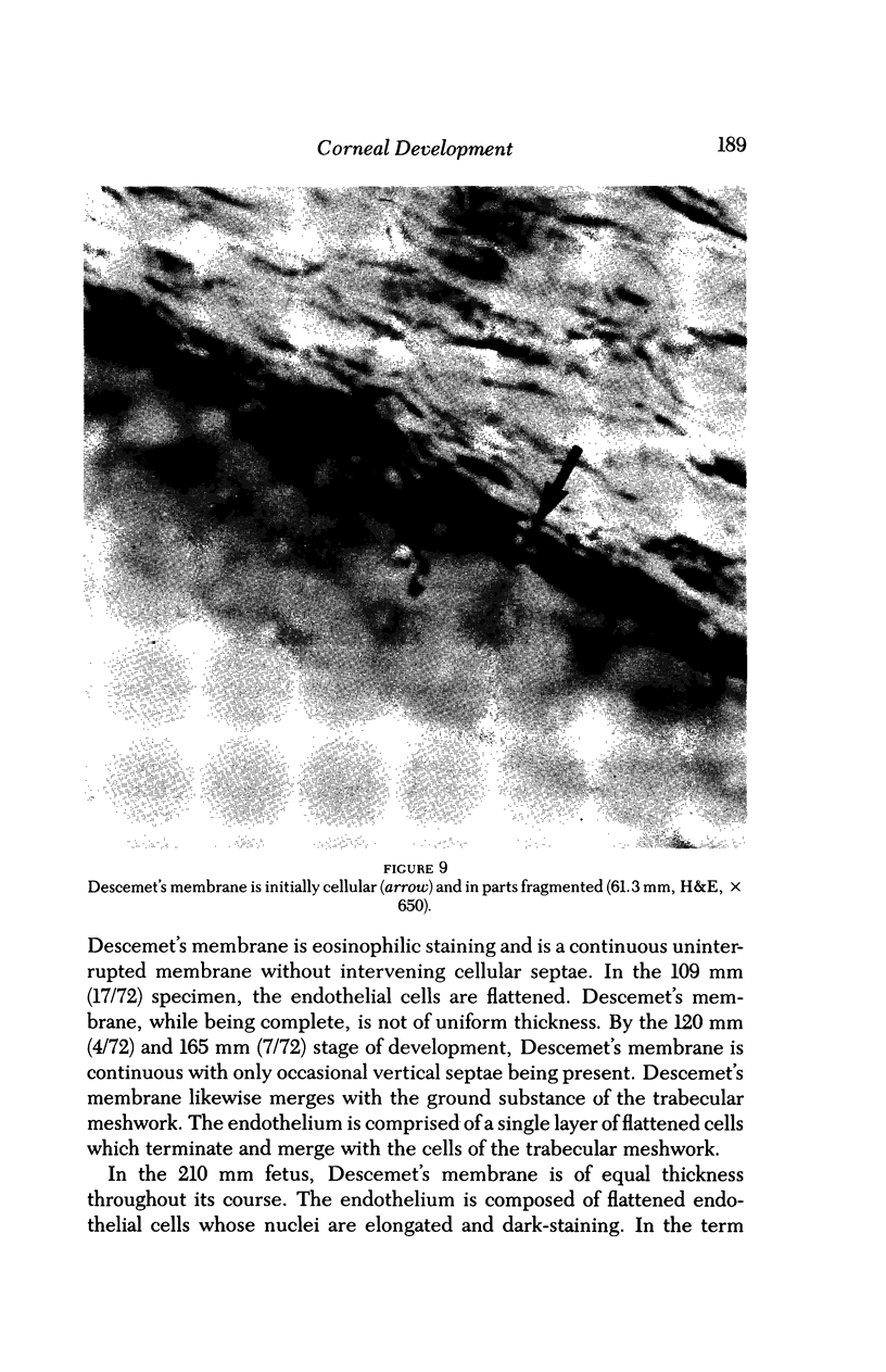
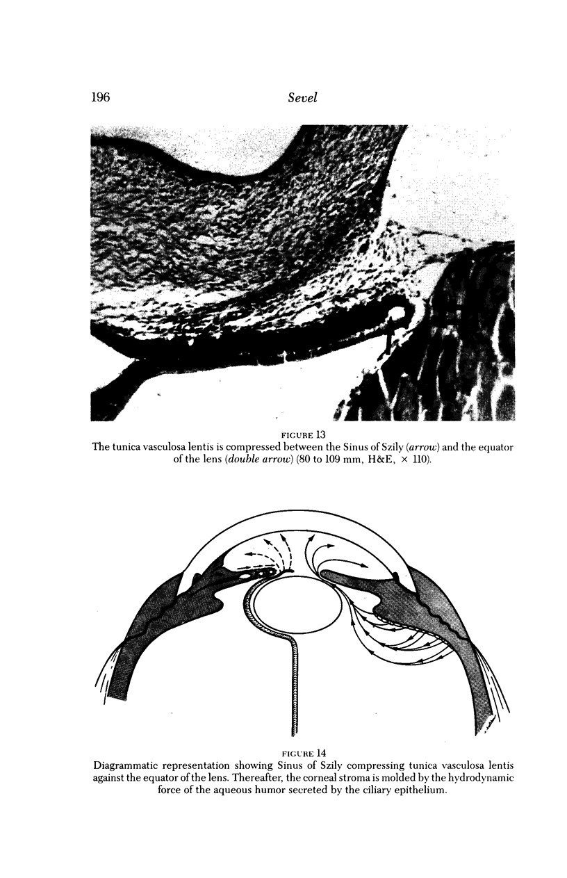
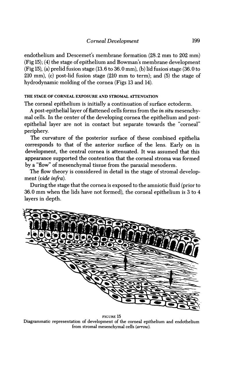
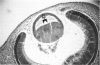
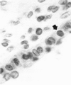
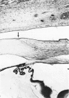
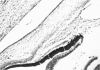
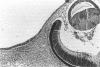
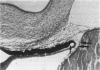
The corneal epithelium is initially a continuation of the surface ectoderm, but later on in development appears to arise from the superficial cells of the corneal stroma. The corneal epithelium varies in thickness depending on the status of the lids, viz either fused or open. When the lids are fused, the epithelium is only 2 to 3 layers in depth. When the lids are separated, a basement membrane is distinguishable and the epithelium is 4 to 5 layers in depth. Bowman's membrane develops from processes of the superficial mesenchymal cells of the stroma that become thickened and are arranged in the long axis of the corneal surface. The corneal stroma develops from in situ mesenchymal tissue and does not migrate from the limbal mesenchymal tissue. The attenuation of the central cornea, early in development, is due to the impingement of the lens against the developing cornea. The central constriction of the cornea has led previous observers to believe that the stroma migrates from the peripheral limbal area towards the center of the cornea. Descemet's membrane arises from processes of the deep mesenchymal cells of the corneal stroma. These processes thicken and become arranged in the long axis of the posterior surface of the cornea. The membrane is initially cellular, and well-defined septae are noted between the cells. With maturity, Descemet's membrane becomes a homogeneous structure. The endothelium is derived from the mesenchymal cells of the posterior stroma. These cells are initially cuboidal but then become flattened. During the early development of the cornea, the tunica vasculosa lentis may play an important role. It is suggested that the anterior chamber is maintained early on by a transudate from the vessels of the tunica vasculosa lentis. The vessels of the tunica vasculosa lentis are compressed by the vanguard of the optic cup against the equator of the lens. With regression of this vascular system, there is a simultaneous development of aqueous humor. The hydrodynamic force of aqueous production assists corneal molding by a vis a tergo affect. Pari passu with this hydrodynamic force, the corneal stromal fibers increase in length and width. The structures involved in aqueous humor production, viz the ciliary epithelium, and the aqueous humor drainage, viz the filtration angle, trabecular meshwork and aqueous veins, develop contemporaneously as the tunica vasculosa lentis regresses. The limiting and fixed stabilizing site is at the limbus, the site of insertion of the rectus muscles. At these sites a dimpling occurs as the cornea is enlarging.
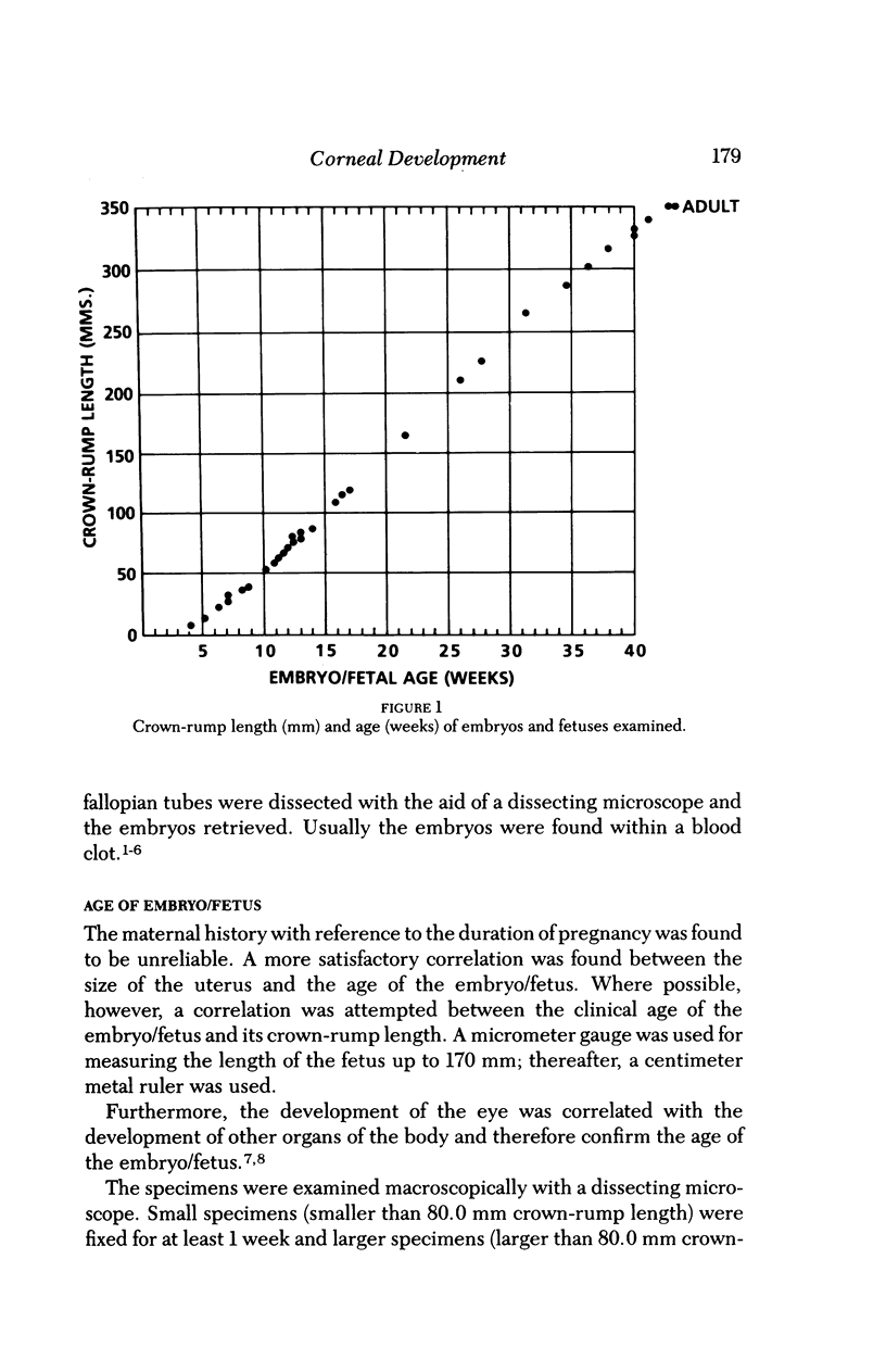
Full text
PDF

Images in this article
Selected References
These references are in PubMed. This may not be the complete list of references from this article.
- COULOMBRE A. J., COULOMBRE J. L. The role of intraocular pressure in the development of the chick eye. IV. Corneal curvature. AMA Arch Ophthalmol. 1958 Apr;59(4):502–506. doi: 10.1001/archopht.1958.00940050058005. [DOI] [PubMed] [Google Scholar]
- Hagedoorn A. THE EARLY DEVELOPMENT OF THE ENDOTHELIUM OF DESCEMET'S MEMBRANE, THE CORNEA AND THE ANTERIOR CHAMBER OF THE EYE. Br J Ophthalmol. 1928 Sep;12(9):479–495. doi: 10.1136/bjo.12.9.479. [DOI] [PMC free article] [PubMed] [Google Scholar]
- Hay E. D. Development of the vertebrate cornea. Int Rev Cytol. 1980;63:263–322. doi: 10.1016/s0074-7696(08)61760-x. [DOI] [PubMed] [Google Scholar]
- Sevel D. A reappraisal of the origin of human extraocular muscles. Ophthalmology. 1981 Dec;88(12):1330–1338. doi: 10.1016/s0161-6420(81)34856-8. [DOI] [PubMed] [Google Scholar]
- Waring G. O., 3rd, Bourne W. M., Edelhauser H. F., Kenyon K. R. The corneal endothelium. Normal and pathologic structure and function. Ophthalmology. 1982 Jun;89(6):531–590. [PubMed] [Google Scholar]