Abstract
The investigation of cell lineages and clonal organization in tissues is facilitated by techniques that allow labelling of clonal cell lineages. Here, we describe a novel transgenic mouse that allows clonal cell lineages to be traced in virtually any tissue. A green fluorescent cell lineage is generated by a random mutation at an enhanced green fluorescent protein gene that carries a premature stop codon, ensuring clonality. The transgenic system allows efficient detection of mutations and stem-cell fate mapping in the epidermis using live mice, as well as in the kidney and liver post-mortem. Cell lineages that descended from single epidermal stem cells were found to be capable of generating three adjacent corneocytes using the system, providing evidence for horizontal migration of epidermal cells between epidermal proliferative units (EPUs), in contrast to the classical EPU model. The transgenic mouse system is expected to provide a novel tool for stem-cell lineage studies.
Keywords: clonal cell lineage, enhanced green fluorescent protein, epidermal stem cell, in vivo imaging, mutation
Introduction
Stem cells are known to produce a variety of somatic cell types needed for periodic tissue renewal and tissue regeneration after injury (Van der Kooy & Weiss, 2000; Ro & Rannala, 2001). Recent research implies that stem cells may even produce cells of developmentally unrelated tissue types in experimentally modified conditions (Brazelton et al, 2000; Toma et al, 2001). Thus, it is important to determine how diverse a range of cell types and how expansive a clonal cell lineage is a single stem cell of a particular tissue able to produce in a mammalian system in vivo. To address these questions, a cell fate mapping technique is needed that allows one to label single cells in a tissue and subsequently trace their clonal cell lineages with minimal experimental perturbations. The clonality of a cell lineage generated by current cell fate mapping techniques, however, is usually not guaranteed (Clarke & Tickle, 1999; Flake, 2001). Methods using tritiated thymidine or bromodeoxyuridine simultaneously label large numbers of cells that are undergoing proliferation making it unlikely that a cell lineage generated by these methods will have originated from a single progenitor cell. Another limitation is that signals from labelled cells are diluted out after multiple cell divisions (Flake, 2001; Stern & Fraser, 2001). Mapping techniques using replication-deficient retroviruses that carry a marker gene can overcome the dilution problem. However, the retrovirus-mediated methods can only label cells that are undergoing proliferation. Also, labelling of single cells is not easily achieved, even when using a very low titre of viruses, hindering analysis of clonal cell lineages (Clarke & Tickle, 1999; Stern & Fraser, 2001). The mouse Dolichos biflorus 1 (Dlb-1) system relies on mutations to label individual cells and has been used successfully for mapping of clonal cell lineages (Winton et al, 1988), but the system can only be applied to the epithelium of the small intestine.
Here, we present a novel transgenic mouse system in which clonal cell lineages can be generated in any tissue. Single cells are labelled by means of random mutations that have very low frequency, and thus the clonality of a cell lineage generated in this system is virtually certain. The transgenic mouse that we have developed carries a premature stop-codon-containing enhanced green fluorescent protein (EGFP) gene as a target gene for mutations. A cell that has undergone a mutation at the premature stop codon and its descendant cell lineage will express EGFP; thus, a clonal cell lineage can be traced by following a green fluorescent colony. Cell fate mapping using the transgenic mouse (referred to as the stop-EGFP mouse) may offer advantages over other mapping techniques in revealing the potential of single stem cells to provide cells to replenish other cellular compartments, or to produce cells of diverse tissue types. Because detection of EGFP expression in individual cells requires no additional substrates and fluorescent imaging is by itself noninvasive, even repeated in vivo analyses of a specific clonal cell lineage over time are feasible.
Results And Discussion
The stop-EGFP gene and the wild-type EBFP gene
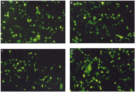
Okabe et al (1997) developed a ‘green mouse' in which the EGFP gene was ubiquitously expressed. We have obtained the plasmid used by Okabe et al and have introduced a premature stop codon (TAG) between the second and third codon position of the EGFP gene. This construct is subsequently referred to as the stop-EGFP gene. The neutrality of the position of the premature stop codon was verified experimentally by means of a cell transfection experiment (Fig 1). A mutation at any one of the three nucleotides of the stop codon will restore EGFP function except in the case of a mutation from G to A (i.e., GC → AT transition) at the third site, which produces another stop codon, TAA. To make up for the inability of our stop-EGFP construct to detect a GC → AT transition mutation at the stop codon, the wild-type enhanced blue fluorescent protein (EBFP) gene was used as another mutational target. The EBFP gene differs from the EGFP gene by only a single nucleotide (Heim et al, 1994); a mutation at the 196th nucleotide of the EBFP gene from C to T (i.e., GC → AT transition) generates the wild-type EGFP gene. A transgenic system carrying both the stop-EGFP gene and the wild-type EBFP gene can therefore detect all possible point mutations arising by either transitions (AT → GC and GC → AT) or transversions (AT → TA, AT → CG, GC → TA and GC → CG).
Figure 1.
Transient expression of revertant forms of the stop-EGFP gene in the NIH3T3 cell line. Three possible revertant forms of the stop-EGFP gene, each containing AAG (encoding lysine), TTG (encoding leucine) or TGG (encoding tryptophan) at the site of the premature stop codon, were generated (see supplementary information online) and used for the transfection experiment. All three revertants, AAG-revertant (A), TTG-revertant (B) and TGG-revertant (C), expressed functional proteins, which were as strongly fluorescent as the wild-type EGFP (D). The result strongly supports the neutrality of the position of the premature stop codon. Green fluorescence from cells transfected with the stop-EGFP gene or the wild-type EBFP gene was comparable with that from a negative control, nontransfected cells (data not shown).
Generation of stop-EGFP mice
Two transgenic founder mice carrying both the stop-EGFP gene and the wild-type EBFP gene have been generated (see supplementary Fig S1 online). Southern blot analysis shows that both transgenic lines carry more than 10 copies of the transgenes (see supplementary Fig S1 online). Expression of both the stop-EGFP gene and the EBFP gene was confirmed by reverse-transcription–PCR (see supplementary Fig S2 online).
In vivo detection of epidermal stem-cell clonal lineages
To investigate a clonal cell lineage that descended from a stem cell in the epidermis, we induced mutations in an 8-week-old stop-EGFP mouse using a potent mutagen, N-ethyl-N-nitrosourea (ENU), and performed in vivo imaging of the dorsal skin at 6 weeks post-ENU administration. The turnover rate of the epidermal tissue is estimated to be about 2 weeks (Potten, 1975); thus, one expects around three epidermal turnovers during the 6-week period, so that cell lineages that originated from nonstem-cell mutants (induced by the ENU exposure) can be expected to have disappeared after that period. Several studies suggest that an interval of 6 weeks after labelling is enough to eliminate epidermal cell lineages that originated from transit amplifying (TA) cells, allowing epidermal stem-cell lineages to be specifically detected (Schneider et al, 2003).
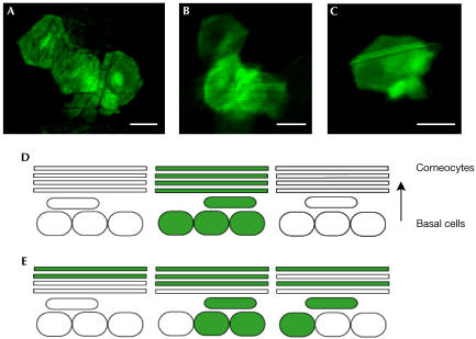
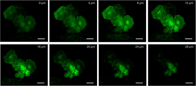
In vivo imaging of the dorsal epidermis of a stop-EGFP mouse at 6 weeks post-ENU administration revealed an epidermal stem-cell clonal lineage containing three adjacent corneocytes (Fig 2A). The hexagonal shape is characteristic of the morphology of a cornified cell (corneocyte) in the outermost layer of the epidermis. This finding is in contrast to the classical model for stem-cell clonal lineages in the mouse dorsal epidermis (Allen & Potten, 1974; Potten & Morris, 1988). The model predicts that around 10–11 basal cells (with a stem cell in the centre) are located beneath one column of corneocytes and the TA basal cells that descended from a central stem cell are thought to move directly upward to differentiate terminally into corneocytes (Fig 2D). These basal cells (i.e., 9–10 TA cells plus one stem cell), along with the stack of differentiated cells above them, are organized in a spatially distinct proliferative compartment, called an ‘epidermal proliferative unit' (EPU). According to this model, if a basal stem cell has undergone a mutation at the premature stop codon of the stop-EGFP gene, only one EPU (occupying the area of a single corneocyte on the skin surface) should become green fluorescent (Fig 2D). However, Fig 2A shows that a cell lineage that descended from a single epidermal stem cell can generate three adjacent corneocytes (i.e., epidermal cells belonging to three adjacent EPUs). The probability that the three corneocytes were generated from more than one stem cell, each having undergone independent mutation at the stop codon of the stop-EGFP gene, is virtually zero because of the low frequency of mutation, ensuring clonality. Thus, the finding suggests that a single stem cell is capable of providing epidermal cells for at least three adjacent EPUs by means of horizontal migration of either basal or suprabasal cells (Fig 2E). Longitudinal optical sectioning of the cell lineage (Fig 3) shows bright green fluorescent signals in the basal layer beneath the hexagonal cells. To demonstrate that the above finding is not restricted to young mice, mutations were also induced in a 7-month-old stop-EGFP mouse by treatment with ENU, and in vivo imaging of the dorsal skin was performed at 13 weeks post-ENU administration (i.e., after around 6.5 epidermal turnovers). This imaging experiment also revealed an epidermal stem-cell clonal lineage containing three neighbouring corneocytes (Fig 2B) with green fluorescent signals in the basal layer (data not shown). No green fluorescent epidermal cells were detected in the dorsal skin of four untreated stop-EGFP mice, suggesting that the green fluorescent cell lineages observed were generated in adult mice exposed to ENU rather than arising because of the spontaneous mutation.
Figure 2.
Green fluorescent mutant colonies in the dorsal epidermis of ENU-treated stop-EGFP mice. (A) In vivo imaging of the dorsal epidermis of a stop-EGFP mouse at 6 weeks post-ENU administration revealed an epidermal stem-cell clonal lineage containing three adjacent corneocytes. Bright fluorescent signals within the hexagonal cells are from a deeper layer (see Fig 3). (B) Mutations were induced in a 7-month-old stop-EGFP mouse by treatment with ENU and imaging of the dorsal skin was performed at 13 weeks post-ENU administration. The in vivo imaging experiment also revealed an epidermal stem-cell clonal lineage containing three adjacent corneocytes. (C) In vivo imaging of the dorsal epidermis of another stop-EGFP mouse at 18 days post-ENU administration revealed a green fluorescent mutant colony containing a single corneocyte on the skin surface. The cell lineage was not detected when the same area of the dorsal epidermis was imaged again at 6 weeks post-ENU administration. (D) Schematic illustration of epidermal proliferative units (EPUs). Adapted from Potten & Morris (1988). The arrow indicates vertical migration of epidermal cells for terminal differentiation. According to the EPU model, a single stem cell having undergone a mutation at the premature stop codon of the stop-EGFP gene should generate only one green fluorescent EPU (shown in green), which occupies the area of a single corneocyte on the skin surface. (E) Schematic representation of an epidermal stem-cell clonal lineage containing three adjacent corneocytes detected in this study (shown in green), suggesting migration of epidermal cells to adjacent EPUs. (A–C) Scale bars, 20 μm.
Figure 3.
Longitudinal optical sectioning of the epidermal cell lineage shown in Fig 2A. The reference depth, 0 μm, was chosen arbitrarily as a depth where a fairly bright signal was detected. The image was scanned at 4 μm intervals of depth, moving the focal plane vertically from the reference depth to successively deeper layers. The brighter fluorescent signals seen in deeper optical sections are presumed to be basal cells or suprabasal cells. Each image was taken using a × 10 objective. Scale bars, 20 μm.
Although the aforementioned EPU model is preferred for describing the cellular proliferative structure in the mouse epidermis, Potten (1981) considers the possibility of horizontal migration of epidermal cells between EPUs. Supporting this idea, Ghazizadeh & Taichman (2001) found that several adjacent corneocytes were labelled when the epidermis was analysed at 37 weeks after infection of replication-deficient lacZ-carrying retrovirus. Kameda et al (2003) also found expanded sizes of epidermal cell lineages at 8–16 weeks after retrovirus infection encoding the lacZ gene. These data strongly suggest that a single stem cell can contribute to multiple EPUs, although the possibility that those cell lineages have originated from multiple stem cells in adjacent EPUs cannot be completely ruled out because retrovirus can infect multiple cells located nearby. That is, labelled multiple corneocytes might be generated from each stem cell in their respective EPUs, which is independently labelled by the retroviral infection.
Repeated imaging of the same area of the epidermis
At 18 days post-ENU administration, another stop-EGFP mouse was anaesthetized and the dorsal skin was investigated. In contrast to the epidermal cell lineage observed at 6 weeks post-ENU administration (Fig 2A), the in vivo imaging experiment revealed a green fluorescent mutant colony containing a single corneocyte on the skin surface (Fig 2C). The same area of the epidermis was imaged again at 6 weeks post-ENU administration, and the green fluorescent cell lineage was not detected. Although we cannot rule out the possibility that we accidentally missed the cell lineage in the second imaging, we speculate that the cell lineage might have originated from a TA cell, and thus had disappeared after epidermal turnovers. Kameda et al (2003) also reported similar observations. They found small epidermal cell lineages at 2–4 weeks after infection of lacZ-carrying retrovirus, but did not observe this type of small cell lineage more than 10 weeks after the retroviral infection. They suggested that these small cell lineages had originated from TA cells and not from stem cells, and thus had disappeared after tissue turnover (Kameda et al, 2003).
The epidermal cell lineage observed at 13 weeks post-ENU administration (shown in Fig 2B) was detected again when the same area of the skin was re-imaged at 16 weeks post-ENU administration (data not shown), demonstrating that it is possible to carry out repeated observations on stem-cell lineages over time in a live mouse. The data from in vivo imaging of the epidermis of ENU-treated stop-EGFP mice are summarized in supplementary Table S1 online. In total, three epidermal cell lineages were identified by in vivo imaging of five ENU-treated stop-EGFP mice.
Detection of green fluorescent mutants in other organs
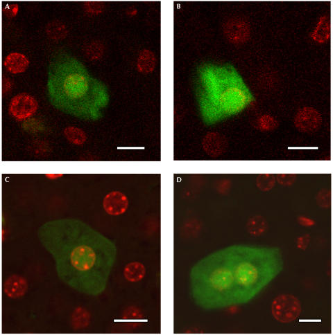
To test the applicability of the stop-EGFP transgenic mouse system to other organs, the kidney and liver were analysed at 5 months post-ENU administration. The organs were fixed in 4% paraformaldehyde and sectioned into slices (200 μm in thickness) using a vibratome (see supplementary information online). Each slice was then analysed under a fluorescent microscope. In an entire kidney from an ENU-treated stop-EGFP mouse, several green fluorescent mutant cells were detected (Fig 4A,B). The mutant cells contained what appeared to be brighter nuclei. The bright spots were confirmed to be nuclei by 4′,6-diamidino-2-phenylindole (DAPI) staining (Fig 4A,B). In a scan of the entire liver of an ENU-treated stop-EGFP mouse, a total of 26 mutations were identified (Fig 4C,D). The number of mutations and estimated total number of cells in each lobe are shown in Table 1. Hepatocytes are normally proliferatively quiescent without induced regeneration (Sell, 2001). In this study, a mutant colony carrying more than two nuclei has not been detected at 5 months post-ENU administration, and this may reflect the slow rate of cell turnover in this tissue.
Figure 4.
Green fluorescent mutant cells in the kidney and the liver. Panels A–D show merged images of EGFP signals and DAPI signals of mutant cells detected in the kidney (A,B) and liver (C,D). The bright green fluorescent signals within these cells are colocalized with the DAPIstained nuclei. Scale bars, 10 μm.
Table 1.
Observed distribution of stop-EGFP revertant mutations in four lobes of the liver
| Lobe | Mutation count | Cell count | Mutation frequency |
|---|---|---|---|
| Left caudal |
12 |
(6.61±2.32) × 107 |
(1.82±3.04) × 10−7 |
| Right anterior |
6 |
(3.06±0.84) × 107 |
(1.96±0.25) × 10−7 |
| Left anterior |
1 |
(2.04±0.32) × 107 |
(0.49±0.25) × 10−7 |
| Right caudal |
7 |
(2.18±0.43) × 107 |
(3.21±0.84) × 10−7 |
| All | 26 | (13.89±1.99) × 107 | (1.87±0.27) × 10−7 |
The cell count is the estimated total number of cells in each lobe and the mutation frequency is the total number of mutations divided by the total number of cells. The total number of cells in each lobe was estimated by multiplying the total cell number in a reference slice by the total number of slices of a similar size sectioned from the organ.
The stop-EGFP transgenic system can be applied to virtually any tissue that undergoes constant renewal (e.g., epithelial tissues of the gastrointestinal tract) or to tissues with episodic regenerative capabilities. For example, if liver regeneration in an ENU-treated stop-EGFP mouse is induced by partial hepatectomy or by treatment with hepatoxic chemicals, clonal cell lineages could be detected in the regenerated liver, which might provide insight into the liver regeneration process. In our system, background mutations were virtually undetectable because of an extremely low rate of spontaneous mutation (e.g., no mutations were detected in the skin, kidney and liver of untreated stop-EGFP mice); thus, cells can be labelled at a specific time point by treatment with a pulse of mutagen. Therefore, imaging experiments carried out after several tissue turnovers in a renewing tissue of adult mice treated with a mutagen will allow one to specifically trace cell lineages that descended from stem cells of the tissue.
The stop-EGFP system also has the potential to be applied to tracing cell fates in developmental studies. For example, embryos could be exposed to ENU in utero by exposing pregnant mice to the mutagen (Winton et al, 1988). With the aim of allowing a clonal cell lineage to be traced in developmental studies, Bonnerot & Nicolas (1993) developed a transgenic mouse in which infrequent (at the rate of 10−6 per cell generation) spontaneous homologous recombination activates lacZ expression in tissues. The clonality of cell lineages generated during development in this system is virtually certain owing to the low frequency of spontaneous recombination events. The transgenic mouse (referred to as the laacZ mouse) provided new insights into the myotome formation and cerebellar development (Nicolas et al, 1996; Mathis & Nicolas, 2003). A problem with the laacZ mouse system is that one cannot determine when during development the recombination events occur, which leads to difficulties in interpreting the data (Sanes, 1994). In the stop-EGFP system, however, an extremely low rate of spontaneous mutation ensures that cells are probably labelled at a specific time point during development (by treatment with a pulse of mutagen), and this may overcome this limitation of the laacZ system.
Speculation
Although we have only performed in vivo imaging of clonal cell lineages in the epidermis in this study, a similar approach could be applied to other organs such as the liver, muscle and brain (Naumov et al, 1999; Chen et al, 2000; Feng et al, 2000). Repeated in vivo imaging of the same GFP-expressing cells over time has recently been successfully performed in these organs (Chen et al, 2000; Feng et al, 2000). If such repeated in vivo imaging of the same tissue sites is performed over time using our transgenic mouse system, one might be able to investigate the dynamics of the clonal development of a cell lineage in many different tissues (e.g., in the regenerating liver).
Methods
Generation of the stop-EGFP and the wild-type EBFP genes. We have obtained the plasmid pCX-EGFP, which was used to generate a ‘green mouse' (Okabe et al, 1997). The stop-EGFP gene was generated from pCX-EGFP by PCR using the primers STOPEGFP5 and 3EGFP (see supplementary information online for primer sequences) and subsequently used to replace the EGFP gene within pCX-EGFP. The new construct is referred to as pCX-stop-EGFP. The wild-type EBFP gene was generated from the wild-type EGFP gene using site-directed mutagenesis (QuickChange™, Stratagene). The resulting construct is referred to as pCX-EBFP.
Cell culture tests. To test the green fluorescence intensity of protein expressed from each construct, a transfection experiment was performed using the NIH3T3 mouse fibroblast cell line. The procedures used followed the manufacturer's guidelines (FuGENE 6 Transfection Reagent, Roche). At 36 h after transfection, the transfected cells were imaged using an inverted fluorescent microscope.
Animal experiments. All experiments using live mice were performed in compliance with the recommendations of the Canadian Council on Animal Care, and have been approved by the Health Sciences Animal Policy and Welfare Committee of the University of Alberta.
Generation of transgenic mice The pCX-stop-EGFP and pCX-EBFP constructs were digested with SalI and BamHI, and a DNA fragment of 3.2 kb was purified. The 3.2 kb fragment from pCX-stop-EGFP was mixed with that from pCX-EBFP, and then microinjected into fertilized oocytes using standard techniques. Genotypes of mice were determined by genomic PCR using primers, EGFPMIDDLE5 and 3EGFP (see supplementary information online for primer sequences).
ENU administration. The procedures followed standard operating protocols developed by the Office of Environmental Health & Safety at the University of Alberta. Briefly, about 1 g of ENU (ISOPAC, Sigma) was dissolved in 10 ml of 95% ethanol and then 90 ml of phosphate-citrate buffer (pH 5.0) was added to the ENU solution. ENU at 150 mg/kg (single dose) was administered intraperitoneally to each mouse.
In vivo detection of epidermal cell lineages. At one week before the in vivo imaging experiment, the dorsal hair in telogen was depilated (about 2.5 cm × 2.5 cm area) using a depilatory agent (Nair, Carter-Wallace Inc.). On the day of the imaging experiment, the mouse was anaesthetized and placed with its dorsal skin on a microscope coverslip on the microscope stage. The depilated area of the epidermis was illuminated by a 50 W mercury lamp and scanned using an inverted laser scanning confocal fluorescent microscope (Zeiss LSM 510) with a × 10 objective and an LP 520 emission filter (Zeiss). Green fluorescent mutant epidermal cells were imaged using the microscope with an Argon laser (488 nm) and a × 10 objective.
Supplementary information is available at EMBO reports online (http://www.nature.com/embor/journal/v5/n9/extref/7400218s1.pdf).
Supplementary Material
Supplementary Information
Acknowledgments
We thank P. Dickie, X. Sun and G. Hipperson for the technical assistance. This research was supported by the 2002 CIHR Peter Lougheed Scholar Award to B.R. from the Canadian Institutes of Health Research and the Peter Lougheed Foundation.
References
- Allen TD, Potten CS (1974) Finestructural identification and organization of the epidermal proliferative unit. J Cell Sci 15: 291–319 [DOI] [PubMed] [Google Scholar]
- Bonnerot C, Nicolas JF (1993) Clonal analysis in the intact mouse embryo by intragenic homologous recombination. C R Acad Sci III 316: 1207–1217 [PubMed] [Google Scholar]
- Brazelton TR, Rossi FM, Keshet GI, Blau HM (2000) From marrow to brain: expression of neuronal phenotypes in adult mice. Science 290: 1775–1779 [DOI] [PubMed] [Google Scholar]
- Chen BE, Lendvai B, Nimchinsky EA, Burbach B, Fox K, Svoboda K (2000) Imaging high-resolution structure of GFP-expressing neurons in neocortex in vivo. Learn Mem 7: 433–441 [DOI] [PubMed] [Google Scholar]
- Clarke JD, Tickle C (1999) Fate maps old and new. Nat Cell Biol 1: E103–E109 [DOI] [PubMed] [Google Scholar]
- Feng G, Mellor RH, Bernstein M, Keller-Peck C, Nguyen QT, Wallace M, Nerbonne JM, Lichtman JW, Sanes JR (2000) Imaging neuronal subsets in transgenic mice expressing multiple spectral variants of GFP. Neuron 28: 41–51 [DOI] [PubMed] [Google Scholar]
- Flake AW (2001) Fate mapping of stem cells. In Stem Cell Biology, Marshark DR, Gardner RL, Gottlieb D (eds) pp 375–397. Cold Spring Harbor, NY: Cold Spring Harbor Laboratory Press [Google Scholar]
- Ghazizadeh S, Taichman LB (2001) Multiple classes of stem cells in cutaneous epithelium: a lineage analysis of adult mouse skin. EMBO J 15: 1215–1222 [DOI] [PMC free article] [PubMed] [Google Scholar]
- Heim R, Prasher DC, Tsien RY (1994) Wavelength mutations and posttranslational autoxidation of green fluorescent protein. Proc Natl Acad Sci USA 91: 12501–12504 [DOI] [PMC free article] [PubMed] [Google Scholar]
- Kameda T, Nakata A, Mizutani T, Terada K, Iba H, Sugiyama T (2003) Analysis of the cellular heterogeneity in the basal layer of mouse ear epidermis: an approach from partial decomposition in vitro and retroviral cell marking in vivo. Exp Cell Res 283: 167–183 [DOI] [PubMed] [Google Scholar]
- Mathis L, Nicolas JF (2003) Progressive restriction of cell fates in relation to neuroepithelial cell mingling in the mouse cerebellum. Dev Biol 258: 20–31 [DOI] [PubMed] [Google Scholar]
- Naumov GN, Wilson SM, MacDonald IC, Schmidt EE, Morris VL, Groom AC, Hoffman RM, Chambers AF (1999) Cellular expression of green fluorescent protein, coupled with high-resolution in vivo videomicroscopy, to monitor steps in tumor metastasis. J Cell Sci 112: 1835–1842 [DOI] [PubMed] [Google Scholar]
- Nicolas JF, Mathis L, Bonnerot C, Saurin W (1996) Evidence in the mouse for self-renewing stem cells in the formation of a segmented longitudinal structure, the myotome. Development 122: 2933–2946 [DOI] [PubMed] [Google Scholar]
- Okabe M, Ikawa M, Kominami K, Nakanishi T, Nishimune Y (1997) ‘Green mice' as a source of ubiquitous green cells. FEBS Lett 407: 313–319 [DOI] [PubMed] [Google Scholar]
- Potten CS (1975) Epidermal transit times. Br J Dermatol 93: 649–658 [DOI] [PubMed] [Google Scholar]
- Potten CS (1981) Cell replacement in epidermis (keratopoiesis) via discrete units of proliferation. Int Rev Cytol 69: 271–318 [DOI] [PubMed] [Google Scholar]
- Potten CS, Morris RJ (1988) Epithelial stem cells in vivo. J Cell Sci 10(Suppl): 45–62 [DOI] [PubMed] [Google Scholar]
- Ro S, Rannala B (2001) Methylation patterns and mathematical models reveal dynamics of stem cell turnover in the human colon. Proc Natl Acad Sci USA 98: 10519–10521 [DOI] [PMC free article] [PubMed] [Google Scholar]
- Sanes JR (1994) Lineage tracing. The latest in lineage. Curr Biol 4: 1162–1164 [DOI] [PubMed] [Google Scholar]
- Schneider TE, Barland C, Alex AM, Mancianti ML, Lu Y, Cleaver JE, Lawrence HJ, Ghadially R (2003) Measuring stem cell frequency in epidermis: a quantitative in vivo functional assay for long-term repopulating cells. Proc Natl Acad Sci USA 100: 11412–11417 [DOI] [PMC free article] [PubMed] [Google Scholar]
- Sell S (2001) Heterogeneity and plasticity of hepatocyte lineage cells. Hepatology 33: 738–750 [DOI] [PubMed] [Google Scholar]
- Stern CD, Fraser SE (2001) Tracing the lineage of tracing cell lineages. Nat Cell Biol 3: E216–E218 [DOI] [PubMed] [Google Scholar]
- Toma JG, Akhavan M, Fernandes KJ, Barnabe-Heider F, Sadikot A, Kaplan DR, Miller FD (2001) Isolation of multipotent adult stem cells from the dermis of mammalian skin. Nat Cell Biol 3: 778–784 [DOI] [PubMed] [Google Scholar]
- Van der Kooy D, Weiss S (2000) Why stem cells? Science 287: 1439–1441 [DOI] [PubMed] [Google Scholar]
- Winton DJ, Blount MA, Ponder BA (1988) A clonal marker induced by mutation in mouse intestinal epithelium. Nature 333: 463–466 [DOI] [PubMed] [Google Scholar]
Associated Data
This section collects any data citations, data availability statements, or supplementary materials included in this article.
Supplementary Materials
Supplementary Information


