Abstract
In Ralstonia pickettii PKO1, a denitrifying toluene oxidizer that carries a toluene-3-monooxygenase (T3MO) pathway, the biodegradation of toluene and trichloroethylene (TCE) by the organism is induced by TCE at high concentrations. In this study, the effect of TCE preexposure was studied in the context of bacterial protective response to TCE-mediated toxicity in this organism. The results of TCE degradation experiments showed that cells induced by TCE at 110 mg/liter were more tolerant to TCE-mediated stress than were those induced by TCE at lower concentrations, indicating an ability of PKO1 to adapt to TCE-mediated stress. To characterize the bacterial protective response to TCE-mediated stress, the effect of TCE itself (solvent stress) was isolated from TCE degradation-dependent stress (toxic intermediate stress) in the subsequent chlorinated ethylene toxicity assays with both nondegradable tetrachloroethylene and degradable TCE. The results of the toxicity assays showed that TCE preexposure led to an increase in tolerance to TCE degradation-dependent stress rather than to solvent stress. The possibility that such tolerance was selected by TCE degradation-dependent stress during TCE preexposure was ruled out because a similar extent of tolerance was observed in cells that were induced by toluene, whose metabolism does not produce any toxic products. These findings suggest that the adaptation of TCE-induced cells to TCE degradation-dependent stress was caused by the combined effects of solvent stress response and T3MO pathway expression.
Trichloroethylene (TCE), a suspected carcinogen (30) and U.S. Environmental Protection Agency priority pollutant (10), is one of the most commonly detected volatile organic contaminants in groundwater. In oxic contaminated subsurface environments, TCE can be degraded by means of a cooxidation reaction catalyzed by microbial oxygenases such as ammonia monooxygenase (2), alkene monooxygenase (8, 9), methane monooxygenase (12, 23), propane monooxygenase (24), toluene dioxygenase (6, 14, 52), and toluene monooxygenase (11, 33, 48). Among these, TCE cooxidation by bacteria carrying toluene oxygenase pathways has been of particular interest in environmental microbiology, since studies have shown that aerobic TCE biodegradation is stimulated in situ when toluene is added (15, 26).
TCE exposure can enhance biodegradation capacity via the induction of toluene oxygenase activity (4, 40). However, TCE exposure may also lead to TCE-mediated toxicity that can limit biodegradation capacity in TCE-degrading toluene oxidizers. Because of these competing effects, optimizing biodegradative capacity in sites contaminated by TCE (especially at high concentrations) may involve finding the TCE exposure level at which the combined effects of TCE-induced biodegradative activity (TCE inducibility) and TCE-mediated toxic effects result in maximum biodegradation capacity.
TCE exposure may lead to two types of toxicity in TCE-degrading oxygenase-containing bacteria. The first is TCE degradation-dependent (or degradation intermediate-mediated) toxicity, which results in the reduction of cellular growth, viability, and respiratory activity (1, 9, 25, 32, 37, 44, 45, 51). Although the exact nature of the destructive intermediate species remains unknown, it has been proposed that acyl chlorides, generated from hydrolysis or rearrangement of TCE epoxide (monooxygenase-catalyzed reaction) or TCE-dioxetane (dioxygenase-catalyzed reactions), cause damage by alkylating various cellular constituents (12, 22, 31, 45). In addition to this by-product-mediated toxicity, TCE itself may cause a solvent stress at high concentrations. It is recognized that organic solvents with a log Kow value between 1.5 and 3 (TCE has a log Kow of 2.61 [13]) may generally cause cytoplasmic membrane-associated toxicity in microorganisms (36, 41). Although the effect of TCE degradation-dependent toxicity on TCE inducibility was previously reported for a toluene dioxygenase strain (Pseudomonas putida TVA8 [40]), little is known about the combined effects of TCE degradation-dependent stress and TCE solvent stress on the induction of biodegradation by TCE in bacteria that carry TCE-degrading oxygenase pathways.
Among toluene-oxidizing bacteria, a toluene monooxygenase-containing bacterial strain, Burkholderia cepacia G4, has been suggested to resist TCE degradation-dependent toxicity (11, 19, 31). Despite such tolerance, this type of organism may become sensitive to TCE degradation-dependent stress when the growth substrate or energy is limited (25). Energy-requiring DNA repair mechanisms (42) are involved in the energy-dependent tolerance observed in strain G4 (50). This suggests that adding a carbon and energy source (primary substrate) would aid the in survival of this type of toluene monooxygenase-containing bacterium when degrading TCE. In groundwater, aerobic metabolism of such added primary substrates by soil microorganisms may result in oxygen limitation, and in turn, the oxygen limitation would result in a decrease in the degradation of TCE by aerobic toluene-oxidizing bacteria that use only oxygen as a terminal electron acceptor (20). Alternatively, it has been proposed that the degradation of TCE by denitrifying toluene oxidizers can be stimulated by adding nitrate to groundwater, since the aqueous solubility of nitrate (660 g/liter) is much greater than that of oxygen (21). Olsen and coworkers previously demonstrated that nitrate enhanced the biodegradation of toluene and TCE by denitrifying toluene oxidizers (18, 21). However, the question whether denitrifying toluene degraders can tolerate TCE-mediated toxicity remains to be examined.
In 1996, Leahy et al. (21) reported that the degradation of TCE by a denitrifying toluene-3-monooxygenase (T3MO)-containing bacterium, Ralstonia pickettii PKO1, was activated after the cells were incubated in the presence of TCE at 2.35 mM (equivalent to 306 mg/liter). This suggested that the denitrifying strain PKO1 was able to tolerate TCE-mediated stresses, and TCE preexposure probably played some role in chemical-stress-resistant responses. In view of the considerations set forth above, this study sought to systematically examine the induction of catabolic enzyme activity by TCE, as well as the cellular stress response to TCE-mediated toxicity, in strain PKO1. The specific objectives of this study were (i) to examine the relationship between the TCE concentration and the kinetics of induction of catabolic enzyme activity and (ii) to characterize the effects of TCE preexposure on cellular responses to both TCE degradation-dependent stress and solvent stress. Using chlorinated ethylene toxicity analysis with the nondegradable solvent PCE and the degradable solvent TCE, the effect of TCE itself (solvent stress) was isolated from TCE degradation-dependent toxicity when cells were exposed to high TCE concentrations. By comparing cellular responses between uninduced and TCE-induced cells, the effects of TCE preexposure in the denitrifying TCE-degrading strain are discussed in the context of the bacterial protective response to TCE-mediated toxicity in this organism.
MATERIALS AND METHODS
Materials.
A basal salts medium (BM [21]) was used for the minimal mineral medium. TNA (tryptone nutrient agar [34]) served as the solid growth medium. Distilled-deionized water from a Corning Millipore D2 system was used for all experiments. For hydrocarbons, HPLC (high performance liquid chromatography)-grade toluene (Aldrich Chemical Co.) and spectrophotometer-grade TCE and tetrachloroethylene (PCE) (Fisher Scientific Co.) were used. The sole carbon and energy source (primary substrate) was sodium dl-lactate (Fisher Scientific Co.), which had the least repressive effect on the induction of toluene degradation among all the ancillary carbon sources that were tested (i.e., succinate, fumarate, adipate, pyruvate, glutamate, and lactate).
Culture conditions.
To prepare uninduced cell suspensions, cells were typically grown on lactate. Cells previously stored at −70°C were grown on TNA solid medium and then subjected to serial dilutions with a 40 mM phosphate buffer. After 3 days of incubation at 30°C, 10 colonies from a TNA plate were suspended in 1 ml of 40 mM phosphate buffer (pH 6.8). The cell suspension was transferred to a 1-liter culture flask containing 100 ml of BM amended with 1,000 mg of sodium dl-lactate per liter and then incubated by shaking (250 rpm) at room temperature (23 to 27°C). After approximately 18 h of incubation, the late-exponential-phase cultures were used as uninduced cell suspensions. The A425 (optical density at 425 nm) values of the cultures were approximately 0.6.
Toluene-induced cell suspensions were prepared to determine the kinetics of TCE degradation by toluene-induced PKO1 cells. For this preparation, cells were grown in 4-liter culture flasks containing 500 ml of BM amended with 2,000 mg of sodium dl-lactate per liter. To ensure adequate enzymatic induction, toluene (saturating) vapor was continuously provided to each culture from 0.5 ml of liquid toluene placed in a 1.8-ml GC vial attached to the inside of the stopper in each 4-liter culture flask. The prepared 4-liter culture flasks were incubated by shaking (250 rpm) in a temperature-controlled room (30°C). After approximately 18 h of incubation, the cells were harvested from the spent medium by centrifugation (9,264 × g at 4°C for 10 min). The cell pellet was washed once with fresh BM and then resuspended in 100 ml of fresh BM prior to use.
To control the exposure of cells to TCE during incubation with TCE plus lactate, the uninduced cells were grown in 160-ml serum bottles containing 10 ml of BM amended with 1,000 mg of sodium dl-lactate per liter and different concentrations of TCE. For this, 1 ml of the uninduced cell suspension, prepared as described above, was transferred into each 160-ml bottle containing 9 ml of preoxygenated BM with 1,110 mg of sodium dl-lactate per liter. A predetermined volume of a TCE stock prepared in N′,N′-dimethylformamide (900 g/liter) was then added to the aqueous phase in each 160-ml serum bottle, using airtight microsyringes. TCE concentrations in the serum bottles are reported on the basis of the initial aqueous phase concentrations. This aqueous concentration was calculated based on the amount of TCE added and the air and aqueous volumes in each serum bottle. For this calculation, a dimensionless TCE Henry constant of 0.472 (47) was used. After the addition of TCE, the serum bottles were crimp-sealed with Teflon-lined butyl septa and vortexed for 30 s. The serum bottles were incubated by shaking (250 rpm) at room temperature. Prior to the measurement of either toluene degradation or TCE degradation, cells were harvested from the spent medium by centrifugation (8,000 rpm at 4°C for 10 min). The cell pellet was washed once with fresh BM and then resuspended in 50 ml of fresh BM prior to use. To attain a biomass greater than A425 of 0.1 in the 50-ml cell suspension, spent medium was collected from several different 160-ml serum bottles, depending on the incubation conditions.
To prepare TCE-induced cells for the chlorinated ethylene toxicity analysis, cells were precultured for 24 h in the presence of 110 mg of TCE per ml, using a 160-ml serum bottle containing BM amended with lactate at an initial concentration of 1,000 mg of sodium dl-lactate per liter. For the 24-h incubation, a serial culture technique was used, transferring 1 ml of a cell suspension from the preincubated 160-ml serum bottle to a newly prepared serum bottle every 8 h.
Analytical techniques.
A viable plate count method was used to quantify the viable and culturable biomass. A single viable cell was assumed to form one colony on TNA plates (i.e., one CFU). Previous microscopic examination has supported the validity of this assumption; i.e., most of the suspended PKO1 cells do not coalesce into groups but, rather, remain separate from one another (35). To quantify the total biomass, cellular protein and A425 were also measured. For the cellular-protein measurement, the Bio-Rad protein assay kit (Bio-Rad Co.) was used with bovine serum albumin as a standard. The cells were digested at 90°C for 30 min in 5 N NaOH.
Toluene was analyzed by reverse-phase HPLC using a Hewlett-Packard 1090 series II system with a hypersil 5C18 column and a UV (210-nm) detector. Lactate was analyzed by HPLC using the same Hewlett-Packard system with a Capcell pak C18UG120 column and a UV (205 nm) detector. Chlorinated ethylenes were analyzed by gas chromatography (GC) using a Hewlett-Packard 6890 system with an electron capture detector. Under these conditions, the detection limits for toluene, lactate, TCE, and PCE were 10 ppb, 10 ppm, 1 ppb, and 1 ppb, respectively. For the HPLC and GC analyses, aqueous portions of 0.5 ml were obtained with airtight 1-ml microsyringes, mixed with an equal volume of methanol in 1.8-ml GC vials, and stored at 4°C until analyzed.
Hydrocarbon degradation measurements.
Initial rates of toluene and TCE degradation were measured to quantify toluene-degradative activity and to determine the kinetics of TCE degradation, respectively. For this, 15-ml completely mixed batch reactors equipped with sampling ports containing GC septa (Alltech Associates, Inc.) were used as described elsewhere (35). Before degradation was measured, cell suspensions in BM were preoxygenated for 30 min. The reactors were filled with cell suspensions without headspace and then crimp-sealed. A predetermined amount of either toluene or TCE was added from a stock in N′,N′-dimethylformamide through the GC septum into each crimp-sealed reactor. The initial aqueous concentration of toluene was 5 mg/liter, and initial TCE concentrations were varied. To obtain initial degradation data, toluene disappearance was monitored either every 15 min for 1 h or every 30 min for 2 h and TCE disappearance was monitored either every 30 min for 2 h or every 60 min for 4 h. Specific (biomass normalized) degradation rates are reported based on viable biomass as well as cellular protein. To examine the ability of T3MO to degrade PCE, the disappearance of PCE during 24 h of incubation was measured with toluene-induced cells under both resting and growing conditions, as described elsewhere (39). For the resting conditions, the 160-ml serum bottles containing 10 ml of BM were used with two initial PCE concentrations (0.1 and 0.3 mg/liter in the aqueous phase). For the growing conditions, the initial aqueous-phase concentration of toluene was 5 mg/liter in the prepared bottles. Abiotic loss of a hydrocarbon was also examined using controls that consisted of the batch reactors (either the 15-ml CMBRs or the 160-ml serum bottles) that contained BM and the hydrocarbon, but no cells.
Chlorinated ethylene toxicity analysis.
The viability of resting cells and the growth rate of lactate-grown cells were measured to explore cellular responses to chlorinated-ethylene exposure. To measure the viability of resting cells, the initial and final concentrations of viable biomass were measured after 9 h of incubation in 16-ml test tubes containing 4 ml of BM amended with various concentrations of chlorinated ethylene. For this incubation, a predetermined amount of a chlorinated ethylene in an N′,N′-dimethylformamide phase was added to the aqueous phase in each test tube. The tubes were immediately capped with Teflon-lined silicon septa and then vortexed for 30 s. The prepared tubes were incubated by shaking (250 rpm) at room temperature. To determine the growth rate in the presence of a chlorinated ethylene, the A425 was measured every hour for 9 h in a separate experimental setup with 16-ml test tubes containing 4 ml of BM amended with lactate (initial concentration, of 1,000 mg of sodium dl-lactate per liter) and a range of chlorinated ethylene concentrations. Since the 16-ml test tubes fit into the spectrophotometer, the A425 could be measured directly. Data obtained from the exponential growth phase were used to estimate the specific growth rate (μ). To report chlorinated-ethylene concentrations in the 16-ml tubes, the initial aqueous concentrations were calculated based on a headspace of 12 ml and the Henry constant of a chlorinated ethylene. For PCE, a dimensionless Henry constant of 0.803 was used (47).
Determination of the half-saturation constant (Kg) for lactate growth.
To determine the kinetics of lactate growth, growth rates were measured for lactate-grown cells in 160-ml serum bottles containing 10 ml of BM amended with different initial lactate concentrations. The specific growth rate for each lactate concentration was quantified based on the natural logarithmic viable biomass data (i.e., ln[X/X0]) obtained after 0, 4, and 8 h of incubation, assuming no lag-phase. To estimate Kg values, a nonlinear regression was performed on the data for specific growth rate versus initial lactate concentration, using an integrated form of the Monod growth kinetic model.
RESULTS
Kinetics of induction of toluene-degradative activity in response to TCE concentration.
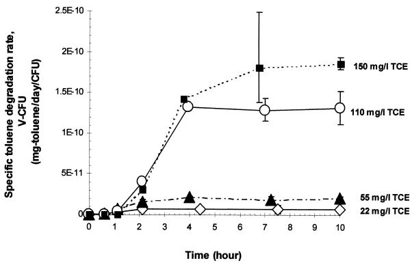
Figure 1 presents the results of a series of batch measurements of toluene degradation under different initial TCE concentrations. The initial lactate concentration was 1,000 mg of sodium dl-lactate per liter for all treatments. The y error bars represent the standard deviations among at least three independent experiments. In these experiments, the specific (viable biomass-normalized) toluene degradation rate increased with time, after a lag period of approximately 1 h. Specific toluene degradation rates reached plateau levels within 3 to 8 h, depending on the incubation conditions.
FIG. 1.
Specific toluene-degradative activity plotted against time in batch incubations with different initial TCE concentrations.
As shown in Fig. 1, both slope and plateau level increased with the level of TCE concentration, demonstrating TCE concentration-dependent induction kinetics. The slope represents the rate of induction of toluene-degradative activity when cells were induced by TCE. Since the slope for 110 mg of TCE per liter was close to that for 150 mg of TCE per liter, the induction rate was probably maximized at or near 110 mg of TCE per liter. The slopes for 22, 55, and 110 mg of TCE per liter (determined after corresponding lag periods) were approximately 21, 47, and 95%, respectively, of the maximum slope (i.e., the slope for 150 mg of TCE per liter), suggesting a linear relationship between the TCE concentration and the induction rate. In contrast, TCE inducibility (i.e., maximum capacity of a specific TCE concentration level to induce toluene degradation), represented by the plateau, did not linearly increase with TCE concentration. A significant increase in TCE inducibility was observed between 55 and 110 mg of TCE per liter, suggesting that a TCE threshold concentration for induction existed between 55 and 110 mg of TCE per liter.
To examine whether the growth substrate was limited in the batch experiments in Fig. 1, lactate concentrations were measured in 160-ml serum bottles at the end of the 10-h incubation and then compared with independently measured half-saturation constant (Kg) values. HPLC analysis showed that the residual lactate concentration was above 200 mg of sodium dl-lactate per liter in all the 160-ml bottles. The experimentally determined Kg value for the cells exposed to no TCE was 28.6 ± 9.6 mg of sodium dl-lactate per liter, and the Kg value for cells exposed to an initial TCE concentration of 110 mg/liter was 62.6 ± 4.6 mg of sodium dl-lactate per liter. Thus, the residual lactate concentrations were at least three times higher than the half-saturation constants, indicating that growth substrate limitation did not occur during the 10-h incubation in the batch experimental system.
TCE inducibility and specific viability in response to TCE concentration.
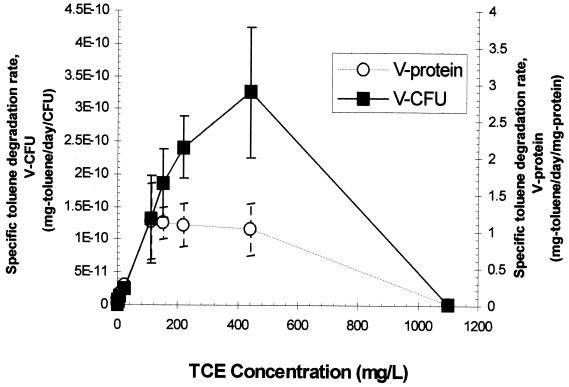
To further explore TCE inducibility in response to a wide range of TCE concentrations, plateau-specific toluene-degradative activity values were compared after incubating the cells for 9 h with different initial TCE concentrations. Figure 2 shows that the viable biomass-normalized degradative activity increased with TCE concentration at levels below and equal to 440 mg/liter. The maximum value of the observed plateau levels shown in Fig. 2 appeared at 440 mg of TCE per liter and was approximately 25% of the specific toluene degradation rate in toluene (saturating vapor)-induced cells shown in Table 1. It was reasonable to compare the maximum value of the observed plateau levels for TCE-induced cells (Fig. 2) with the specific toluene degradation rate attained from toluene (saturating vapor)-induced cells (Table 1), since toluene degradation rates for TCE-induced cells and for toluene-induced cells reached plateau levels in 9 and 72 h, respectively. The induction of toluene degradation by TCE was completely inhibited at the aqueous solubility limit of TCE (1,100 mg/liter at 25°C [47]). The TCE concentration at which the induction of toluene degradation was inhibited in strain PKO1 (i.e., between 440 and 1,100 mg of TCE per liter) was higher than the result previously reported by Shingleton et al. (40) for the toluene dioxygenase-containing strain TVA8 (i.e., 10 mg/liter). This suggests a tolerance in strain PKO1.
FIG. 2.
Plateau levels of specific toluene-degradative activity plotted against TCE concentrations in 9-h incubations.
TABLE 1.
Induction of toluene-degradative activity in R. pickettii PKO1 under different culture conditions
| Inducing agent (initial concn) | Growth substrate | Length of incubation period (h)a | Specific toluene degradation rateb (mg of toluene/day/1010 CFU) |
|---|---|---|---|
| None | Lactated | 9 | 0.01 ± 0.01 |
| Spent mediumc | Lactated | 9 | 0.14 ± 0.02 |
| Phenylalanine (>20 mg/liter) | Lactated | 9 | 0.17 ± 0.03 |
| TCE (110 mg/liter) | None | 9 | 0.17 ± 0.03 |
| TCE (110 mg/liter) | Lactated | 9 | 1.31 ± 0.34 |
| Toluene (saturating vapor) | Toluene (saturating vapor) | 72 | 13.05 ± 4.11 |
Steady-state toluene-degradative activity levels were attained within the reported incubation periods.
The specific degradation rate represents the steady-state degradative-activity level. The reported error is the standard deviation of at least three measurements for each culture condition.
The spent medium was collected from 160-ml serum bottles that were incubated for 9 h with lactate (initial concentration, 1,000 mg of sodium dl-lactate per liter) and 220 mg of TCE per liter). The collected spent medium was aseptically purged with air for 30 min and filtered through a 0.2-μm-pore-size membrane to remove remaining TCE and cells from the spent medium.
Initial concentration (1,000 mg of sodium dl-lactate/liter).
To measure TCE-mediated toxicity, specific viability (i.e., CFU per cellular protein) was calculated based on the viable biomass-normalized and cellular protein-normalized toluene-degradative activity values in Fig. 2. At TCE concentrations higher than 110 mg/liter, there was a significant deviation between cellular protein-normalized degradation activity and viable biomass-normalized activity. The specific viability values for 150, 220, and 440 mg of TCE per liter were approximately 68, 54, and 41%, respectively, of that for the control (i.e., no TCE exposure). This indicates that exposure to such TCE concentrations resulted in a reduction in specific viability and suggests that only a certain portion of the population could survive during incubation with high TCE concentrations.
Because of the observed decrease in specific viability at high TCE concentrations (Fig. 2), it was expected that some intracellular molecules would be released from cells that were exposed to such high solvent concentrations (7). Such released molecules, in turn, could act as an additional inducer during the 9-h incubation. To examine this possibility, the capacity of a TCE-free medium to induce toluene-degradative activity was measured using spent medium collected from 160-ml bottles with cells that had been incubated for 9 h with 220 mg of TCE per liter, purged with air for 30 min, and filtered through a 0.2-μm-pore-size membrane to remove the remaining TCE and cells. For this, a TCE concentration higher than 110 mg/liter (i.e., 220 mg/liter) was used because a more pronounced reduction of specific viability was observed at 220 mg of TCE per liter than at 110 mg/liter. As shown in Table 1, when initially uninduced PKO1 cells were incubated in 160-ml bottles containing 10 ml of the TCE-free spent medium amended with lactate (1,000 mg of sodium dl-lactate per liter), the viable biomass-normalized toluene-degradative activity was 0.14 ± 0.02 mg of toluene/day/1010 CFU, which is 11% of that for the cells incubated with TCE (110 mg/liter) plus lactate. The possibility that intracellular constituents, such as aromatic amino acids, may act as inducing agents was also examined by using fresh BM amended with phenylalanine at 20, 100, and 200 mg/liter. For all of the phenylalanine concentrations tested, the measured toluene-degradative activity was 0.17 ± 0.03 mg of toluene/day/1010 CFU, which is 13% of that for the cells incubated with TCE plus lactate. Thus, even though toluene-degradative activity could be induced by TCE-free spent medium and by phenylalanine, the results clearly indicate that TCE was the major inducer in these experiments.
Table 1 also compares TCE inducibility in different growth states (i.e., resting conditions versus growing conditions). The TCE inducibility for resting cells was 13% of that for growing cells. The similarity of this result to those for growing cells induced by TCE-free spent medium and by phenylalanine was probably a coincidence, since the TCE inducibility for resting cells represents the endogenous capacity to induce the T3MO pathway in the presence of TCE, which is different from the induction of toluene degradation by less effective inducing agents under growing conditions.
Inactivation of TCE degradation in response to TCE preexposure.
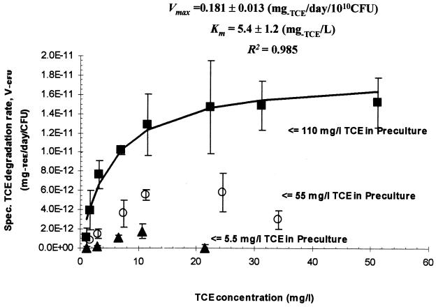
Figure 3 presents rate data for TCE degradation by resting PKO1 cells that were previously incubated for 9 h with different initial TCE concentrations (5.5, 55, and 110 mg/liter) and an initial lactate concentration of 1,000 mg/liter. The y error bars are the standard deviations among at least two independent measurements for each TCE concentration. The specific rate of degradation increased with the level of prior TCE preculture exposure. The results from Fig. 3 show that the inactivation of TCE degradation in TCE-induced PKO1 cells depended on the prior TCE concentration in preculture. TCE degradation rates decreased at TCE concentrations higher than 11 and 25 mg/liter for the cells preincubated at 5.5 and 55 mg of TCE per liter, respectively. In contrast, the specific rate for the cells precultured with 110 mg of TCE per liter did not decrease even when the precultured resting cells were subsequently exposed to 50 mg of TCE per liter. These findings indicate that the cells previously exposed to higher TCE concentrations could tolerate higher TCE exposure, exhibiting an adaptation to TCE exposure.
FIG. 3.
Specific TCE degradation rate (V−CFU) plotted against TCE concentration in batch experiments with TCE-induced PKO1 resting cells. The symbols represent observations, and the line represents the Michaelis-Menten model fit.
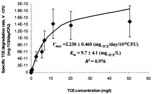
To examine whether the observed adaptive response to TCE exposure was attributable to TCE degradation-dependent stress (reactive intermediates) in the preculture, the results in Fig. 3 were compared with TCE degradation rate data from cells in which the T3MO pathway was expressed by preculturing cells with saturating toluene vapor (Fig. 4). For the purpose of this examination, the use of cells preincubated with saturating toluene vapor was feasible, since the intermediates of toluene degradation (m-cresol, 3- and 4-methylcatechol, and the alpha-keto acids derived from muconate semialdehyde [33]) are not toxic to PKO1, although toluene is an inducing agent for expression of the T3MO pathway (4) as well as a hydrocarbon solvent with a log Kow value of 2.73 (13), similar to that of TCE. Figure 4 presents the results of TCE degradation rate determination for cells that were previously toluene induced. The specific TCE degradation rate did not decrease when the toluene-exposed cells were subsequently exposed to 50 mg of TCE per liter under resting conditions. This result is consistent with that for TCE (110 mg/liter)-induced cells. Note that it was difficult to detect TCE degradation in the initial rate measurements when the TCE concentrations used for the initial rate measurements were higher than 50 mg/liter. Because of this, it was not clear whether TCE degradation for TCE-induced cells was more resistant to subsequent TCE exposure than that for toluene-induced cells. Nevertheless, the findings from the comparison between TCE-induced and toluene-induced cells rule out the possibility that the adaptive response to TCE exposure was caused by TCE degradation-dependent stress in the TCE preexposure.
FIG. 4.
Specific TCE degradation rate (V−CFU) plotted against TCE concentration in batch experiments with toluene (saturating-vapor)-induced PKO1 resting cells. The symbols represent observations, and the line represents the Michaelis-Menten model fit.
The kinetics of TCE degradation were compared for TCE-induced and toluene-induced cells. For kinetic characterization of TCE degradation, Michaelis-Menten kinetic parameters (Vmax and Km) were determined from TCE degradation rate data that exhibited no inhibition. For TCE-induced cells, kinetic parameters were determined from data for cells that had been incubated at 110 mg of TCE per liter (Fig. 3). For toluene-induced cells, the data in Fig. 4 were used to estimate the kinetic parameters. To estimate the parameters, a nonlinear regression analysis was performed with the averaged degradation rate data, using a differentiated form of the Michaelis-Menten model. Based on the CFU-normalized degradation rate data, the Vmax value for TCE-induced cells is 8.1% of the Vmax value for toluene-induced cells. For the kinetic data in Fig. 3 and 4, cellular protein assays were also conducted for biomass measurement. When normalized by the cellular protein, the Vmax value for TCE-induced cells was 14.8% of the Vmax value for toluene-induced cells, which is greater than the result obtained from the CFU-normalized degradation rate data. This difference may be attributed to a lower specific viability (i.e., CFU per cellular protein) in the toluene-induced population than in the TCE-induced population. It was difficult to draw any conclusion from a comparison of affinity (Km) values for TCE- and toluene-induced cells because of the large confidence intervals associated with this parameter.
Table 2 compares kinetic parameters for TCE degradation by PKO1 with values reported from other studies that examined either purified toluene oxygenase enzymes or bacterial strains carrying toluene oxygenase pathways. For the purpose of this comparison, the units of nanomoles of TCE per minute per milligram of protein and micromole per liter are used to report Vmax and Km values, respectively. The Vmax value for toluene-induced PKO1 cells was smaller than the values reported for other toluene oxygenase-associated systems, with the exception of recombinant Escherichia coli FM5/pKY287 containing tmoABCDE of Pseudomonas mendocina KR1. Among Km values, the half-saturation constant for PKO1 was higher than values reported for other toluene oxygenase-associated systems. For TCE (110 mg/liter)-induced PKO1 cells, the Vmax was also smaller than values reported for other toluene oxygenase-associated systems and the Km value was also large, being somewhat similar to the Km value for toluene-induced P. putida B2 carrying a toluene dioxygenase pathway.
TABLE 2.
Reported Michaelis-Menten kinetic parameters for aerobic degradation of TCE by pure cultures of bacterial strains containing toluene oxygenases or a purified toluene oxygenase
| Bacterial strain and enzyme | Inducer (concn) | Vmax (nmol of TCE/min/mg of protein) | Km (μM) | Source or reference |
|---|---|---|---|---|
| B. cepacia G4, toluene 2-monooxygenase | Phenol (460 mg/liter) | 7.9 | 3 | 11 |
| B. cepacia G4, toluene 2-monooxygenase | Toluene (NRa)b | 10.0c | 6 | 19 |
| B. cepacia PR123, toluene 2-monooxygenase | Constitutive | 15-18 | 13-17 | 43 |
| Purified toluene 2-monooxygenase | 37d | 12 | 31 | |
| R. pickettii PKO1, toluene 3-monooxygenase | Toluene (saturating vapor) | 4.01 ± 0.82 | 74.6 ± 29.8 | This study |
| R. pickettii PKO1, toluene 3-monooxygenase | TCE (110 mg/liter) | 0.59 ± 0.04 | 41.5 ± 9.1 | This study |
| P. mendocina KR1, toluene-4-monooxygenase | Toluene (0.4%, vol/vol) | 20 | 10 | 43 |
| E. coli FM5/pKY287, a recombinant containing tmoABCDE of KR1 | Toluene (saturating vapor) | 1-2 | NR | 49 |
| P. putida F1, toluene dioxygenase | Toluene (0.4%, vol/vol) | 8 | 5 | 43 |
| P. putida B2, toluene dioxygenase | Toluene (50 mg/liter) | 13.9e | 49.2 | 17 |
NR, not reported.
Cells were grown on toluene in a chemostat at dilution rates of 0.07 to 0.09 liter/h.
Converted from reported units of nanomoles of TCE per minute per milligram of dried cell mass, assuming that 50% of the dried cell mass is protein.
Reported on the basis of milligrams of hydroxylase component.
Converted from reported units of milligrams of TCE per day per milligram of dried cell mass, assuming that 50% of the dried cell mass is protein.
Cellular responses of uninduced and TCE-induced PKO1 cells to TCE and PCE exposure.
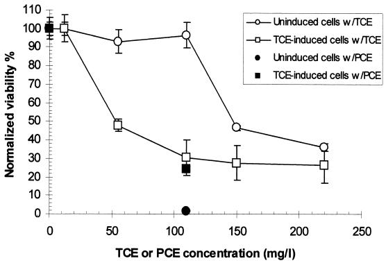
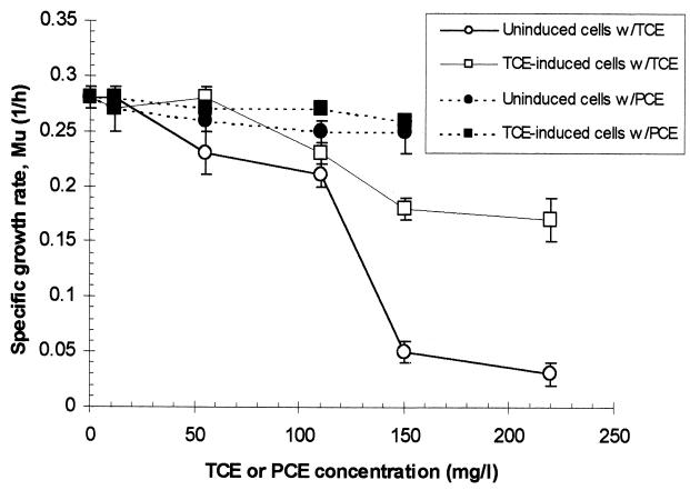
To further characterize the effect of TCE preexposure on cellular adaptation to TCE-mediated stress, the viability and growth rate of uninduced and TCE-induced PKO1 cells were compared. Figures 5 and 6 present the cellular response of PKO1 to chlorinated-ethylene (TCE and PCE) exposure under resting and growing conditions, respectively. For TCE-induced cells, a two-step incubation process was used: in the first incubation, cells were exposed to 110 mg of TCE per liter for 24 h under growing conditions (lactate growth), and in the second incubation, the TCE-induced cells were exposed to different chlorinated-ethylene concentrations for 9 h under either no-carbon conditions or lactate-growing conditions. For the uninduced cells, the same 9-h incubation conditions with chlorinated ethylene were used to examine the effect of chlorinated-ethylene exposure on uninduced cells (lactate-grown cells). To isolate the effect of TCE itself from that of overall TCE-mediated toxicity at high TCE concentrations, cellular responses to nondegradable PCE and degradable TCE exposures were also compared. PCE was chosen as the organic solvent to examine T3MO activity-independent solvent effects on PKO1 because PCE is not transformed by T3MO despite its similar chemical structure to TCE. In addition, PCE has a log Kow of 2.88 at 25°C (13), which is comparable to that for TCE. The nondegradability of PCE by PKO1 was confirmed in a separate experiment conducted to examine PCE-degradative activity by PKO1 cells induced by toluene as well as by TCE.
FIG. 5.
Normalized viability in uninduced and TCE (110 mg/liter)-induced cells plotted against chlorinated-ethylene (TCE or PCE) concentration in 9-h incubations under carbon starvation conditions.
FIG. 6.
Specific growth rate in uninduced and TCE (110 mg/liter)-induced cells plotted against chlorinated-ethylene (TCE or PCE) concentration in 9-h incubations under lactate growing conditions.
The cellular response to chlorinated-ethylene exposure under resting conditions was quantified by measuring viability (plate counts) at the end of each 9-h incubation with uninduced and TCE-induced cells. Figure 5 presents the viability data. The initial viable-biomass values for uninduced and TCE-induced cells were 1.42 × 10⁹ ± 0.05 × 10⁹ and 1.90 × 10⁹ ± 0.05 × 10⁹ CFU/ml, respectively. The x-axis values are the concentrations of TCE and PCE exposure during the 9-h incubations. The y-axis represents the viability normalized by the viability in the control (i.e., no chlorinated-ethylene exposure). The y error bar represents the standard deviation among at least three independent measurements. After exposure to 110 mg of PCE per liter, the viability of uninduced and TCE-induced cells decreased to 1.7% ± 0.1% and 24.5% ± 1.3%, respectively, of the viability in the control (Fig. 5), indicating significant solvent toxicity. The fact that the TCE-induced cells exhibited less reduction of viability than did the uninduced cells under resting conditions indicates that TCE preexposure led to increased tolerance to solvent toxicity under resting conditions. Figure 5 also shows the results from cells exposed to different TCE concentrations. For uninduced cells, no significant reduction of viability was observed at concentrations of 110 mg of TCE per liter and below. At concentrations of 150 and 220 mg of TCE per liter, viability decreased to 48.0% ± 2.1% and 35.9% ± 2.1%, respectively, of the viability in the control. The reduced viability can be attributed to TCE degradation-independent solvent stress (TCE itself) rather than stress resulting from the production of intermediates of TCE degradation, because the induction of T3MO activity by TCE would have been insignificant under resting conditions (Table 1). In the TCE-induced cells, a gradual reduction of viability was detected at a lower TCE concentration (between 12 and 55 mg of TCE per liter) than was seen for uninduced cells (Fig. 5). This indicates that TCE preexposure resulted in a more sensitive response to TCE exposure. Because the result obtained with the uninduced cells indicates that solvent toxicity (TCE itself) occurred at a concentration higher than 110 mg of TCE per liter, the reduced viability of TCE-induced cells observed between 12 and 110 mg of TCE per liter can be attributed to TCE degradation-dependent toxicity rather than to solvent toxicity.
The cellular response to chlorinated-ethylene exposure under growing conditions was quantified by measuring the specific growth rate (lactate growth) during each 9-h incubation with the same uninduced and TCE-induced cells described above. Figure 6 presents specific growth rates, determined from A425 data obtained during the 9-h incubation with chlorinated ethylene. The incubation conditions used for growth rate measurement with TCE were similar to those used to obtain the data plotted in Fig. 1. Note that the cells were initially uninduced; however, the T3MO pathway would be gradually expressed during the incubation, as observed in Fig. 1. The y error bar represents the standard deviation among at least three independent measurements. After exposure to PCE over a concentration range from 0 to 100% of its aqueous solubility (maximum solubility, 150 mg of PCE per liter at 25°C [47]), neither the uninduced nor TCE-induced cells were sensitive to solvent stress under growing conditions (Fig. 6). This indicates that TCE preexposure did not affect the cellular response to solvent stress under growing conditions. Compared to resting cells (Fig. 5), growing cells exhibited a more tolerant response to solvent stress. This energy-dependent solvent stress response is consistent with previous reports for solvent-tolerant Pseudomonas strains (36, 41). When exposed to different TCE concentrations, both uninduced and TCE-induced cells were sensitive to TCE-mediated stress (Fig. 6). Because cells could tolerate PCE exposure (solvent stress) under growing conditions, the reduction in specific growth rate observed in the cells that were exposed to TCE can be attributed to TCE degradation-dependent toxicity rather than solvent toxicity. The reduction in growth rates for TCE-induced cells was smaller than that for uninduced cells. This provides evidence that TCE preexposure led to an increased tolerance to TCE degradation-dependent toxicity.
DISCUSSION
R. pickettii PKO1 provides a good model system for understanding the basis of tolerance to TCE-mediated stress observed among toluene monooxygenase-containing bacteria. In TCE (110 mg/liter)-induced and toluene (saturating-vapor)-induced PKO1 cells, TCE degradation was not inhibited at concentrations near 50 mg of TCE per liter, resulting in a level of tolerance similar to that seen in Ralstonia eutropha JMP134 and B. cepacia G4, which are the most TCE tolerant among previously reported TCE-degrading oxygenase-containing bacteria (3, 11, 19). The level of tolerance observed in strain PKO1 was greater than that found in bacteria containing other TCE-degrading oxygenase systems, such as toluene dioxygenase (46), ammonia monooxygenase (16), soluble methane monooxygenase (32), propane monooxygenase (24), and propene monooxygenase (38). In addition, because the roles of TCE and lactate may be decoupled in this organism, the lactate-TCE system used in this study enabled us to examine the relationship between the concentration of a cometabolically transformed substrate and the induction of a toluene monooxygenase pathway. The roles of TCE and lactate may be decoupled in the strain because (i) TCE, which does not support growth in the strain (21), is an inducer for T3MO pathway expression (4), and (ii) lactate has the least repressive effect on the induction of toluene degradative activity (5 to 10%) among all of the ancillary carbon sources tested (i.e., succinate, fumarate, adipate, pyruvate, glutamate, and lactate). The findings from the system reveal that as a consequence of this tolerance in strain PKO1, both toluene- and TCE-degradative activities were induced by TCE even at high concentrations where a certain portion of the population was damaged (i.e., reduction of specific viability) by TCE degradation-dependent stress as well as TCE itself (solvent stress).
Strain PKO1 exhibited an adaptive response to TCE-mediated stress. The results of chlorinated ethylene toxicity analysis reveal that TCE preexposure (110 mg of TCE per liter for 24 h) led to the selection of cells that could tolerate TCE degradation-dependent stress rather than solvent stress. Although the exact mechanisms responsible for the adaptation to TCE exposure were not identified, the comparison of TCE degradation rate data between TCE (110 mg/liter)-induced and toluene (saturating-vapor)-induced cells (Fig. 3 and 4) enables us to rule out the possibility that TCE degradation-dependent stress during TCE preexposure led to tolerance to its own toxicity (i.e., TCE degradation-dependent toxicity). Because both TCE and toluene are hydrocarbon solvents as well as inducers for the T3MO pathway, the tolerance observed in TCE-induced cells could have resulted from either a bacterial solvent stress response, T3MO pathway expression, or both. In P. putida strain DOT-T1E, a set of genes encoding a solvent efflux pump was linked with the activation of the toluene dioxygenase operon (29). Because the rate of induction of toluene degradation significantly increased at TCE concentrations at which TCE itself exhibited solvent toxicity (Fig. 5), it is likely that the solvent stress response was also linked to the regulation of T3MO pathway expression in strain PKO1. These findings suggest that the combined effects of solvent stress response and T3MO pathway expression led to the tolerance observed in TCE-induced cells.
In continuous-treatment systems such as continuous-flow bioreactors and in situ bioremediation scenarios, cellular growth is a key factor to successful treatment, especially when the by-products of degradation are toxic to microbes (5). Because of this, the observed increase in the rate of growth of TCE-induced PKO1 cells even at high TCE concentrations (Fig. 6) has a potential benefit in the biological removal of TCE in hazardous-waste treatment systems. In the context of bioremediation, however, the addition of lactate might be inappropriate since the utilization of lactate by autochthonous soil microbes would result in oxygen limitation in the subsurface environment. An alternative strategy may be the coaddition of lactate with nitrate since nitrate can be used as an alternative electron acceptor that stimulates hypoxic biodegradation by soil denitrifying toluene monooxygenase bacteria such as R. pickettii PKO1 (18, 20, 28). Furthermore, because of the ability of toluene monooxygenase bacteria to adapt to TCE degradation-dependent toxicity, toluene monooxygenase bacteria can survive in the presence of high TCE concentrations and the survivors can be used to remove TCE via cometabolism. Thus, the use of denitrifying toluene monooxygenase bacteria may be beneficial to TCE bioremediation, especially when TCE concentrations are high.
Acknowledgments
This research was supported by Superfund Basic Research Program grant P42-ES-04911 from the National Institute of Environmental Health Sciences.
The technical assistance of Aksara Putthividhya is gratefully appreciated, and we also thank Juliana Malinverni for critical discussions.
REFERENCES
- 1.Alvarez-Cohen, L., and P. L. McCarty. 1991. Effects of toxicity, aeration, and reductant supply on trichloroethylene transformation by a mixed methanotrophic culture. Appl. Environ. Microbiol. 57:228-235. [DOI] [PMC free article] [PubMed] [Google Scholar]
- 2.Arciero, D., T. Vannelli, M. Logan, and A. B. Hooper. 1989. Degradation of trichlorethylene by the ammonia-oxidizing bacterium Nitrosomonas europaea. Biochem. Biophys. Res. Commun. 159:640-643. [DOI] [PubMed] [Google Scholar]
- 3.Ayoubi, P. J., and A. R. Harker. 1998. Whole-cell kinetics of trichloroethylene degradation by phenol hydroxlase in a Ralstonia eutropha JMP134 derivative. Appl. Environ. Microbiol. 64:4353-4356. [DOI] [PMC free article] [PubMed] [Google Scholar]
- 4.Byrne, A. M., and R. H. Olsen. 1996. Cascade regulation of the toluene-3-monooxygenase operon (tbuA1UBVA2C) of Burkholderia pickettii PKO1: role of the tbuA1 promoter (PtbuA1) in the expression of its cognate activator, TbuT. J. Bacteriol. 178:6327-6337. [DOI] [PMC free article] [PubMed] [Google Scholar]
- 5.Chang, H.-L., and L. Alvarez-Cohen. 1995. Model for the cometabolic biodegradation of chlorinated organics. Environ. Sci. Technol. 29:2357-2367. [DOI] [PubMed] [Google Scholar]
- 6.Dabrock, B., M. Klebeler, B. Averhoff, and G. Gottschalk. 1994. Identification and characterization of a transmissible linear plasmid from Rhodococcus erythropolis BD2 that encondes isopropylbenzene and trichloroethylene catabolism. Appl. Environ. Microbiol. 60:853-860. [DOI] [PMC free article] [PubMed] [Google Scholar]
- 7.de Smet, M.-J., J. Kingma, and B. Witholt. 1978. The effect of toluene on the structure and permeability of the outer and cytoplasmic membranes of Escherichia coli. Biochim. Biophys. Acta 506:64-80. [DOI] [PubMed] [Google Scholar]
- 8.Ensign, S. A. 1996. Aliphatic and chlorinated alkenes and epoxides as inducers of alkene monooygenase and epoxidase activities in Xanthobacter strain Py2. Appl. Environ. Microbiol. 62:61-66. [DOI] [PMC free article] [PubMed] [Google Scholar]
- 9.Ensign, S. A., M. R. Hyman, and D. J. Arp. 1992. Cometabolic degradation of chlorinated alkenes by alkene monooxygenase in a propylene-grown Xanthobacter strain. Appl. Environ. Microbiol. 58:3038-3046. [DOI] [PMC free article] [PubMed] [Google Scholar]
- 10.Environmental Protection Agency. 1980. Ambient water quality criteria for trichloroethylene. Publication 440/5-80-073. National Technical Information Service, Springfield, Va.
- 11.Folsom, B. R., P. J. Chapman, and P. H. Pritchard. 1990. Phenol and trichloroethylene degradation by Pseudomonas cepacia G4: kinetics and interactions between substrates. Appl. Environ. Microbiol. 56:1279-1285. [DOI] [PMC free article] [PubMed] [Google Scholar]
- 12.Fox, B. G., J. G. Borneman, L. P. Wackett, and J. D. Lipscomb. 1990. Haloalkene oxidation by the soluble methane monooxygenase from Methylosinus trichosporium OB3b: mechanistic and environmental implications. Biochemistry 29:6419-6427. [DOI] [PubMed] [Google Scholar]
- 13.Hansch, G., A. Leo, and D. Hoekman. 1995. Exploring QSAR: hydrophobic, electronic, and steric constants. American Chemical Society professional reference book. American Chemical Society, Washington, D.C.
- 14.Heald, S., and R. O. Jenkins. 1994. Trichloroethylene removal and oxidation toxicity mediated by toluene dioxygenase of Pseudomonas putida. Appl. Environ. Microbiol. 60:4634-4637. [DOI] [PMC free article] [PubMed] [Google Scholar]
- 15.Hopkins, G. D., and P. L. McCarty. 1995. Field evaluation of in situ aerobic cometabolism of trichloroethylene and three dichloroethylene isomers using phenol and toluene as the primary substrates. Environ. Sci. Technol. 29:1628-1637. [DOI] [PubMed] [Google Scholar]
- 16.Hyman, M. R., S. A. Russell, R. L. Ely, K. J. Williamson, and D. J. Arp. 1995. Inhibition, inactivation, and recovery of ammonia-oxidizing activity in cometabolism of trichloroethylene by Nitrosomonas europaea. Appl. Environ. Microbiol. 61:1480-1487. [DOI] [PMC free article] [PubMed] [Google Scholar]
- 17.Kelly, C. J., P. R. Bienkowski, and G. S. Sayler. 2000. Kinetic analysis of a tod-lux bacterial reporter for toluene degradation and trichloroethylene cometabolism. Biotechnol. Bioeng. 69:256-265. [DOI] [PubMed] [Google Scholar]
- 18.Kukor, J. J., and R. H. Olsen. 1996. Catechol 2,3-dioxygenase functional in oxygen-limited (hypoxic) environments. Appl. Environ. Microbiol. 62:1728-1740. [DOI] [PMC free article] [PubMed] [Google Scholar]
- 19.Landa, A. S., E. M. Sipkema, J. Weijma, A. A. C. M. Beenackers, J. Dolfing, and D. B. Janssen. 1994. Cometabolic degradation of trichloroethylene by Pseudomonas cepacia G4 in a chemostat with toluene as the primary substrate. Appl. Environ. Microbiol. 60:3368-3374. [DOI] [PMC free article] [PubMed] [Google Scholar]
- 20.Leahy, J. G., and R. H. Olsen. 1997. Kinetics of toluene degradation by toluene-oxidizing bacteria as a function of oxygen concentration, and the effect of nitrate. FEMS Microbiol. Ecol. 23:23-30. [Google Scholar]
- 21.Leahy, J. G., A. M. Byrne, and R. H. Olsen. 1996. Comparison of factors influencing trichloroethylene degradation by toluene-oxidizing bacteria. Appl. Environ. Microbiol. 62:825-833. [DOI] [PMC free article] [PubMed] [Google Scholar]
- 22.Li, S., and L. P. Wackett. 1992. Trichloroethylene oxidation by toluene dioxygenase. Biochem. Biophys. Res. Commun. 185:443-451. [DOI] [PubMed] [Google Scholar]
- 23.Lontoh, S., and J. D. Semrau. 1998. Methane and trichloroethylene degradation by Methylosinus trichosporium OB3b expressing particulate methane monooxygenase. Appl. Environ. Microbiol. 64:1106-1114. [DOI] [PMC free article] [PubMed] [Google Scholar]
- 24.Malachowsky, K. J., T. J. Phelps, A. B. Teboli, D. E. Minnikin, and D. C. White. 1994. Aerobic mineralization of trichloroethylene, vinyl chloride, and aromatic compounds by Rhodococcus species. Appl. Environ. Microbiol. 60:542-548. [DOI] [PMC free article] [PubMed] [Google Scholar]
- 25.Mars, A. E., J. Houwing, J. Dolfing, and D. B. Janssen. 1996. Degradation of toluene and trichloroethylene by Burkholderia cepacia G4 in growth-limited fed-batch culture. Appl. Environ. Microbiol. 62:886-891. [DOI] [PMC free article] [PubMed] [Google Scholar]
- 26.McCarty, P. L., M. N. Goltz, G. D. Hopkins, M. E. Dolan, J. P. Allan, B. T. Kawakami, and T. J. Carrothers. 1998. Full-scale evaluation of in situ cometabolic degradation of trichloroethylene in groundwater through toluene injection. Environ. Sci. Technol. 32:88-100. [Google Scholar]
- 27.McClay, K., S. H. Streger, and R. J. Steffan. 1995. Induction of toluene oxidation activity in Pseudomonas mendocina KR1 and Pseudomonas sp. strain ENVPC5 by chlorinated solvents and alkanes. Appl. Environ. Microbiol. 61:3479-3481. [DOI] [PMC free article] [PubMed] [Google Scholar]
- 28.Mikesell, M. D., J. J. Kukor, and R. H. Olsen. 1993. Metabolic diversity of aromatic hydrocarbon-degrading bacteria from a petroleum-contaminated aquifer. Biodegradation 4:249-259. [DOI] [PubMed] [Google Scholar]
- 29.Mosqueda, G., and J.-L. Ramos. 2000. A set of genes encoding a second toluene efflux system in Pseudomonas putida DOT-T1E is linked to the tod genes for toluene metabolism. J. Bacteriol. 182:937-943. [DOI] [PMC free article] [PubMed] [Google Scholar]
- 30.National Cancer Institute. 1976. Carcinogenesis bioassay of trichloroethylene. CAS no. 79-016. U.S. Department of Health, Education, and Welfare publication (NIH) 76-802. U.S. Department of Health, Education, and Welfare, Washington, D.C.
- 31.Newman, L. M., and L. P. Wackett. 1997. Trichloroethylene oxidation by purified toluene 2-monooxygenase: products, kinetics, and turnover-dependent inactivation. J. Bacteriol. 179:90-96. [DOI] [PMC free article] [PubMed] [Google Scholar]
- 32.Oldenhuis, R., J. Y. Oedzes, J. J. van der Waarde, and D. B. Janssen. 1991. Kinetics of chlorinated hydrocarbon degradation by Methylosinus trichosporium OB3b and toxicity of trichloroethylene. Appl. Environ. Microbiol. 57:7-14. [DOI] [PMC free article] [PubMed] [Google Scholar]
- 33.Olsen, R. H., J. J. Kukor, and B. Kaphammer. 1994. A novel toluene-3-monooxygenase pathway cloned from Pseudomonas pickettii PKO1. J. Bacteriol. 176:3749-3756. [DOI] [PMC free article] [PubMed] [Google Scholar]
- 34.Olsen, R. H., and J. Hansen. 1976. Evolution and utility of a Pseudomonas aeruginosa drug resistance factor. J. Bacteriol. 125:837-844. [DOI] [PMC free article] [PubMed] [Google Scholar]
- 35.Park, J., Y.-M. Chen, J. J. Kukor, and L. M. Abriola. 2002. Influence of substrate exposure history on biodegradation in a porous medium. J. Contam. Hydrol. 51:233-256. [DOI] [PubMed]
- 36.Ramos, J. L., E. Duque, J.-J. Rodriguez-Herva, P. Godoy, A. Haidour, F. Reyes, and A. Fernandez-Barrero. 1997. Mechanisms for solvent tolerance in bacteria. J. Biol. Chem. 272:3887-3890. [DOI] [PubMed] [Google Scholar]
- 37.Rasche, M. E., M. R. Hyman, and D. J. Arp. 1991. Factors limiting aliphatic chlorocarbon degradation by Nitrosomonas europaea: cometabolic inactivation of ammonia monooxygenase and substrate specificity. Appl. Environ. Microbiol. 57:2986-2994. [DOI] [PMC free article] [PubMed] [Google Scholar]
- 38.Reij, M. W., J. Kieboom, J. A. M. de Bont, and S. Hartmans. 1995. Continuous degradation of trichloroethylene by Xanthobacter sp. strain Py2 during growth on propene. Appl. Environ. Microbiol. 61:2936-2942. [DOI] [PMC free article] [PubMed] [Google Scholar]
- 39.Ryoo, D., H. Shim, K. Canada, P. Barbieri, and T. K. Wood. 2000. Aerobic degradation of teterachloroethylene by toluene-o-xylene monooxygenase of Pseudomonas stutzeri OX1. Nat. Biotechnol. 18:775-778. [DOI] [PubMed] [Google Scholar]
- 40.Shingleton, J. T., B. M. Applegate, A. C. Nagel, P. R. Bienkowski, and G. S. Sayler. 1998. Induction of the tod operon by trichloroethylene in Pseudomonas putida TVA8. Appl. Environ. Microbiol. 64:5049-5052. [DOI] [PMC free article] [PubMed] [Google Scholar]
- 41.Sikkema, J., J. A. M. de Bont, and B. Poolman. 1995. Mechanisms of membrane toxicity of hydrocarbons. Microbiol. Rev. 59:201-222. [DOI] [PMC free article] [PubMed] [Google Scholar]
- 42.Snyder, L., and W. Champness. 1997. Molecular genetics of bacteria. American Society for Microbiology, Washington, D.C.
- 43.Sun, A. K., and T. K. Wood. 1996. Trichloroethylene degradation and minerialization by pseudomonads and Methylosinus trichosporium OB3b. Appl. Microbiol. Biotechnol. 45:248-256. [DOI] [PubMed] [Google Scholar]
- 44.van Hylckama Vlieg, J. E. T., W. de Koning, and D. B. Janssen. 1997. Effect of chlorinated ethene conversion on viability and activity of Methylosinus trichosporium OB3b. Appl. Environ. Microbiol. 63:4961-4964. [DOI] [PMC free article] [PubMed] [Google Scholar]
- 45.Wackett, L. P., and S. R. Householder. 1989. Toxicity of trichloroethylene to Pseudomonas putida F1 is mediated by toluene dioxygenase. Appl. Environ. Microbiol. 55:2723-2725. [DOI] [PMC free article] [PubMed] [Google Scholar]
- 46.Wackett, L. P., and D. T. Gibson. 1988. Degradation of trichloroethylene by toluene dioxygenase in whole-cell studies with Pseudomonas putida F1. Appl. Environ. Microbiol. 54:1703-1708. [DOI] [PMC free article] [PubMed] [Google Scholar]
- 47.Weber, W. J., Jr., and F. A. DiGiano. 1996. Process dynamics in environmental systems. John Wiley & Sons, Inc., New York, N.Y.
- 48.Whited, G. M., and D. T. Gibson. 1991. Toluene-4-monooxygenase, a three-component enzyme system that catalyzes the oxidation of toluene to p-cresol in Pseudomonas mendocina KR1. J. Bacteriol. 173:3010-3016. [DOI] [PMC free article] [PubMed] [Google Scholar]
- 49.Winter, R. B., K. Yen, and B. D. Ensley. 1989. Efficient degradation of trichloroethylene by a recombinant Escherichia coli. Bio/Technology 7:282-285. [Google Scholar]
- 50.Yeager, C. M., P. J. Bottomley, and D. J. Arp. 2001. Requirement of DNA repair mechanisms for survival of Burkholderia cepacia G4 upon degradation of trichloroethylene. Appl. Environ. Microbiol. 67:5364-5391. [DOI] [PMC free article] [PubMed] [Google Scholar]
- 51.Yeager, C. M., P. J. Bottomley, and D. J. Arp. 2001. Cytotoxicity associated with trichloroethylene oxidation in Burkholderia cepacia G4. Appl. Environ. Microbiol. 67:2107-2115. [DOI] [PMC free article] [PubMed] [Google Scholar]
- 52.Zylstra, G. J., L. P. Wackett, and D. T. Gibson. 1989. Trichloroethylene degradation by Escherichia coli containing the cloned Pseudomonas putida F1 toluene dioxygenase genes. Appl. Environ. Microbiol. 55:3162-3166. [DOI] [PMC free article] [PubMed] [Google Scholar]


