Abstract
To study the mechanism of β1-integrin function in vivo, we have generated transgenic mouse expressing a dominant negative mutant of β1-integrin under the control of mouse mammary tumor virus (MMTV) promoter (MMTV-β1-cyto). Mammary glands from MMTV-β1-cyto transgenic females present significant growth defects during pregnancy and lactation and impaired differentiation of secretory epithelial cells at the onset of lactation. We report herein that perturbation of β1-integrin function in involuting mammary gland induced precocious dedifferentiation of the secretory epithelium, as shown by the premature decrease in β-casein and whey acidic protein mRNA levels, accompanied by inactivation of STAT5, a transcription factor essential for mammary gland development and up-regulation of nuclear factor-κB, a negative regulator of STAT5 signaling. This is the first study demonstrating in vivo that cell–extracellular matrix interactions involving β1-integrins play an important role in the control of milk gene transcription and in the maintenance of the mammary epithelial cell differentiated state.
INTRODUCTION
Integrins are adhesive transmembrane heterodimer receptors composed of an α and a β subunit. They bind to extracellular matrix (ECM) proteins via their extracellular domain and interact with cytoskeletal and signaling molecules via their cytoplasmic domain. Integrins function as signaling receptors, stimulating various intracellular signaling cascades. This enables them to modulate important cellular functions such as proliferation, survival, and gene expression (for review, see Giancotti and Ruoslahti, 1999).
To study the mechanisms of β1-integrin function in vivo, we have generated transgenic mice expressing a dominant negative mutant of β1-integrin under the control of the MMTV promoter (MMTV-β1-cyto) in the mammary gland epithelium (Faraldo et al., 1998). The transgene encodes a chimeric protein consisting of the cytoplasmic and transmembrane domains of β1-integrin and the extracellular domain of CD4, a molecule not involved in cell-ECM adhesion. This chimeric molecule does not bind to ECM proteins or to integrin α subunits. However, in vitro, in adherent cells, it was localized at focal contact sites, and its overexpression induced cell detachment and prevented adhesion to ECM proteins (Lukashev et al., 1994). Mammary glands of the MMTV-β1-cyto females at midpregnancy and early lactation exhibited growth defects (i.e., reduced proliferation and increased apoptosis rates), resulting from a lack of mitogen-activated protein kinase activation via the Shc and phosphoinositide 3-kinases pathways (Faraldo et al., 1998, 2001). In addition, β-casein and whey acidic protein (WAP) gene expression was diminished in 2-d-lactating transgenic glands, indicating impaired differentiation of secretory epithelial cells.
The activity of a number of transcription factors is known to be regulated during mammary gland development. One of these factors, STAT5, has been shown to play an important role in milk gene expression in cultured mammary epithelial cells (Groner and Gouilleux, 1995). In addition, in STAT5A-deficient mice, lobuloalveolar development during pregnancy was severely impaired, and expression of WAP and to a lesser extent that of β-casein gene was decreased (Liu et al., 1997; Teglund et al., 1998). On activation of the prolactin receptor, STAT5 is phosphorylated by the receptor-associated kinase JAK2 and translocated to the nucleus. Experiments carried out with mammary epithelial cells in culture have suggested that the activation of STAT5 via the prolactin receptor pathway requires interaction with a reconstructed basement membrane (Edwards et al., 1998).
The expression and activity of the transcription factor nuclear factor-κB (NFκB) is also tightly regulated during mammary gland development, being relatively high in the virgin and pregnant gland, below the detection threshold during lactation, and strongly up-regulated in involution (Brantley et al., 2000; Clarkson et al., 2000). Regulation of the NFκB pathway involves degradation of the IκB inhibitor in response to extracellular signals followed by the release and nuclear translocation of NFκB (Barkett and Gilmore, 1999). Several recent studies have reported negative cross talk between the NFκB and STAT pathways (Luo and Yu-Lee, 2000; Musikacharoen et al., 2001). In particular, NFκB was shown to inhibit the activation of β-casein gene transcription induced by the prolactin-STAT5 signal, suggesting the involvement of NFκB in control of the differentiation of the secretory epithelium of the mammary gland (Geymayer and Doppler, 2000). In addition, NFκB has been shown to regulate the growth and branching of mammary epithelia in vivo (Brantley et al., 2001) and to play a role in protecting mammary epithelial cells from apoptosis (Clarkson et al., 2000).
The MMTV-β1-cyto mouse line is a unique in vivo model system that allows to examine the role of cell–ECM interactions in mammary gland development. In this study, we made use of this transgenic mouse line to analyze the effects of perturbation of β1-integrin function on mammary gland involution, an important tissue-remodeling process taking place after weaning. The mammary gland involution is characterized by the induction of programmed cell death in epithelial cells and the dedifferentiation of the secretory epithelium, i.e., the cessation of milk gene expression (Walker et al., 1989; Strange et al., 1992; Li et al., 1997). We found that, although apoptosis rates in involuting glands were similar in transgenic and wild-type animals, the expression of milk genes was down-regulated more rapidly in transgenic animals. This premature dedifferentiation of secretory epithelial cells in the transgenic glands was accompanied by a decrease in STAT5 nuclear levels and an increase in NFκB activity. These data prove the involvement of cell–ECM interactions in the maintenance of the mammary epithelial cell differentiation state in vivo.
MATERIALS AND METHODS
Transgenic Mice and Mammary Tissue Preparation for Morphological Analysis
The transgenic mouse line expressing a dominant negative mutant of β1-integrin under the control of the MMTV promoter has been described elsewhere (Faraldo et al., 1998). In all cases, only mice with litters of seven to eight pups were used. For involution studies, pups were removed from their mothers after 1 wk of lactation, and the mammary glands were analyzed at various times after weaning. For whole-mount staining, mammary glands were flattened on microscope slides, fixed overnight in Carnoy's solution (75% ethanol, 25% acetic acid), and stained with carmine alum as described previously (Sympson et al., 1994). For histological analysis and apoptosis assays, the fourth (inguinal) glands were fixed overnight in 4% paraformaldehyde, dehydrated in ethanol, cleared in chloroform, and embedded in paraffin.
Immunostaining and Terminal Deoxynucleotidyl Transferase dUTP Nick-End Labeling Assay
For immunohistochemistry, sections were deparaffinized and incubated in 1% H2O2 to block endogenous peroxidase activity. Rabbit polyclonal antibodies against STAT5A (sc-1081) and the p65 subunit of NFκB (sc-109) (Santa Cruz Biotechnology, Santa Cruz, CA) were used at a concentration of 5 μg/ml, and positive nuclei were revealed with the Envision system (Dako, Cambridge, United Kingdom). After light counterstaining with hematoxylin, 1000–1500 nuclei/sample were counted. For immunofluorescence staining, mammary glands were embedded in Tissue-Tek (Miles Diagnostic Division, Elkhart, IN) immediately after dissection and frozen in isopentane cooled by liquid nitrogen. Before staining, 5–7-μm cryosections were fixed for 10 min in acetone at −20°C and incubated with 10% fetal calf serum for 20 min at room temperature. The sections were then incubated with primary antibodies for 1 h at 37°C, washed with phosphate-buffered saline, and treated with fluorescein isothiocyanate- or Texas Red-conjugated secondary antibodies (Jackson Immunoresearch, West Grove, PA) diluted 1:100, for 30 min at 37°C. Monoclonal rat anti-mouse CD4 antibody (BD PharMingen, San Diego, CA) was used at a concentration of 10 μg/ml; rabbit polyclonal antibody raised against mouse laminin 1 (provided by Dr. H. Feracci, Institut Curie, Paris, France) was diluted 1:250. To detect apoptotic nuclei, paraformaldehyde-fixed paraffin sections were analyzed by TdT digoxygenin nick-end labeling with Apoptag Plus (Oncor, Gaithersburg, MD) following manufacturer's instructions. After counterstaining with methyl green 2500–3000 nuclei/sample were counted.
DNA Ladders
DNA was extracted from 50 mg of mammary gland tissue samples isolated from 7-d-lactating, 2- and 4-d-involuting glands by using QIAamp DNA kit (QIAGEN, Hilden, Germany) according to manufacturer's instructions. Equal amounts of DNA were separated on 1.2% agarose gels, stained with ethidium bromide, and photographed.
RNA Isolation and Analysis
The third (thoracic) gland was used for RNA extraction. Total RNA was isolated using RNA-plus reagent (Bioprobe Systems, Montreuil-Sous-Bois, France) following manufacturer's instructions. Twenty micrograms of total RNA was separated on 1% agarose/formaldehyde gels, transferred to nylon membranes (Hybond-N; Amersham Biosciences, Little Chalfont, Buckinghamshire, United Kingdom), and hybridized with [32P]dCTP random-primed labeled cDNA probes. The cDNA probe for mouse WAP (Campbell et al., 1984) was kindly provided by Dr. R. Jaggi, (Laboratory for Clinical and Experimental Research, Bern, Switzerland) and that for mouse β-casein (Gupta et al., 1982) was provided by Dr. J. Rosen (Baylor College of Medicine, Baylor, TX). The blots were exposed to the high-performance autoradiography film (Amersham Biosciences). Quantitative analysis of the results was performed using PhosphorImager (Molecular Dynamics, Sunnyvale, CA) and the ImageQuant program.
Protein Analysis
Protein lysates were prepared from the fourth and fifth mammary glands as follows. Pieces of gland tissue were weighted and homogenized at 125 mg/ml in lysis buffer (40 mM Tris-HCl, pH 8.0, 276 mM NaCl, 20% glycerol, 2% NP-40, 4 mM EDTA, 20 mM NaF, 2 mM sodium orthovanadate, 40 μg/ml phenylmethylsulfonyl fluoride, 10 μg/ml aprotinin, 10 μg/ml leupeptin). The extracts were cleared by centrifugation at 10,000 × g, 4°C for 15 min. The immunoprecipitation assays were performed with 2 mg of protein extract, as described previously (Faraldo et al., 2001). Rabbit polyclonal anti-STAT5A (R & D Systems, Wiesbaden-Nordenstadt, Germany) and anti-JAK2 (Upstate Biotechnology, Lake Placid, NY) antibodies were used for immunoprecipitation. Immunoblotting was performed with polyvinylidene difluoride membranes treated according to the manufacturer's instructions. The following primary antibodies were used: rabbit polyclonal antibodies against phospho-STAT5, JAK2, phospho-JAK2 (Upstate Biotechnology), β1-integrin cytoplasmic domain (Marcantonio and Hynes, 1988), and mouse monoclonal antibodies against STAT5 (BD Transduction Laboratories, Heidelberg, Germany). To normalize the loading, the blots were probed with anti-β-actin (Sigma-Aldrich, Steinheim, Germany) monoclonal antibody. Immunoblots were developed using the enhanced chemiluminescence detection system (Amersham Biosciences).
Preparation of Nuclear Extracts and Band Shift Assay
Frozen mammary gland tissue was ground to a powder, and nuclear extracts were prepared as described previously (Shapiro et al., 1988). For protein–DNA interaction assays, 1 μg of nuclear extract was incubated for 15 min with 20,000 cpm of 32P-labeled duplex oligonucleotide probe in 20 mM HEPES, pH 7.9, 60 mM KCl, 2 mM MgCl2, 1 mM dithiothreitol, 10% (vol/vol) glycerol, and 100 ng/μl poly(dI-dC). The oligonucleotides used were as follows: 5′-TCGAGATTCCGGGAACCGCGT (STAT5 binding site), 5′-TCGATCTTTGGCTTGAAGCCAATA (NF1 binding site), and 5′-GGTCGAGGGGACTTTCCCTAGC (NFκB binding site). In competition experiments, unlabeled oligonucleotides were incubated for 15 min with the nuclear extract before adding labeled probe. For supershift assays, 1 μg of antibody against phosphoSTAT5 (Upstate Biotechnology), or against the p50 or p65 subunits of NFκB (Santa Cruz Biotechnology) was used. Protein–DNA complexes were subjected to electrophoresis in 6% native acrylamide gels and visualized by autoradiography.
Transfections and Reporter Gene Assays
COS7 cells were grown in medium supplemented with 10% fetal calf serum (Seromed; Biochrom KG, Berlin, Germany), 2 mM l-glutamine, and penicillin-streptomycin (Invitrogen, Carlsbad, CA). Transfections were performed with GenePorter reagent (Gene Therapy Systems, San Diego, CA) following manufacturer's instructions. Cells were split into 12-well dishes at a density of 1.5 × 10⁵ cells/well. Twenty-four hours later cells were transfected with 250 ng of mouse prolactin receptor (PRLR) expression vector, 65 ng of STAT5A expression vector (Mayr et al., 1998), 100 ng of Renilla reporter construct (Promega, Madison, WI), and 650 ng of β-casein-luciferase reporter containing the −344 to −1 sequences of the rat β-casein promoter or 650 ng of NFκB-luciferase reporter, containing a NFκB binding site (kindly provided by Dr. W.Doppler, Universität Innsbruck, Innsbruck, Austria). In addition, when mentioned, cells were cotransfected with 1.2 μg of the MMTV-β1-cyto-CD4 plasmid (Faraldo et al., 1998) or MMTV-CD4 construct (containing the transmembrane domain of the β1-integrin and the extracellular domain of CD4), or psp72-MMTV plasmid. Transfection medium was removed after 18 h, and medium containing 2% fetal calf serum and 2 μM dexamethasone was added for 24 h to induce transgene expression. For hormone induction, cells were further incubated for 24 h with 5 μg/ml ovine prolactin (30 U/mg; Sigma-Aldrich), 2 μM dexamethasone, and 5 μg/ml insulin.
To estimate firefly and Renilla luciferase activities, cells were scrapped in 100 μl of Pasive lysis buffer (Promega) and after two freeze/thaw cycles, lysates were cleared by centrifugation at 10,000 × g, 4°C for 2 min. Luciferase activity in 10 μl of lysate aliquots was measured in a Lumat 9501 (Berthold, Pforzheim, Germany), by using firefly and Renilla substrates (Promega). Values obtained for firefly luciferase were normalized to Renilla luciferase activity. At least three independent experiments were performed in each case.
RESULTS
Mammary Glands of MMTV-β1-cyto Mice Achieve Fully Differentiated Phenotype at Peak Lactation
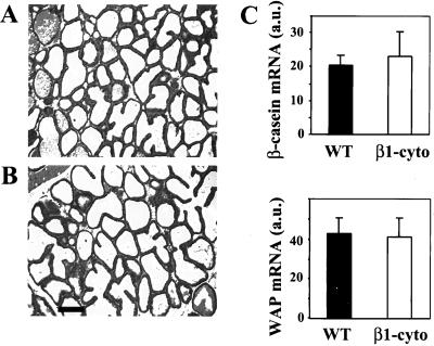
In previous studies, we found that the mammary glands of MMTV-β1-cyto animals were less developed than those of their wild-type littermates during the first days of lactation and that the mammary epithelium presented differentiation defects, as revealed by low milk protein mRNA levels (Faraldo et al., 1998). However, as estimated by histological analyses, after 7 d of lactation, transgenic glands seemed to be similar to those of wild-type animals, with functional alveoli and almost no fat pad stroma (Figure 1, A and B). To determine whether the differentiation defects persisted in the mammary epithelium of MMTV-β1-cyto mice at peak lactation, we isolated RNA from 7-d-lactating wild-type and transgenic mammary glands and evaluated the expression of WAP and β-casein genes. Quantitative analysis showed no significant difference between wild-type and transgenic mice (Figure 1C). Thus, by the 7th d of lactation, the secretory epithelial cells of transgenic mammary glands had gained a fully differentiated phenotype similar to that of the wild-type animals.
Figure 1.
Analysis of wild-type and MMTV-β1-cyto transgenic mammary glands at peak lactation. (A and B) Hematoxylin-eosin–stained sections of mammary glands from 7-d-lactating wild-type (A) and transgenic females (B). Bar, 100 μm. (C) Analyses of β-casein and WAP mRNA levels in 7-d-lactating glands by Northern blot. Diagrams present the values obtained for five animals in each case, normalized to GAPDH, mean ± SEM.
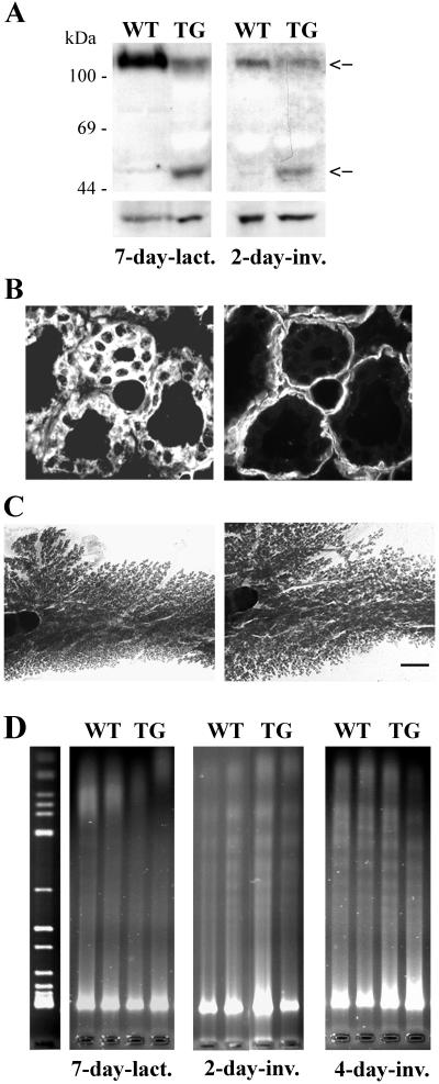
The MMTV promoter is known to be expressed at high rates throughout lactation (Pattengale et al., 1989). Accordingly, in our previous study, we have demonstrated by Northern blotting the presence of a high amount of the transgene transcript in the extracts of transgenic mammary glands at peak lactation (Faraldo et al., 1998). Using an antibody recognizing the β1-integrin cytoplasmic domain, we estimated the levels of transgenic protein and of endogenous β1-integrin in extracts from 7-d-lactating glands by Western blotting. The data presented in Figure 2A show that the transgene expression in lactating mammary gland resulted in a significant decrease of endogenous β1-integrin content compared with that in wild-type glands. Furthermore, quantitative analysis of the immunoblots has revealed that in transgenic gland extracts, the amount of transgenic protein was twofold greater than that of endogenous β1-integrin.
Figure 2.
Transgene expression and analysis of DNA integrity in lactating and involuting mammary glands. (A) Western blotting performed with protein extracts from 7-d-lactating and 2-d-involuting mammary glands by using an antibody against the β1-integrin cytoplasmic domain. Arrows indicate positions of endogenous β1-integrin and transgenic protein in the gel. β-Actin was used as loading control (bottom). (B) Double indirect immunofluorescence staining of a section from a 2-d-involuting transgenic gland with an anti-CD4 monoclonal antibody (left) and a polyclonal anti-laminin antibody (right). (C) Whole-mount staining of 4-d-involuting wild-type (left) and transgenic (right) glands. Bar, 25 μm for B; 2 mm for C. (D) Analysis of DNA integrity in 7-d-lactating, 2- and 4-d-involuting wild-type and transgenic glands. On the left, 1-kb DNA ladder (Invitrogen) is shown.
Epithelial Cell Apoptosis Rates Are Not Altered in Involuting MMTV-β1-cyto Glands
After weaning, the mammary gland undergoes major remodeling, including the dedifferentiation of secretory epithelial cells and apoptosis. To examine the mammary involution process in MMTV-β1-cyto transgenic mice, we removed the pups from their mothers after 1 wk of lactation and analyzed the mammary glands at various times after weaning. Although the MMTV promoter is progressively down-regulated during involution, at its early stages, rather high levels of transgene expression were detected in the transgenic glands by immunoblotting and immunofluorescence techniques (Figure 2, A and B). Similarly to lactating glands, in involution, the amount of endogenous β1-integrin was lower in transgenic than in wild-type animals, and the ratio of transgenic protein to endogenous β1-integrin in transgenic gland extract, as estimated by quantitative analysis of the blot, was of 1.5. However, an important amount of the transgenic protein was found in the cytosol, making difficult the estimation of its effective concentration (Figure 2B). Whole-mount staining of mammary glands at 2 and 4 d of involution showed no significant difference between wild-type and transgenic animals (Figure 2C; our unpublished data). Histological analysis of the glands confirmed this result. Consistently, terminal deoxynucleotidyl transferase dUTP nick-end labeling assays showed the rates of apoptosis to be similar in wild-type and transgenic mammary glands at 2 and 4 d of involution (Table 1). Furthermore, to compare the levels of apoptosis in involuting wild-type and transgenic glands, DNA integrity was analyzed in mammary tissue samples. No DNA fragmentation could be revealed in 7-d-lactating glands, whereas at 2 and 4 d of involution, the extent of apoptosis as assessed by DNA laddering was similar in wild-type and transgenic mammary glands (Figure 2D).
Table 1.
Apoptosis rates in involuting mammary glands of wild-type and MMTV-β1-cyto transgenic mice
| Stages | Apoptotic nuclei, %
|
|
|---|---|---|
| Wild-type | MMTV-β1-cyto | |
| 2-d-Involuting | 3.35 ± 0.6 (5) | 2.97 ± 0.7 (5) |
| 4-d-Involuting | 4.1 ± 0.9 (7) | 3.91 ± 0.65 (8) |
Values are means ± SEM; 2500 to 3000 nuclei/specimen were counted. The number of animals analyzed is indicated in parentheses. No statistically significant difference was observed between wild-type and transgenic glands.
Secretory Epithelial Cells in Involuting MMTV-β1-cyto Mammary Glands Undergo a Premature Dedifferentiation
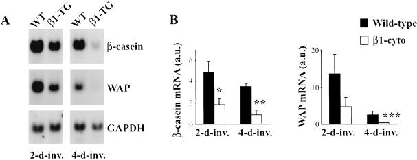
Normal involution is accompanied by the gradual cessation of milk production, as the secretory epithelial cells undergo dedifferentiation. Transcripts for milk proteins are, however, detectable several days after weaning. We used Northern blotting to analyze the expression of WAP and β-casein genes in a group of wild-type and transgenic animals at various times during involution (Figure 3). Levels of β-casein mRNA were lower in transgenic glands at 2 and 4 d of involution compared with those found in wild-type glands. Similarly, WAP mRNA levels were significantly lower in transgenic glands. These results indicate a precocious dedifferentiation of milk-producing epithelial cells in involuting MMTV-β1-cyto mammary glands.
Figure 3.
Analysis of milk protein gene expression in involuting wild-type and MMTV-β1-cyto mammary glands by Northern blotting. (A) Representative experiment. Twenty micrograms of total RNA from 2-d and 4-d-involuting glands was loaded per lane. (B) Quantitative analysis of the blots. Diagrams represent the values obtained for four or five animals in each case, normalized to GAPDH, mean + SEM. *p < 0.02; **p < 0.002; ***p < 0.05.
Epithelial Cells of Involuting MMTV-β1-cyto Mammary Glands Contain Less Functional STAT5 Than Wild-Type Glands
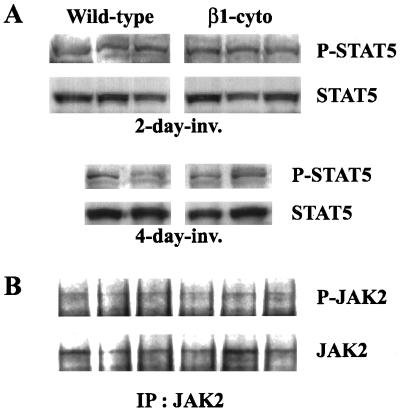
The transcription factor STAT5 has been implicated in the regulation of milk protein gene expression and is thought to be responsible for integrating prolactin and ECM signals (Streuli et al., 1995; Edwards et al., 1998). Therefore, to study the mechanisms underlying the early dedifferentiation of mammary epithelial cells expressing the transgene during involution, we analyzed the phosphorylation/activation status of molecules belonging to the STAT5 signaling pathway. At first, we compared the levels of STAT5 phosphorylation in involuting transgenic and wild-type glands. Western blotting analysis of mammary gland protein extracts showed no difference in levels of STAT5 phosphorylation on the 2nd and 4th d of involution (Figure 4). Similarly, levels of STAT5A phosphorylation were similar in extracts from wild-type and transgenic involuting glands after immunoprecipitation with anti-STAT5A antibody (our unpublished data). We also analyzed the phosphorylation status of JAK2, the kinase known to phosphorylate STAT5 in response to prolactin in mammary epithelial cells (Groner and Gouilleux, 1995). The signal obtained in immunoprecipitation/Western blotting assay performed with the 2-d-involuting gland protein extracts by using anti-phospho-JAK2 and anti-JAK2 antibodies, was rather weak (Figure 4B). However, the data presented in Figure 4B suggest that the levels of phosphorylation of JAK2 were similar in involuting wild-type and transgenic glands. The amount of phosphorylated JAK2 in protein extracts from 4-d-lactating glands was below detection level.
Figure 4.
STAT5 and JAK2 phosphorylation levels in wild-type and MMTV-β1-cyto transgenic glands. (A) Protein extracts of 2- and 4-d-involuting mammary glands (100 μg of protein/lane) were analyzed by Western blotting for phospho-STAT5 and total STAT5 content. (B) Two milligrams of 2-d-involuting mammary gland protein extracts were immunoprecipitated with anti-JAK2 polyclonal antibody and analyzed by Western blotting for phospho-JAK2 and total JAK2 content.
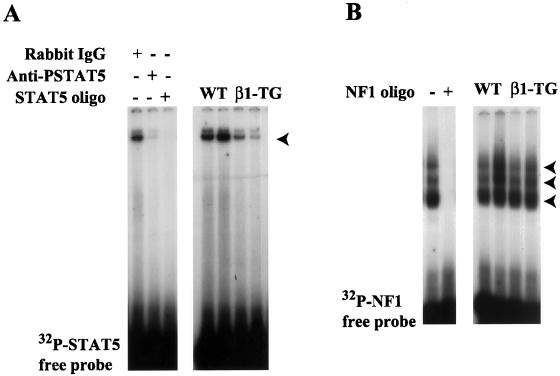
To determine the amount of activated STAT5 capable of interacting with DNA, we isolated nuclear proteins from involuting glands and performed a gel mobility shift assay, by using a fragment of the β-casein promoter containing the STAT5 binding site as a probe. A specific complex containing STAT5 was detected in nuclear extracts from 2-d involuting wild-type glands (Figure 5A). The binding of STAT5 to DNA was inhibited by adding an excess of unlabeled STAT5 oligonucleotides or anti-phospho-STAT5 antibody. Comparative analysis of nuclear extracts from wild-type and transgenic glands showed that transgenic glands contained significantly smaller amounts of STAT5 able to complex with DNA (Figure 5B). To determine whether the observed effect was specific to STAT5, we compared the DNA-binding activities of another transcription factor active in involuting mammary gland, NF1, in the nuclear extracts prepared from wild-type and transgenic glands. The levels of NF1 able to bind to DNA were not altered in transgenic glands (Figure 5B).
Figure 5.
Detection of STAT5 and NF1 in the nuclear extracts from wild-type and MMTV-β1-cyto mammary glands by band-shift analysis. (A) Left, 1 μg of nuclear extract from a 2-d wild-type-involuting gland was incubated with either 1 μg of rabbit IgG, 1 μg of anti-phospho-STAT5 antibody, or a 50-fold excess of unlabeled STAT5 oligonucleotide, before adding the 32P-labeled STAT5 probe. Right, 1 μg of nuclear extracts from two wild-type and two MMTV-β1-cyto transgenic glands was incubated with the 32P-labeled STAT5 probe. The arrowhead indicates the STAT5-DNA complex. (B) Left, 1 μg of nuclear extract from a 2-d wild-type-involuting gland was incubated with the 32P-labeled NF1 probe either in the presence (+) or absence (−) of a 50-fold excess of unlabeled NF1 oligonucleotide. Right, 1 μg of nuclear extracts from two wild-type and two MMTV-β1-cyto transgenic glands was incubated with the 32P-labeled NF1 probe. As described previously, three major NF1-DNA complexes indicated by arrowheads were detected in the mammary gland nuclear extracts (Furlong et al., 1996).
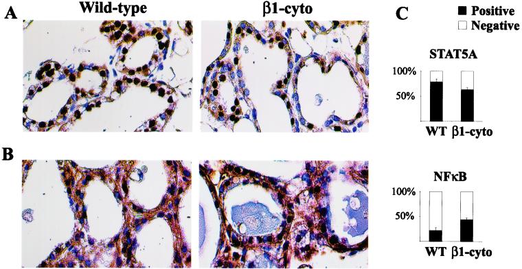
Immunohistochemical localization of STAT5A in 2-d involuting glands (Figure 6) revealed a small but statistically significant decrease in the amount of positive nuclei in transgenic animals compared with controls (63.9 ± 6.5 and 79.1 ± 5.4%, respectively; p ≤ 0.04). These results are consistent with the data obtained in gel mobility shift assay and indicate an early decrease in STAT5 activity in involuting MMTV-β1-cyto glands.
Figure 6.
Detection of STAT5A and NFκB in involuting mammary glands by immunohistochemical technique. (A and B) Paraffin sections of 2-d-involuting mammary glands were labeled with polyclonal antibodies to STAT5A (A) or the p65 subunit of NFκB (B) and counterstained with hematoxylin. (C) Content of the STAT5A and NFκB positive nuclei in involuting glands. The bars represent the relative content of positive nuclei, mean ± SEM, p < 0.04. Three wild-type and five transgenic animals were analyzed.
NF-κB Transcription Factor Is Prematurely Activated in Involuting MMTV-β1-cyto Mammary Glands
Numerous studies have shown that NFκB activity is subject to modulation by adhesion signals (Qwarnstrom et al., 1994; Ramarli et al., 1998; de Fougerolles et al., 2000). In addition, this transcription factor has been shown to inhibit STAT5-mediated β-casein gene expression (Geymayer and Doppler, 2000). We therefore examined the localization of the p65 subunit of NFκB in involuting transgenic and wild-type mammary glands. As reported previously (Brantley et al., 2000; Clarkson et al., 2000), we found that in the initial phase of involution, NFκB accumulated in the nuclei of a subpopulation of epithelial cells with some cytoplasmic staining also detected (Figure 6). Quantitative analysis showed that transgenic glands contained significantly more epithelial cell nuclei positive for p65 than did wild-type glands on day 2 of involution (44 ± 3.8 and 22.4 ± 4.8%, respectively; p ≤ 0.02).
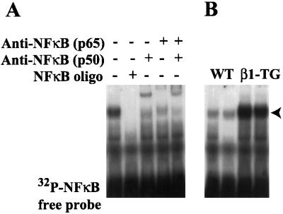
We evaluated the active NFκB content of nuclear extracts of wild-type and transgenic animals, by using an oligonucleotide containing the consensus NFκB element from the human immunodeficiency virus-long terminal repeat as a probe. Consistent with previous studies (Brantley et al., 2000; Clarkson et al., 2000), one major complex was detected in nuclear extracts from 2-d-involuting glands. The formation of this complex was inhibited by adding an excess of unlabeled NFκB oligonucleotide to the incubation mixture or by adding antibodies against the p50 and p65 subunits of NFκB (Figure 7), thus proving the specificity of the interaction. Importantly, analysis of the nuclear extracts of day 2 involuting glands by this method showed that significantly more NFκB was present in transgenic glands than in wild-type glands, confirming the data obtained by immunohistochemical methods (Figure 7B).
Figure 7.
Detection of nuclear NFκB by band-shift analysis. (A) Two micrograms of nuclear extract from a 2-d wild-type-involuting gland was incubated with either 1 μg of polyclonal antibodies to p50, p65, or the combination of both or a 50-fold excess of unlabeled NFκB oligonucleotide, before adding the 32P-labeled NFκB probe. (B) Two micrograms of nuclear extract from two wild-type and two MMTV-β1-cyto transgenic glands was incubated with the 32P-labeled NFκB probe. The arrowhead indicates the NFκB–DNA complex.
Expression of β1-CD4 Chimera in Cultured Cells Interferes with Prolactin/STAT5 Signaling Pathway, but Does Not Alter NFκB Activity
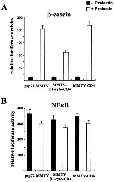
The effects of the chimeric proteins similar to that used in this study, i.e., containing β1-integrin cytoplasmic domain, on the adhesion-associated events were extensively examined in cultured cells. However, so far, the impact of the β1-integrin cytoplasmic domain overexpression on the prolactin/STAT5 and the NFκB signaling pathways was not analyzed. We therefore addressed this question by using a transient transfection approach. COS7 cells that have been used in these experiments are devoid of PRLR and STAT5; however, the prolactin/STAT5 signaling pathway can be reconstructed by cotransfection of the plasmids encoding PRLR and STAT5 and activated by prolactin (Pfitzner et al., 1998). To assess the effects of β1-CD4 chimera on activation of the prolactin/STAT5 signaling pathway, COS7 cells were transiently cotranfected with the required pathway components and the MMTV-β1-cyto plasmid. β-Casein gene promoter-luciferase reporter was used to monitor the prolactin/STAT5 pathway activation. Expression of β1-CD4 chimera resulted in a twofold decrease of prolactin-induced transcription from the reporter plasmid (Figure 8A). These data prove that expression of the transgenic protein β1-CD4 interferes with the activation of the prolactin/STAT5 signaling pathway.
Figure 8.
Effects of β1-CD4 expression on the activation of the prolactin/STAT5 and the NFκB signaling pathways in COS7 cells. Cells were transiently cotransfected with plasmids encoding PRLR and STAT5A, and, when mentioned, with the psp72-MMTV, MMTV-β1-CD4, or MMTV-CD4 plasmids. (A) β-Casein gene promoter-luciferase reporter activity. (B) NFκB-luciferase reporter activity. In each case, the data shown are the results of a representative experiment performed in duplicate. Mean values are presented with SD indicated by bars.
NFκB-luciferase reporter plasmid was used to analyze the effect of β1-CD4 expression on the activation status of the NFκB pathway. Transfection of the MMTV-β1-cyto plasmid did not change NFκB activity levels in prolactin-treated or untreated cells (Figure 8B). Moreover, HC11 mammary epithelial cells stably transfected with MMTV-β1-cyto plasmid exhibited NFκB activity levels similar to those detected in mock-transfected cells (our unpublished data). These data suggest that under the conditions of the experiment, expression of β1-CD4 did not additionally activate the NFκB pathway.
DISCUSSION
Our previous work has shown that the mammary epithelium of MMTV-β1-cyto mice presents defects in growth and differentiation during pregnancy and early (2-d) lactation (Faraldo et al., 1998, 2001). However, in this study, we have found that later, at peak lactation (7 d), the morphology of transgenic and wild-type mammary glands was similar, with secretory epithelial cells achieving a fully differentiated phenotype, as assessed by the expression of milk protein genes. Thus, the defects in growth and differentiation observed in MMTV-β1-cyto glands were temporary, and the expression of a β1-integrin dominant negative mutant in the mammary epithelium delayed mammary gland development during lactation.
The mammary gland begins to undergo involution after cessation of the suckling stimulus and resulting milk stasis at weaning. To study the effects of the perturbation of the cell–ECM interactions in the mammary epithelium of MMTV-β1-cyto mice on involution, pups were removed from their mothers after 1 wk of lactation, when the transgenic mammary glands had already attained the wild-type phenotype. We found that the expression of the β1-integrin dominant negative mutant resulted in the precocious dedifferentiation of milk-producing cells, because transgenic glands contained less mRNA encoding WAP and β-casein on the 2nd and 4th d of involution than did wild-type glands. This precocious dedifferentiation of secretory epithelial cells in the transgenic glands was accompanied by a decrease in nuclear STAT5 levels and an increase in NFκB activity.
STAT5 is an essential regulator of the mammary gland development and function (Liu et al., 1997; Teglund et al., 1998). In this study, we have demonstrated that expression of β1-CD4 chimera in cultured cells diminished prolactin/STAT5-dependent transcription from the β-casein promoter-luciferase reporter plasmid, and thus, interfered with prolactin/STAT5 pathway activation. Consistently, the STAT5 activity was impaired in involuting (this study) and, according to our preliminary data, in 2-d-lactating MMTV-β1-cyto glands (not shown). Together, these results suggest that in vivo, perturbation of cell–ECM interactions affects the prolactin/STAT5 signaling pathway resulting in deficient mammary gland development.
It is thought that STAT5 activity is regulated mainly by phosphorylation induced by prolactin or other factors. Surprisingly, the levels of STAT5 and JAK2 phosphorylation were similar in transgenic and wild-type glands. However, the amount of active STAT5 localized in the mammary epithelial cell nuclei was significantly smaller in precociously dedifferentiated MMTV-β1-cyto glands. These data are in agreement with the observation that Tyr phosphorylation of STAT5 does not grant its nuclear translocation (Ali, 1998) and suggest that adhesion-associated events may participate in the control of the STAT5 function regulating its intracellular distribution. Similarly, a recent study has shown that cell-ECM adhesion can regulate extracellular signal-regulated kinase activity by affecting its transport to the nucleus (Aplin et al., 2001).
Numerous studies have shown that NFκB is activated by adhesion (Qwarnstrom et al., 1994; Ramarli et al., 1998; de Fougerolles et al., 2000). Paradoxically, the results of this work show that the perturbation of β1-integrin–mediated adhesion in involuting mammary glands led to precocious NFκB activation. However, the regulation of NFκB activity is complex and involves many factors not necessarily directly relevant to adhesion. Interestingly, NFκB activity is maximal at the onset of the second phase of involution (Clarkson et al., 2000), when the basement membrane is degraded and cell–matrix interactions are disrupted (Talhouk et al., 1992; Lund et al., 1996). We could not reveal any stimulation of NFκB in cultured cells expressing the β1-CD4 chimera. Therefore, the premature NFκB activation observed in MMTV-β1-cyto glands may result from some other alterations induced by perturbation of cell–ECM interactions, particular to the specific tissue context, i.e., involuting mammary gland, and difficult to approach in vitro. In conclusion, our data suggest that in transgenic glands, the perturbation of cell–ECM interactions involving β1-integrins may cause precocious activation of the processes normally involved in the later stages of involution accompanied by important ECM degradation.
At peak lactation, in the absence of ductal and alveolar growth, when the fully differentiated secretory cells have ceased proliferating, and apoptosis is an extremely rare event, MMTV-β1-cyto glands were similar to those from wild-type animals. The transgenic mice exhibited a clearly distinct mammary phenotype only at dynamic stages of mammary development, such as growth phase in pregnancy and at the beginning of lactation, or gland remodeling during involution. All these stages of mammary development require matrix metalloproteinases whose expression and activity is tightly controlled by cell–ECM interactions and thus may be altered in transgenic glands (for review, see Sternlicht and Werb, 2001). This issue remains to be investigated.
Our previous studies have shown that the transgene expression during mammary gland growth phase at midpregnancy and at the beginning of lactation resulted in moderately elevated mammary epithelial cell apoptosis rates (Faraldo et al., 1998). However, we did not observe any significant increase of the apoptosis levels in involuting transgenic glands. These data are in agreement with the observation that conditional ablation of β1-integrin in epidermis did not lead to increased apoptosis rates (Brakebusch et al., 2000). Furthermore, the phosphorylation level and the amount of extracellular signal-regulated kinase, c-Jun NH2-terminal kinase, focal adhesion kinase, and Akt, the molecules implicated in transmission of the cell survival signal triggered by integrin-mediated adhesion in involuting MMTV-β1-cyto glands, were similar to those in wild-type glands (our unpublished data).
In the MMTV-β1-cyto mice, the content of β1-integrin in the mammary epithelium was significantly decreased compared with that in wild-type glands. Expression of the similar chimeric proteins in cultured cells was reported to alter the activation status of endogenous integrins, rather than their expression levels (Bodeau et al., 2001). We suggest that down-regulation of β1-integrin expression observed in MMTV-β1-cyto glands may result from the perturbation of cell-ECM adhesion induced by the long-term transgene expression in vivo. A progressive loss of β1-integrin from differentiating keratinocytes, when they move out from the basal layer of the epidermis and loose contact with the basement membrane provides an example of such negative regulation (Adams and Watt, 1990).
In addition to β1-family, α6β4 is the major integrin ECM receptor expressed by mammary epithelial cells. A study by Muschler et al. (1999) carried out with cultured mammary epithelial cells reported that the signals from both β1 and β4-integrins are required for β-casein expression and suggested that these integrins may act in concert in the control of the mammary epithelial cell differentiation. Furthermore, two recent reports have demonstrated that conditional ablation of β1-integrin in the epidermis resulted in a reduced expression of α6β4 integrin in basal keratinocytes (Brakebusch et al., 2000; Raghavan et al., 2000). These results suggest that the perturbation of β1-integrin function may subsequently alter the function and signaling events associated with α6β4. Accordingly, in the MMTV-β1-cyto lactating glands, we have observed an abnormal localization of the β4-integrin chain at the lateral surfaces of the alveolar epithelial cells (Faraldo et al., 1998).
Experiments performed with cultured cells suggested that the chimeric proteins similar to that used in this study, i.e., containing β1-integrin cytoplasmic domain, may also interfere with β3 and β5-integrin function (Lukashev et al., 1994). These β-chains interact with αv to form an integrin dimer. Although the αv chain is expressed by many mammary epithelial cell lines, in mammary gland, this integrin chain was not found in luminal epithelial cells, and only a small amount of it was detected on the basal surface of myoepithelial cells (Zutter et al., 1990; Clezardin et al., 1993; Anbazhagan et al., 1995). Similar to β1-integrin, αvβ3 was implicated in the control of cell survival (Brooks et al., 1994). However, using immunofluorescence methods, we did not observe any compensatory increase of αvβ3 or other αv-containing integrins in MMTV-β1-cyto glands (unpublished data).
Briefly, in this study, we show that expression of a β1-integrin dominant negative mutant in involuting mammary gland did not alter the kinetics of apoptosis but resulted in the premature dedifferentiation of secretory epithelium; affected the prolactin/STAT5 signaling pathway, essential for mammary development; and induced the precocious activation of NFκB transcription factor. These results reveal an important role of cell–ECM interactions mediated by β1-integrins in the control of milk gene expression and in maintenance of the mammary epithelial cell differentiated state in vivo.
ACKNOWLEDGMENTS
We particularly thank Drs. I. Cerutti and C. Gouget, and the personnel of the Service d'Expérimentation Animale et de Transgenèse, Villejuif, especially R. Duchâteau and A. Loeuillet, for taking care of the transgenic mice. We also greatly appreciate advice of Drs. W. Doppler, P. Furth, D. Medina, and J. Teulière, and donation of reagents by Drs. W. Doppler, R. Hynes, R. Jaggi, and J. Rosen. This work was supported by the Association pour la Recherche contre le Cancer (ARC 4440). M.M.F. was supported by fellowships from the Institut Curie and from the Fondation pour la Recherche Médicale. M.-A.D. is Chargé Recherche, and M.A.G. is Directeur de Recherche at the Institut de la Santé et de la Recherche Médicale.
Abbreviations used:
- MMTV
mouse mammary tumor virus
- ECM
extracellular matrix
- WAP
whey acidic protein
Footnotes
Article published online ahead of print. Mol. Biol. Cell 10.1091/mbc.E02–02–0086. Article and publication date are at www.molbiolcell.org/cgi/doi/10.1091/mbc.E02–02–0086.
REFERENCES
- Adams JC, Watt FM. Changes in keratinocyte adhesion during terminal differentiation: reduction in fibronectin binding precedes α5β1 integrin loss from the cell surface. Cell. 1990;63:425–435. doi: 10.1016/0092-8674(90)90175-e. [DOI] [PubMed] [Google Scholar]
- Ali S. Prolactin receptor regulates Stat5 tyrosine phosphorylation and nuclear translocation by two separate pathways. J Biol Chem. 1998;273:7709–7716. doi: 10.1074/jbc.273.13.7709. [DOI] [PubMed] [Google Scholar]
- Anbazhagan R, Bartkova J, Stamp G, Pignatelli M, Gusterson B, Bartek J. Expression of integrin subunits in the human infant breast correlates with morphogenesis and differentiation. J Pathol. 1995;176:227–232. doi: 10.1002/path.1711760304. [DOI] [PubMed] [Google Scholar]
- Aplin AE, Stewart SA, Assoian RK, Juliano RL. Integrin-mediated adhesion regulates ERK nuclear translocation and phosphorylation of Elk-1. J Cell Biol. 2001;153:273–283. doi: 10.1083/jcb.153.2.273. [DOI] [PMC free article] [PubMed] [Google Scholar]
- Barkett M, Gilmore TD. Control of apoptosis by Rel/NF-κB transcription factors. Oncogene. 1999;18:6910–6924. doi: 10.1038/sj.onc.1203238. [DOI] [PubMed] [Google Scholar]
- Bodeau AL, Berrier AL, Mastrangelo AM, Martinez R, LaFlamme SE. A functional comparison of mutations in integrin β cytoplasmic domains: effects on the regulation of tyrosine phosphorylation, cell spreading, cell attachment and β1 integrin conformation. J Cell Sci. 2001;114:2795–2807. doi: 10.1242/jcs.114.15.2795. [DOI] [PubMed] [Google Scholar]
- Brakebusch C, et al. Skin and hair follicle integrity is crucially dependent on β1 integrin expression on keratinocytes. EMBO J. 2000;19:3990–4003. doi: 10.1093/emboj/19.15.3990. [DOI] [PMC free article] [PubMed] [Google Scholar]
- Brantley DM, Chen CL, Muraoka RS, Bushdid PB, Bradberry JL, Kittrell F, Medina D, Matrisian LM, Kerr LD, Yull FE. Nuclear factor-κB (NF-κB) regulates proliferation and branching in mouse mammary epithelium. Mol Biol Cell. 2001;12:1445–1455. doi: 10.1091/mbc.12.5.1445. [DOI] [PMC free article] [PubMed] [Google Scholar]
- Brantley DM, Yull FE, Muraoka RS, Hicks DJ, Cook CM, Kerr LD. Dynamic expression and activity of NF-κB during post-natal mammary gland morphogenesis. Mech Dev. 2000;97:149–155. doi: 10.1016/s0925-4773(00)00405-6. [DOI] [PubMed] [Google Scholar]
- Brooks PC, Montgomery AM, Rosenfeld M, Reisfeld RA, Hu T, Klier G, Cheresh DA. Integrin αvβ3 antagonists promote tumor regression by inducing apoptosis of angiogenic blood vessels. Cell. 1994;79:1157–1164. doi: 10.1016/0092-8674(94)90007-8. [DOI] [PubMed] [Google Scholar]
- Campbell SM, Rosen JM, Hennighausen LG, Strech-Jurk U, Sippel AE. Comparison of the whey acidic protein genes of the rat and mouse. Nucleic Acids Res. 1984;12:8685–8697. doi: 10.1093/nar/12.22.8685. [DOI] [PMC free article] [PubMed] [Google Scholar]
- Clezardin P, Frappart L, Clerget M, Pechoux C, Delmas PD. Expression of thrombospondin (TSP1) and its receptors (CD36 and CD51) in normal, hyperplastic, and neoplastic human breast. Cancer Res. 1993;53:1421–1430. [PubMed] [Google Scholar]
- Clarkson RW, Heeley JL, Chapman R, Aillet F, Hay RT, Wyllie A, Watson CJ. NF-κB inhibits apoptosis in murine mammary epithelia. J Biol Chem. 2000;275:12737–12742. doi: 10.1074/jbc.275.17.12737. [DOI] [PubMed] [Google Scholar]
- de Fougerolles AR, Chi-Rosso G, Bajardi A, Gotwals P, Green CD, Koteliansky VE. Global expression analysis of extracellular matrix-integrin interactions in monocytes. Immunity. 2000;13:749–758. doi: 10.1016/s1074-7613(00)00073-x. [DOI] [PubMed] [Google Scholar]
- Edwards GM, Wilford FH, Liu X, Hennighausen L, Djiane J, Streuli CH. Regulation of mammary differentiation by extracellular matrix involves protein-tyrosine phosphatases. J Biol Chem. 1998;273:9495–9500. doi: 10.1074/jbc.273.16.9495. [DOI] [PubMed] [Google Scholar]
- Faraldo MM, Deugnier MA, Lukashev M, Thiery JP, Glukhova MA. Perturbation of β1-integrin function alters the development of murine mammary gland. EMBO J. 1998;17:2139–2147. doi: 10.1093/emboj/17.8.2139. [DOI] [PMC free article] [PubMed] [Google Scholar]
- Faraldo MM, Deugnier MA, Thiery JP, Glukhova MA. Growth defects induced by perturbation of β1-integrin function in the mammary gland epithelium result from a lack of MAPK activation via the Shc and Akt pathways. EMBO Rep. 2001;2:431–437. doi: 10.1093/embo-reports/kve086. [DOI] [PMC free article] [PubMed] [Google Scholar]
- Furlong EEM, Keon NK, Thornton FD, Rein T, Martin F. Expression of a 74-kDa nuclear factor 1 (NF1) protein is induced in mouse mammary gland involution. Involution-enhanced occupation of a twin NF1 binding element in the testosterone-repressed prostate message-2/clusterin promoter. J Biol Chem. 1996;271:29688–29697. doi: 10.1074/jbc.271.47.29688. [DOI] [PubMed] [Google Scholar]
- Geymayer S, Doppler W. Activation of NF-κB p50/p65 is regulated in the developing mammary gland and inhibits STAT5-mediated β-casein gene expression. FASEB J. 2000;14:1159–1170. doi: 10.1096/fasebj.14.9.1159. [DOI] [PubMed] [Google Scholar]
- Giancotti FG, Ruoslahti E. Integrin signaling. Science. 1999;285:1028–1032. doi: 10.1126/science.285.5430.1028. [DOI] [PubMed] [Google Scholar]
- Groner B, Gouilleux F. Prolactin-mediated gene activation in mammary epithelial cells. Curr Opin Genet Dev. 1995;5:587–594. doi: 10.1016/0959-437x(95)80027-1. [DOI] [PubMed] [Google Scholar]
- Gupta P, Rosen JM, D'Eustachio P, Ruddle FH. Localization of the casein gene family to a single mouse chromosome. J Cell Biol. 1982;93:199–204. doi: 10.1083/jcb.93.1.199. [DOI] [PMC free article] [PubMed] [Google Scholar]
- Li M, Liu X, Robinson G, Bar-Peled U, Wagner KU, Young WS, Hennighausen L, Furth PA. Mammary-derived signals activate programmed cell death during the first stage of mammary gland involution. Proc Natl Acad Sci USA. 1997;94:3425–3430. doi: 10.1073/pnas.94.7.3425. [DOI] [PMC free article] [PubMed] [Google Scholar]
- Liu X, Robinson GW, Wagner KU, Garrett L, Wynshaw-Boris A, Hennighausen L. Stat5a is mandatory for adult mammary gland development and lactogenesis. Genes Dev. 1997;11:179–186. doi: 10.1101/gad.11.2.179. [DOI] [PubMed] [Google Scholar]
- Lukashev ME, Sheppard D, Pytela R. Disruption of integrin function and induction of tyrosine phosphorylation by the autonomously expressed β1 integrin cytoplasmic domain. J Biol Chem. 1994;269:18311–18314. [PubMed] [Google Scholar]
- Lund LR, Romer J, Thomasset N, Solberg H, Pyke C, Bissell MJ, Dano K, Werb Z. Two distinct phases of apoptosis in mammary gland involution: proteinase-independent and -dependent pathways. Development. 1996;122:181–193. doi: 10.1242/dev.122.1.181. [DOI] [PMC free article] [PubMed] [Google Scholar]
- Luo G, Yu-Lee L. Stat5b inhibits NF-κB-mediated signaling. Mol Endocrinol. 2000;14:114–123. doi: 10.1210/mend.14.1.0399. [DOI] [PubMed] [Google Scholar]
- Marcantonio EE, Hynes RO. Antibodies to the conserved cytoplasmic domain of the integrin β1 subunit react with proteins in vertebrates, invertebrates, and fungi. J Cell Biol. 1988;106:1765–1772. doi: 10.1083/jcb.106.5.1765. [DOI] [PMC free article] [PubMed] [Google Scholar]
- Mayr S, Welte T, Tomasi JK, Bruno P, Cairns C, Gustafsson J, Doppler W. Selective coupling of STAT factors to the mouse prolactin receptor. Eur J Biochem. 1998;258:784–793. doi: 10.1046/j.1432-1327.1998.2580784.x. [DOI] [PubMed] [Google Scholar]
- Muschler J, Lochter A, Roskelley CD, Yurchenco P, Bissell MJ. Division of labor among the α6β4 integrin, β1 integrins, and an E3 laminin receptor to signal morphogenesis and β-casein expression in mammary epithelial cells. Mol Biol Cell. 1999;10:2817–2828. doi: 10.1091/mbc.10.9.2817. [DOI] [PMC free article] [PubMed] [Google Scholar]
- Musikacharoen T, Matsuguchi T, Kikuchi T, Yoshikai Y. NF-κB, and STAT5 play important roles in the regulation of mouse Toll-like receptor 2 gene expression. J Immunol. 2001;166:4516–4524. doi: 10.4049/jimmunol.166.7.4516. [DOI] [PubMed] [Google Scholar]
- Pattengale PK, Stewart TA, Leder A, Sinn E, Muller W, Tepler I, Schmidt E, Leder P. Animal models of human disease. Pathology and molecular biology of spontaneous neoplasms occurring in transgenic mice carrying and expressing activated cellular oncogenes. Am J Pathol. 1989;135:39–61. [PMC free article] [PubMed] [Google Scholar]
- Pfitzner E, Jahne R, Wissler M, Stoecklin E, Groner B. p300/CREB-binding protein enhances the prolactin-mediated transcriptional induction through direct interaction with the transactivation domain of Stat5, but does not participate in the Stat5-mediated suppression of the glucocorticoid response. Mol Endocrinol. 1998;12:1582–1593. doi: 10.1210/mend.12.10.0180. [DOI] [PubMed] [Google Scholar]
- Qwarnstrom EE, Ostberg CO, Turk GL, Richardson CA, Bomsztyk K. Fibronectin attachment activates the NF-κB p50/p65 heterodimer in fibroblasts and smooth muscle cells. J Biol Chem. 1994;269:30765–30768. [PubMed] [Google Scholar]
- Raghavan S, Bauer C, Mundschau G, Li Q, Fuchs E. Conditional ablation of β1 integrin in skin. Severe defects in epidermal proliferation, basement membrane formation, and hair follicle invagination. J Cell Biol. 2000;150:1149–1160. doi: 10.1083/jcb.150.5.1149. [DOI] [PMC free article] [PubMed] [Google Scholar]
- Ramarli D, Scupoli MT, Fiorini E, Poffe O, Brentegani M, Villa A, Cecchini G, Tridente G, Marchisio PC. Thymocyte contact or monoclonal antibody-mediated clustering of α3β1 or α6β4 integrins activate interleukin-6 (IL-6) transcription factors (NF-κB and NF-IL6) and IL-6 production in human thymic epithelial cells. Blood. 1998;92:3745–3755. [PubMed] [Google Scholar]
- Shapiro DJ, Sharp PA, Wahli WW, Keller MJ. A high-efficiency HeLa cell nuclear transcription extract. DNA. 1988;7:47–55. doi: 10.1089/dna.1988.7.47. [DOI] [PubMed] [Google Scholar]
- Sternlicht MD, Werb Z. How matrix metalloproteinases regulate cell behavior. Annu Rev Cell Dev Biol. 2001;17:463–516. doi: 10.1146/annurev.cellbio.17.1.463. [DOI] [PMC free article] [PubMed] [Google Scholar]
- Strange R, Li F, Saurer S, Burkhardt A, Friis RR. Apoptotic cell death and tissue remodeling during mouse mammary gland involution. Development. 1992;115:49–58. doi: 10.1242/dev.115.1.49. [DOI] [PubMed] [Google Scholar]
- Streuli CH, Edwards GM, Delcommenne M, Whitelaw CB, Burdon TG, Schindler C, Watson CJ. Stat5 as a target for regulation by extracellular matrix. J Biol Chem. 1995;270:21639–21644. doi: 10.1074/jbc.270.37.21639. [DOI] [PubMed] [Google Scholar]
- Sympson CJ, Talhouk RS, Alexander CM, Chin JR, Clift SM, Bissell MJ, Werb Z. Targeted expression of stromelysin-1 in mammary gland provides evidence for a role of proteinases in branching morphogenesis and the requirement for an intact basement membrane for tissue-specific gene expression. J Cell Biol. 1994;125:681–693. doi: 10.1083/jcb.125.3.681. [DOI] [PMC free article] [PubMed] [Google Scholar]
- Talhouk RS, Bissell MJ, Werb Z. Coordinated expression of extracellular matrix-degrading proteinases and their inhibitors regulates mammary epithelial function during involution. J Cell Biol. 1992;118:1271–1282. doi: 10.1083/jcb.118.5.1271. [DOI] [PMC free article] [PubMed] [Google Scholar]
- Teglund S, McKay C, Schuetz E, van Deursen JM, Stravopodis D, Wang D, Brown M, Bodner S, Grosveld G, Ihle JN. Stat5a and Stat5b proteins have essential and nonessential, or redundant, roles in cytokine responses. Cell. 1998;93:841–850. doi: 10.1016/s0092-8674(00)81444-0. [DOI] [PubMed] [Google Scholar]
- Walker NI, Bennett RE, Kerr JF. Cell death by apoptosis during involution of the lactating breast in mice and rats. Am J Anat. 1989;185:19–32. doi: 10.1002/aja.1001850104. [DOI] [PubMed] [Google Scholar]
- Zutter MM, Mazoujian G, Santoro SA. Decreased expression of integrin adhesive protein receptors in adenocarcinoma of the breast. Am J Pathol. 1990;137:863–870. [PMC free article] [PubMed] [Google Scholar]


