Abstract
The immune response to Borrelia burgdorferi, the causative agent of Lyme disease, is complex. We studied the immunoglobulin M (IgM) and IgG antibody response to N40Br, a sensu stricto strain, in the rhesus macaque(nonhuman primate [NHP]) model of infection to identify the spirochetal protein targets of specific antibody. Antigens used in enzyme-linked immunosorbent assays were whole-cell sonicates of the spirochete and recombinant proteins of B. burgdorferi. Immunoblotting with a commercially available strip and subsequent quantitative densitometry of the bands were also used. Sera from four different groups of NHPs were used: immunocompetent, transiently immunosuppressed, extended immunosuppressed, and uninfected. In immunocompetent and transiently immunosuppressed NHPs, there was a strong IgM and IgG response. Major proteins for the early IgM response were P39 and P41 and recombinant BmpA and OspC. Major proteins for the later IgG response were P39, P41, P18, P60, P66, and recombinant BmpA and DbpA. There was no significant response in the NHPs to recombinant OspA or to Arp, a 37-kDa protein that elicits an antibody response during infection in mice. Most antibody responses, except for that to DbpA, were markedly diminished by prolonged dexamethasone treatment. This study supports the hypothesis that recombinant proteins may provide a useful adjunct to current diagnostic testing for Lyme borreliosis.
Lyme borreliosis is a protean, multisystemic disease caused by infection with the spirochete Borrelia burgdorferi, transmitted via tick bites (30). The nonhuman primate (NHP) model is particularly useful for evaluating neurological involvement (8, 10, 23, 27) but is also helpful for analyzing the humoral immune response to the spirochete in an animal phylogenetically similar to humans. Within a few weeks after the NHP is subjected to intradermal inoculation, B. burgdorferi can be found in the blood by PCR or culture. Spirochetes disseminate to tissues, including the heart, bladder, and central and peripheral nervous systems (22). Pathogen-specific immunoglobulins with the aid of complement and macrophages are able to limit spirochetal number but not to eradicate the pathogen in chronic infection (26).
In infected humans or experimental animals, the spirochete must evade a vigorous host innate and acquired immune response, and selective gene expression by the spirochete may improve its survival. Antigens expressed by B. burgdorferi change during the course of infection (3, 13, 36), but it is unclear whether this is the primary mechanism of immune evasion. What is clear, however, is that antibodies with new specificities, directed at the newly appearing antigens, seem to be produced at various times after infection. Some spirochetal antigens expressed in vivo during infection are expressed in limited amounts or not at all during in vitro culture so that assays of antibodies utilizing in vitro cultured spirochetes, such as enzyme-linked immunosorbent assays (ELISAs) or immunoblotting with sonicates of cultured spirochetes, are not ideal for defining the full range of anti-B. burgdorferi antibodies produced by the host. To address this problem, recombinant proteins of B. burgdorferi are beginning to be used as antigens in either ELISAs or immunoblot assays (18, 19). The use of recombinant proteins in characterization of the humoral response has not been extensively characterized in humans or in experimental models. We compared recombinant proteins to sonicates from in vitro-cultured spirochetes as sources of antigens in ELISAs and immunoblotting.
MATERIALS AND METHODS
Animals.
Male rhesus macaques, Macaca mulatta, 3 to 4 years of age, were anesthetized and underwent phlebotomies as previously described (23). Housing and care were in accordance with the Animal Welfare Act and the Guide for the Care and Use of Laboratory Animals in facilities accredited by the Association for Assessment and Accreditation of Laboratory Animal Care International. The study was reviewed and approved by the New Jersey Medical School and the University of California, Davis, Animal Care and Use Committees. Complete blood count and chemistry data were obtained on all animals at each phlebotomy for monitoring purposes. All NHPs had received standard vaccinations, including measles and tetanus, as infants, and received constant veterinary clinical surveillance.
NHPs inoculated intradermally with B. burgdorferi and not treated with dexamethasone were considered immunocompetent (IC). The identification numbers for these animals were DES9, E78, U328, 80T11, E520, 23Z, E680, TO21, TO26, TO32, PAX35, and PAX40. Another group of NHPs were labeled as transiently immunosuppressed (TISP). These animals, 3 days prior to inoculation, received dexamethasone, 2 mg/kg, once daily, which was continued until 28 days post-infection (p.i.), at which time the 1-mg/kg dose was administered once daily for 1 week, after which no more dexamethasone was given. This dose of dexamethasone is considered relatively mild in NHPs, and no steroid-induced side effects (e.g., fluid retention manifested as weight gain, glucose intolerance, change in appearance, and change in behavior, etc.) resulted from this protocol. The TISP group included the following animals: 30099, 30177, 30199,30389, 30154, 30192, 30211, and 30242. In other NHPs which were more strongly immunosuppressed and labeled as immunosuppressed (IS), dexamethasone was continued for the duration of the experiment. The following animals were in this group: 1538, 1614, PAX219, and Z1.
Two types of normal control (NC) sera were used: baseline sera prior to inoculation were available from all animals, and sera from eight animals which had never been infected (i.e., 20127, 21887, 22318, 24037, 27460, 27363, 28842, and 27620) were also available.
Inoculation.
Inoculations were performed intradermally with a total volume of 1 ml, in multiple aliquots of about 0.1 ml each (containing a total of 1 million N40Br strain spirochetes/NHP) along the dorsal thoracic midline, as in previous studies (23, 24, 25) with the exception of the TISP animals 30154, 30192, 30211, and 30242, which were inoculated by allowing infected ticks to feed on them, as previously described (26). The tick-inoculated animals were indistinguishable from the needle-inoculated animals by all measures of infection, immunity, and inflammation (26); thus, the antibody responses of all TISP animals have been analyzed together as a group. N40Br is a B. burgdorferi sensu stricto strain isolated initially from ticks and subsequently isolated from the brain of infected mice (20). All inoculated NHPs were confirmed to be infected by PCR with multiple tissues, including cardiac and skeletal muscle, bladder, and peripheral nerve tissue (2, 22, 24, 25, 27).
Recombinant proteins.
Proteins derived by screening a B. burgdorferi N40 genomic expression library with sera from infected mice were obtained as previously described (5, 11, 12). Some of these proteins represented known, well-characterized proteins of B. burgdorferi, i.e., OspA (p31), OspC (p23), DbpA (p22), BmpA (p39), flagellin (p41), and Arp (a p37 protein). Other recombinants which were tested included proteins of no known function, i.e., p19-23, p23-T2, p27, p29, p31(not OspA), p37-42, p44, p45, and p61.
Antibody to B. burgdorferi. (i) ELISA.
Serum antibody ELISAs were performed essentially as previously described (20, 23, 25). Units for each assay measuring immunoglobulin G (IgG) responses to whole-cell sonicates (WCS) were calculated using a high-titer positive control serum sample to assign units of binding. This serum was diluted in such a way that 100 U represented binding at the top of the linear portion of the curve. The technique was tested on sera to ensure that readings in the linear portion of the curve (generally an optical density [OD] of 0.2 to 1.0), gave internally consistent units. A standard curve was run each time, and units were assigned according to calculation from the standard curve. The numbers shown in the results below represent means of the values for all the NHPs in each group.
For the measurement of antibody to recombinant proteins by ELISA, a panel of sera from four representative NHPs in each group was used: for the IC group, sera from DES9, E78, TO32, and PAX40; for the TISP group, sera from 30099, 30154, 30177, and 30242; for the IS, group, PAX219, Z1,1538, and 1614. Preinfection samples were considered to be NCs in this group.
(ii) Immunoblotting.
Commercial B. burgdorferi sensu stricto nitrocellulose strips (Microbiology Reference Laboratory, Cypress, Calif.) were used as previously described (25) according to the instructions of the diagnostic kit. The strain used for preparation of these blots was a B. burgdorferi sensu stricto strain, called CB, an isolate from an erythema migrans lesion from a patient at New York Medical College in Valhalla, N.Y.
Quantitative analysis of band density for immunoblotting.
Densities of bands were determined by calculating a ratio referenced to a positive control used for all blots, similar to a procedure previously described for evaluating immunoblots in human Lyme neuroborreliosis (21). Specifically, the immunoblot was captured digitally with a Kodak DC120 digital camera onto Kodak 1D imaging software (Kodak Scientific Imaging Systems, New Haven, Conn.). For each band of interest, a ratio was calculated as follows: band density of relevant band/band density of reference band × 100. For IgG the reference band was the 60-kDa band of the high-titer positive control serum, and for IgM the reference band was the 39-kDa band of the high-titer positive control serum. The above bands were chosen because the positive control sera reacted moderately strongly with these bands in a linear range of signal development.
RESULTS
Anti-B. burgdorferi ELISA with N40 WCS as antigen. (i) IgM.
The IgM results were variable within each group of animals; i.e., the range of values at each time point varied up to 100% above and below the mean. However, the general patterns of the curves for each animal were similar within a group. Thus, the results for IC animals peaked in the first month and were back to baseline by week 8. In the TISP animals, the IgM levels continued to rise until approximately week 6, at which time they began to drop. The IS animals had IgM levels which continued to rise until necropsy and did not fall at any point, a finding which confirmed our earlier data (26). The levels of IgM antibody to the spirochete in the IS animals were much higher than the peak in the IC or TISP NHPs. The height of the IgM levels of animals within a group did not predict their IgG levels.
(ii) IgG.
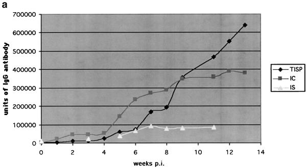
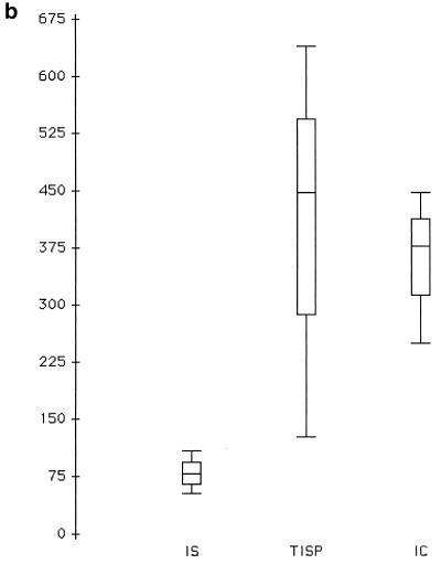
The units of IgG antibody were averaged for each group and each time after inoculation (Fig. 1). The range of values at each point was approximately 20% above and below the mean. The IC animals developed their IgG response earlier, but the amplitude of the antibody was eventually higher in the TISP group. The IgG response in the IS group was not completely suppressed, being clearly above baseline, which was the mean of the NC group, or 4 × 10³ units.
FIG. 1.
(a) Total IgG anti-B. burgdorferi antibody response. Amplitude of IgG anti-B. burgdorferi response, expressed as a mean, as a function over time p.i. for the three groups of infected NHPs. Units of amplitude were determined by comparison with a standard NHP anti-B. burgdorferi antiserum as described in Materials Methods. (b) Comparison of IgG antibody among groups at week 11 p.i. Week 11 was chosen as the date for comparison because it was the latest time p.i. available for all groups. The y axis represents units, and the x axis shows the three groups of NHPs in panel a. The hatched areas are 95% confidence intervals, and the lines of the box represent medians, range from 25th to 75th percentile, and the total range.
Immunoblotting. (i) IgM immunoblotting.
The strongest IgM response in IC and TISP NHPs was to p39 and p41. An anti-p23, presumably anti-OspC, response was present in only 2 of the 14 IC and TISP NHPs, and the band was weak. Other commonly identified proteins in the IgM response in these animals were p58 and p93. As predicted from previously published data (25), the IgM response in the TISP group was increased both in magnitude and duration, and thus the above responses were stronger in TISP animals than in IC animals. Sera from all IC and TISP animals within the first month after infection met criteria for IgM positivity.
(ii) IgG immunoblotting.
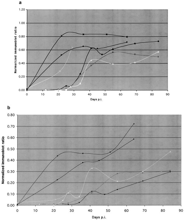
Similar results were obtained for the IC and TISP groups, except for a delay of approximately 1 to 2 weeks in the development of some bands by the TISP NHPs relative to the IC NHPs in the first 2 months of infection and the development of stronger immunoblot reactivity than the IC NHPs in the 3rd month of infection. Table 1 shows the predominant bands which were present by immunoblotting in the IC NHPs. Other bands present in many of the NHPs were 23, 31, 34, and 58 kDa. The immunoblot bands become increasingly strong with increasing time after inoculation, as shown in a representative manner for the P39 protein in Fig. 2a. In addition, new bands continued to appear with increasing time after inoculation, so that by the time of necropsy, 3 months after infection, sera from IC and TISP NHPs contained 15 to 20 bands.
TABLE 1.
Bands on immunoblot identified by the sera from NHPs with chronic Lyme borreliosisa
| Band size (kDa) | Demonstrationb of band by specimen from NHP:
|
Onset wk | Peak level of band density | |||||
|---|---|---|---|---|---|---|---|---|
| DE59 | E78 | U328 | E520 | E680 | 23Z | |||
| 18 | + | + | + | + | + | + | 3-6 | 0.8 |
| 21 | 0 | + | + | + | + | + | 3-5 | 0.8 |
| 30 | + | + | + | + | + | + | 2-3 | 1.0 |
| 39 | + | + | + | + | + | + | 3-5 | 0.8 |
| 41 | + | + | + | + | + | + | 3-5 | 0.6 |
| 60 | + | + | + | + | + | + | 3-5 | 0.7 |
| 66 | 0 | + | 0 | + | 0 | + | 6-8 | 0.3 |
| 75 | + | + | + | 0 | + | 0 | 6-8 | 0.4 |
| 93 | + | 0 | + | 0 | + | 0 | 6-8 | 0.2 |
See Materials and Methods for details.
+, band present; 0, band absent.
FIG. 2.
Reactivity of NHP sera on immunoblots. Reactivities of the 39-kDa (a) and 60-kDa (b) bands on the immunoblots of sera from six IC NHPs are shown as a function of time p.i. Band reactivity is expressed as an index of band density relative to the 60-kDa band of a standard high-titer positive control serum.
The kinetics of the response to a particular protein varied depending on the protein and the animal tested, but there were patterns in the variability. For instance, the response to some proteins plateaued during the course of infection, as in the response to p39. For the response to other proteins, such as to p60 (Fig. 2b), presumably the groEL heat shock protein, the response steadily climbed without reaching a plateau. Immune responses which had kinetics similar to anti-p60 were the anti-p66 and anti-p93.
All IC and TISP NHPs also had an anti-p41 response, presumably antiflagellin, the p41 protein of B. burgdorferi. The IgG response to the p66 protein, another heat shock protein, was present in 12 of the 14 combined IC and TISP animals and was a later response, occurring only after 50 days after inoculation. The response to the p93 protein was present in 12 of 14 NHPs tested but began to develop only after 50 days p.i. Sera from all IC and TISP NHPs met criteria for positive IgG anti-B. burgdorferi immunoblots (9) by the middle or end of the 2nd month p.i. In contrast, none of the IS or NC NHPs met these criteria, although IS NHPs began to develop an anti-p39 IgG response at the beginning of the 2nd month of infection, increasing in amplitude to the levels of the IC and TISP groups by the end of the 3rd month. The most common bands identified by NC animals were p41, p60, and p66, but the frequency of any one of the bands in the normal population was less than 10%. All IC and TISP NHPs had IgG immunoblots which were positive by Dressler criteria (9), used for humans with Lyme borreliosis, by the 8th week postinfection.
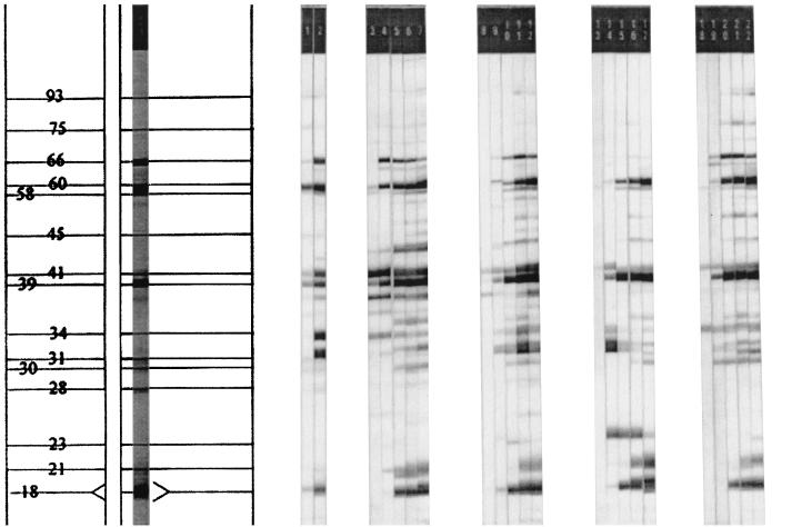
The IgG immunoblots for four representative TISP animals at various times after inoculation are shown below (Fig. 3) to demonstrate how bands developed and in some cases disappeared through the course of the infection.
FIG. 3.
Increasing number of antigens recognized with increasing time p.i. as demonstrated by IgG immunoblot in TISP NHPs. Lanes 1 and 2 contain a low- and a high-titer positive control, respectively. Sera in lanes 3 to 7, 8 to 12, 13 to 17, and 18 to 22 are baseline and day 21, 43, 55, and 93 p.i. for NHPs 30099, 30177, 30199, and 30383, respectively, all of which are TISP animals.
ELISA with recombinant antigens. (i) IgG response.
The following recombinant proteins corresponding to well-characterized Borrelia proteins were utilized to further characterize the antibody response in the NHPs: OspA (p31), OspC (p23), DbpA (p22), BmpA (p39), flagellin (p41), and Arp (a p37 protein). There was good correlation between the ELISA using recombinant antigens and the immunoblot data analysis measuring the densities of the representative band for BmpA (p39) and flagellin (p41). BmpA reactivity in the recombinant ELISA was present in all IC and TISP NHPs, and amplitude-time curves appeared to correlate well with the immunoblot density readings. Recombinant 41 (flagellin) immunoreactivity was present in the sera of five of eight IC and TISP NHPs tested, and its presence appeared to correlate well with a clear p41 band on the immunoblot. However, some sera from IC and TISP animals with absence of p41 reactivity by ELISA with the recombinant p41 had a band on immunoblotting slightly above the p39 band but a bit lower than p41. This band may identify a protein around 40 kDa that is neither BmpA (p39) nor flagellin (p41).
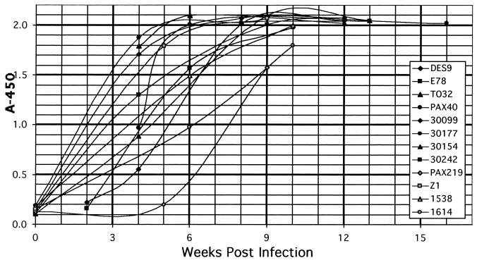
The recombinant protein ELISA was more sensitive for DbpA, a 22-kDa protein. ELISA of IC and TISP sera against this antigen revealed very strong reactivity within weeks in all sera for both IgG and IgM. Yet, there was no reactivity to any 22-kDa bands in any immunoblots. The IgG response to this protein was unique in that it did not depend on the immune status of the infected NHPs; i.e., all infected groups had a strong response to DbpA (Fig. 4). For all other recombinant proteins the IgG response of the IS group was much lower than for the IC and TISP groups.
FIG. 4.
Anti-DbpA IgG response. OD readings of sera at 1:1,000 dilution are shown. NHPs DES9, E678, TO32, and PAX40 were IC animals. NHPs 30099, 30177, 30154, and 30242 were TISP animals. NHPs PAX219, Z1, 1538, and 1614 were IS animals.
There was no reactivity by ELISA to two recombinant proteins tested. The first was recombinant OspA, a finding which correlated well with the immunoblots. The second was Arp, a p37 protein implicated in spirochete dissemination in the mouse (12). Although some sera had a p37 band on immunoblotting, this band may have been representative of antibody directed, not at Arp, but at the FlaA protein of B. burgdorferi (14), also a p37 protein; this appears likely, given the absence of reactivity of these sera to recombinant Arp (see above).
(ii) IgM response.
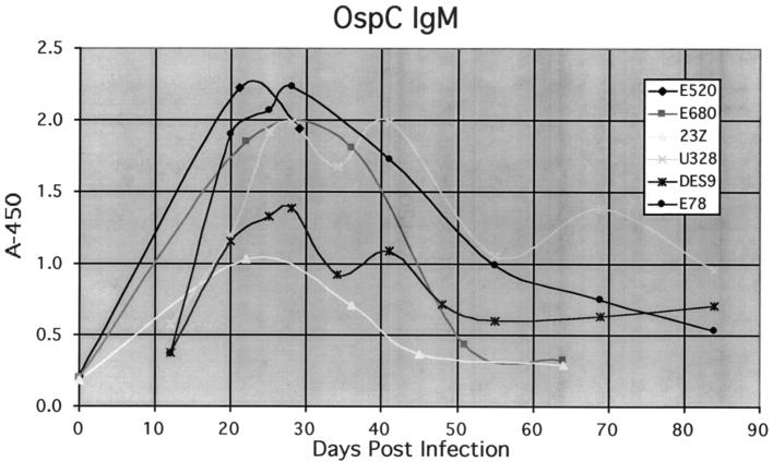
The IgM response to recombinant proteins was generally greater than that to proteins from the immunoblots. An example was OspC. In ELISA using recombinant OspC, all TISP and IC NHPs had strong reactivity, peaking at 5 to 6 weeks p.i. in the TISP animals and at 4 weeks p.i. in the IC animals, and dropping progressively in the 3rd month p.i. (Fig. 5). In contrast, only one of six IC NHPs and three of q8 of the TISP NHPs had OspC IgM immunoreactivity by immunoblot. Another example was DbpA. A strong anti-DbpA IgM response was seen in the recombinant ELISA in all infected animals, including the IS NHPs, yet there was no p22 band on the IgM immunoblot.
FIG. 5.
Anti-OspC IgM response. OD readings of sera at 1:1,000 dilution are shown. All NHPs shown were IC animals.
An exception to the above was p41, flagellin. As noted above, p41 was a prominent band for the IgM response on immunoblots, yet there was no IgM reactivity to the recombinant protein.
Probing for immunoreactive DbpA on the immunoblot strips with anti-DbpA antiserum.
Due to the observation of the absence of detection of a p22 band on the immunoblot strips despite a strong antibody response to recombinant N40 DbpA in the infected NHPs, the immunoblot strips were probed with an anti-DbpA (N40) polyclonal mouse antiserum. There was an absence of signal, indicating the absence of DbpA on the Microbiology Reference Laboratory immunoblot strips.
ELISA of NHP sera with other recombinants as antigen.
We next tested IgG and IgM reactivity of a panel of NHP sera to other recombinant proteins without known function, i.e., P24-13, P19-23, P23-T2, P27-L5, P29-23, P31, P37-42, P41G(197-273), P45-13, and P61. The only significant signals at a 1:1,000 test dilution were for P29, P45, and P61, with only one animal's response being more than 0.4 above baseline, i.e., that to P61. These data, demonstrating weak reactivity in some of the recombinants, were consistent with the results observed in the mouse model (data not shown.)
DISCUSSION
The NHP model of Lyme borreliosis is an excellent model of the human disease (23, 8, 28, 27). After inoculation of B. burgdorferi spirochetes into the skin by either the bite of infected ticks or injection with cultured organisms, the organism, the N40Br strain of the genospecies B. burgdorferi sensu stricto (20), spreads through the skin and enters the circulation with subsequent dissemination to multiple sites (26). The anti-B. burgdorferi antibody response increases over time in amplitude and complexity. Analysis of IgG and IgM serotypes is the cornerstone of diagnosis in the human (9), and these isotypes were studied in these experiments. One of our hypotheses at the onset of this study was that the NHP model would mimic human infection, providing insights important to human infection.
This work represents the first comprehensive analysis of humoral immunity in this model. We studied three different groups of infected NHPs: IC, TISP, and IS. Preinfection sera from the infected NHPs as well as sera from other uninfected NHPs served as negative controls. We began by utilizing a standard approach to analysis of the humoral response: ELISA and immunoblotting using bacterial WCS as the antigen, with isotype(IgG or IgM)-specific conjugates. Early immunosuppression in the TISP group resulted in a temporary delay in the development of a robust IgG response, while prolonged immunosuppression in the IS group resulted in highly diminished development of a spirochete specific antibody response.
Within each similarly treated group, there was variability among animals in the amplitude and time course of the development of the IgG and IgM response to WCS by ELISA. This had been previously noted in this model (26) and is due to the fact that these are outbred animals. This variability was more dramatic when responses to individual proteins of the spirochete were analyzed by immunoblotting and response to recombinant proteins; e.g., some IC NHPs had a strong response to flagellin, while others had a barely detectable response. On the other hand, the responses to other recombinant proteins was uniformly strongly positive (DbpA, OspC, or BmpA) or uniformly negative (Arp).
The variability of the antibody response among infected humans is due to at least three factors. First, there are considerable differences at the genomic level among the B. burgdorferi sensu lato strains which infect humans. Three major genospecies of pathogenic B. burgdorferi have been identified; even within the same genospecies, especially for B. garinii, there is substantial variation (17, 34) in amino acid sequence. Thus, the strain used to prepare antigen to detect antibodies in a serum may not have adequate homology with the infecting strain. Second, during the course of infection expression of antigens by the spirochete occurs at variable times; e.g., OspC expression occurs early in infection, and then fades, while p93 expression occurs late. Thus, assays using only one recombinant protein with the correct sequence may not detect antibodies at each stage of infection. Third, the NHPs used in this study and humans are outbred, and individuals will react to the same strain, and identical recombinant proteins, with different temporal and amplitude patterns of IgG and IgM responses because of these immunogenetic differences.
This work represents the first report of the testing of a panel of recombinant proteins to immune sera in an experimental model of Lyme borreliosis. The development of responses to recombinants in the mouse model have been limited to analyses of single responses, including DbpA (11, 15), BmpA (29, OspC 16), OspA, flagellin, and Arp (12). The data from the NHP model presented in this work are generally consistent with those from the mouse model in identifying OspC and flagellin as early antigens, DbpA and BmpA as prominent antigens throughout the infection, and OspA as an antigen which is not significantly expressed in vivo. However, there are also differences between the mouse and NHP models. Arp (short for arthritis-related protein) is highly expressed in murine disease (12) but is likely not expressed in NHP infection with N40 based on the absence of reactivity to recombinant N40 Arp. DbpA is expressed in infection in mice, monkeys, and humans (6, 7)
This work also confirms previous data from other laboratories working on Lyme disease in finding recombinant proteins particularly useful for serodiagnosis. Not only are many strains of B. burgdorferi highly variable in their sequences of some of the major proteins, but there are also strain differences in which proteins are highly expressed in vitro. Thus, the use of sonicated in vitro-cultivated organism may not be ideal as the source for antigen for immunoblots in the diagnosis of infection caused by a spectrum of strains. Wilske et al. (33, 34, 35) have developed immunoblots composed of recombinant proteins for diagnosis of Lyme disease in Europe, where genetic heterogeneity of the pathogenic strains is a major problem. Wilske's immunoblots are composed of immunodominant proteins from various strains. In our work, the NHPs were infected with N40, and the recombinant proteins were from N40, eliminating concern about strain differences in the recombinant proteins. The fact that immunoblotting did not recognize as many proteins as the recombinant ELISA may be due, at least in part, to the possibility that antibodies to N40 proteins in the NHPs did not recognize proteins in the immunoblots which were from a different B. burgdorferi sensu stricto strain, i.e., CB, an erythema migrans isolate, while all of the recombinants were from N40. An example of this may have been OspC, for which the recombinant protein analysis showed a strong IgM response to N40 OspC while the immunoblots revealed a very weak response to CB OspC. The OspC sequence is known to be highly variable among strains (4, 32).
Many of the recombinant proteins used in this study are considered to be primarily in vivo expressed. These proteins are expressed by cultured spirochetes to various degrees, but expression is increased upon growth and dissemination in vivo (1, 5, 13, 31). Spirochetes cultured in vitro are the source of antigens for most ELISAs and immunoblots. Therefore, recombinant proteins are the preferred antigens for determination of the antibody response to in vivo-expressed antigens. The use of these recombinant proteins provided an understanding of the temporal course of expression in vivo for some of these in vivo-expressed antigens which could not be obtained by using the immunoblots. For instance, OspC and flagellin antibodies were seen early in infection and then tapered off, presumably because these proteins were expressed transiently early in the course of infection. Anti-DbpA antibodies, in contrast, appeared early and continued to be present throughout the 3 months of infection. N40 DbpA is not highly expressed in cultured spirochetes, and the CB immunoblots lacked N40-like DbpA when probed for N40 DbpA antigen using well-characterized anti-N40 DbpA mouse serum. This finding, presumably due to lack of homology for DbpA between the immunoblot strain, CB, and the infecting strain, N40, explains the observation that the strong anti-DbpA response in the NHPs could only be found by using recombinant DbpA in the ELISA. DbpA is a highly diverse molecule, and closely related B. burgdorferi strains can have significantly different DbpAs (15a).
There were a number of surprising finding in the results, including isotype preferences for some of the recombinants and variability among the recombinants in the degree to which responses were suppressed by corticosteroid administration. OspC elicited primarily an IgM-oriented response. BmpA in contrast was IgG dominant, and DbpA induced both an IgM and an IgG response. The response to most recombinants was moderately to markedly suppressed in the IS group, which received corticosteroids throughout infection. The exception to this rule was DbpA, in which all three groups had a strong response, the amplitude and timing of which was virtually indistinguishable between groups.
In summary, these data, the first comprehensive analysis of anti-B. burgdorferi humoral immunity in the NHP model, demonstrate the complexity of the antibody response to the whole spirochete and the difficulties that may be encountered in detecting antibody responses in humans in whom the infecting strain is unknown.
Acknowledgments
This work was supported by grants NO1-AI 95358 and RO1-NS34715 to A.R.P.
REFERENCES
- 1.Akins, D. R., S. F. Porcella, T. G. Popova, D. Shevchenko, S. I. Baker, M. Li, M. V. Norgard, and J. D. Radolf. 1995. Evidence for in vivo but not in vitro expression of a Borrelia burgdorferi outer surface protein F (OspF) homologue. Mol. Microbiol. 18:507-520. [DOI] [PubMed] [Google Scholar]
- 2.Amemiya, K., H. Schaefer, and A. R. Pachner. 1999. Isolation of DNA after extraction of RNA To detect the presence of Borrelia burgdorferi and expression of host cellular genes from the same tissue sample. J. Clin. Microbiol. 37:2087-2089. [DOI] [PMC free article] [PubMed] [Google Scholar]
- 3.Anguita, J., S. Samanta, B. Revilla, K. Suk, S. Das, S. W. Barthold, and E. Fikrig. 2000. Borrelia burgdorferi gene expression in vivo and spirochete pathogenicity. Infect. Immun. 68:1222-1230. [DOI] [PMC free article] [PubMed] [Google Scholar]
- 4.Baranton, G., G. Seinost, G. Theodore, et al. 2001. Distinct levels of genetic diversity of Borrelia burgdorferi are associated with different aspects of pathogenicity. Res. Microbiol. 152:149-156. [DOI] [PubMed] [Google Scholar]
- 5.Bockenstedt, L. K., E. Hodzic, S. Feng, K. W. Bourrel, A. de Silva, R. R. Montgomery, E. Fikrig, J. D. Radolf, and S. W. Barthold. 1997. Borrelia burgdorferi strain-specific Osp C-mediated immunity in mice. Infect. Immun. 65:4661-4667. [DOI] [PMC free article] [PubMed] [Google Scholar]
- 6.Cassatt, D. R., N. K. Patel, N. D. Ulbrandt, and M. S. Hanson. 1998. DbpA, but not OspA, is expressed by Borrelia burgdorferi during spirochetemia and is a target for protective antibodies. Infect. Immun. 66:5379-5387. [DOI] [PMC free article] [PubMed] [Google Scholar]
- 7.Cinco, M., M. Ruscio, and F. Rapagna. 2000. Evidence of Dbps (decorin binding proteins) among European strains of Borrelia burgdorferi sensu lato and in the immune response of LB patient sera. FEMS Microbiol. Lett. 183:111-114. [DOI] [PubMed] [Google Scholar]
- 8.Coyle, P. K. 1995. Neurological Lyme disease: is there a true animal model? Ann. Neurol. 38:560-562. [DOI] [PubMed] [Google Scholar]
- 9.Dressler, F., J. A. Whalen, B. N. Reinhardt, and A. C. Steere. 1993. Western blotting in the serodiagnosis of Lyme disease. J. Infect. Dis. 167:392-400. [DOI] [PubMed] [Google Scholar]
- 10.England, J. D., R. P. Bohm, Jr., E. D. Roberts, and M. T. Philipp. 1997. Lyme neuroborreliosis in the rhesus monkey. Semin. Neurol. 17:53-56. [DOI] [PubMed] [Google Scholar]
- 11.Feng, S., E. Hodzic, B. Stevenson, and S. W. Barthold. 1998. Humoral immunity to Borrelia burgdorferi N40 decorin binding proteins during infection of laboratory mice. Infect. Immun. 66:2827-2835. [DOI] [PMC free article] [PubMed] [Google Scholar]
- 12.Feng, S., E. Hodzic, and S. W. Barthold. 2000. Lyme arthritis resolution with antiserum to a 37-kilodalton Borrelia burgdorferi protein. Infect. Immun. 68:4169-4173. [DOI] [PMC free article] [PubMed] [Google Scholar]
- 13.Fikrig, E., S. W. Barthold, W. Sun, W. Feng, S. R. Telford III, and R. A. Flavel. 1997. Borrelia burgdorferi P35 and P37 proteins, expressed in vivo, elicit protective immunity. Immunity 6:531-539. [DOI] [PubMed] [Google Scholar]
- 14.Gilmore, R. D., Jr., R. L. Murphree, A. M. James, S. A. Sullivan, and B. J. Johnson. 1999. The Borrelia burgdorferi 37-kilodalton immunoblot band (P37) used in serodiagnosis of early Lyme disease is the flaA gene product. J. Clin. Microbiol. 37:548-552. [DOI] [PMC free article] [PubMed] [Google Scholar]
- 15.Hanson, M. S., D. R. Cassatt, B. P. Guo, N. K. Patel, M. P. McCarthy, D. W. Dorward, and M. Hook. 1998. Active and passive immunity against Borrelia burgdorferi decorin binding protein A (DbpA) protects against infection. Infect. Immun. 66:2143-2153. [DOI] [PMC free article] [PubMed] [Google Scholar]
- 15a.Heikkila, T., I. Seppala, H. Saxen, J. Panelius, H. Yrjanainen, and P. Lahdenner. 2002. Species-specific serodiagnosis of Lyme arthritis and neuroborreliosis due to Borrelia burgdorferi sensu stricto, B. afzelii, and B. garinii by using decorin binding protein A. J. Clin. Microbiol. 40:453-460. [DOI] [PMC free article] [PubMed] [Google Scholar]
- 16.Ikushima, M., K. Matsui, F. Yamada, et al. 2000. Specific immune response to a synthetic peptide derived from outer surface protein C of Borrelia burgdorferi predicts protective borreliacidal antibodies. FEMS Immunol. Med. Microbiol. 29:15-21. [DOI] [PubMed] [Google Scholar]
- 17.Jauris-Heipke, S., R. Fuchs, M. Motz, et al. 1993. Genetic heterogeneity of the genes coding for the outer surface protein C (OspC) and the flagellin of Borrelia burgdorferi. Med. Microbiol. Immunol (Berlin) 182:37-50. [DOI] [PubMed] [Google Scholar]
- 18.Kaiser, R., and S. Rauer. 1999. Advantage of recombinant borrelial proteins for serodiagnosis of neuroborreliosis. J. Med. Microbiol. 48:5-10. [DOI] [PubMed] [Google Scholar]
- 19.Magnarelli, L. A., J. W. Ijdo, S. J. Padula, et al. 2000. Serologic diagnosis of Lyme borreliosis by using enzyme-linked immunosorbent assays with recombinant antigens. J. Clin. Microbiol. 38:1735-1739. [DOI] [PMC free article] [PubMed] [Google Scholar]
- 20.Pachner, A. R., and A. Itano. 1990. Borrelia burgdorferi infection of the brain: characterization of the organism and response to antibiotics and immune sera in the mouse model. Neurology 40:1535-1540. [DOI] [PubMed] [Google Scholar]
- 21.Pachner, A. R., and N. S. Ricalton. 1992. Western blotting in evaluating Lyme seropositivity and the utility of a gel densitometric approach. Neurology 42:2185-2192. [DOI] [PubMed] [Google Scholar]
- 22.Pachner, A. R., E. Delaney, and T. O'Neill. 1995. Neuroborreliosis in the nonhuman primate: Borrelia burgdorferi persists in the central nervous system. Ann. Neurol. 38:667-669. [DOI] [PubMed] [Google Scholar]
- 23.Pachner, A. R., E. Delaney, T. O'Neill, and E. Major. 1995. Inoculation of nonhuman primates with the N40 strain of Borrelia burgdorferi leads to a model of Lyme neuroborreliosis faithful to the human disease. Neurology 45:165-172. [DOI] [PubMed] [Google Scholar]
- 24.Pachner, A. R., W. F. Zhang, H. Schaefer, S. Schaefer, and T. O'Neill. 1998. Detection of active infection in nonhuman primates with Lyme neuroborreliosis: comparison of PCR, culture, and a bioassay. J. Clin. Microbiol. 36:3243-3247. [DOI] [PMC free article] [PubMed] [Google Scholar]
- 25.Pachner, A. R., K. Amemiya, M. Bartlett, H. Schaefer, K. Reddy, and W. F. Zhang. 2001. Lyme borreliosis in rhesus macaques: effects of corticosteroids on spirochetal load and isotype switching of anti-Borrelia burgdorferi antibody. Clin. Diagn. Lab. Immunol. 8:225-232. [DOI] [PMC free article] [PubMed] [Google Scholar]
- 26.Pachner, A. R., D. Cadavid, G. Shu, et al. 2001. Central and peripheral nervous system infection, immunity, and inflammation in the NHP model of Lyme borreliosis. Ann. Neurol. 50:330-338. [PubMed] [Google Scholar]
- 27.Pachner, A. R. 2001. The rhesus model of lyme neuroborreliosis. Immunol. Rev. 183:186-204. [DOI] [PubMed] [Google Scholar]
- 28.Philipp, M. T., and B. J. B. Johnson. 1994. Animal models of Lyme disease: pathogenesis and immunoprophylaxis. Trends Microbiol. 2:431-437. [DOI] [PubMed] [Google Scholar]
- 29.Roessler, D., U. Hauser, and B. Wilske. 1997. Heterogeneity of BmpA (P39) among European isolates of Borrelia burgdorferi sensu lato and influence of interspecies variability on serodiagnosis. J. Clin. Microbiol. 35:2752-2758. [DOI] [PMC free article] [PubMed] [Google Scholar]
- 30.Steere, A. C. 2001. Lyme disease. N. Engl. J. Med. 345:115-125. [DOI] [PubMed] [Google Scholar]
- 31.Suk, K., S. Das, W. Sun, et al. 1995. Borrelia burgdorferi genes selectively expressed in the infected host. Proc. Natl. Acad. Sci. USA 92:4269-4273. [DOI] [PMC free article] [PubMed] [Google Scholar]
- 32.Wang, I. N., D. E. Dykhuizen, W. Qiu, et al. 1999. Genetic diversity of ospC in a local population of Borrelia burgdorferi sensu stricto. Genetics 151:15-30. [DOI] [PMC free article] [PubMed] [Google Scholar]
- 33.Wilske, B., V. Fingerle, V. Preac-Mursic, et al. 1994. Immunoblot using recombinant antigens derived from different genospecies of Borrelia burgdorferi sensu lato. Med. Microbiol. Immunol. (Berlin) 183:43-59. [DOI] [PubMed] [Google Scholar]
- 34.Wilske, B., U. Hauser, G. Lehnert, and S. Jauris-Heipke. 1998. Genospecies and their influence on immunoblot results. Wien. Klin. Wochenschr. 110:882-885. [PubMed] [Google Scholar]
- 35.Wilske, B., C. Habermann, V. Fingerle, et al. 1999. An improved recombinant IgG immunoblot for serodiagnosis of Lyme borreliosis. Med. Microbiol. Immunol. (Berlin) 188:139-144. [DOI] [PubMed] [Google Scholar]
- 36.Zhang, J. R., and S. J. Norris. 1998. Kinetics and in vivo induction of genetic variation of vlsE in Borrelia burgdorferi. Infect. Immun. 66:3689-3697. [DOI] [PMC free article] [PubMed] [Google Scholar]


