Eukaryotic cells exhibit a wide range of actin-based motilities, including the movement of neurites during modeling of the nervous system, fibroblast migration in wound healing, and the chemotactic movement of immune cells. Furthermore, several bacterial pathogens have evolved the capacity to hijack the actin system of their host cells for their own movement. At the heart of these forms of movement is actin treadmilling, a process involving the dynamic cycling of actin between a monomeric or globular form (G-actin) and a polymeric or filamentous form (F-actin). Treadmilling is characterized by the net association of ATP-actin and dissociation of ADP-actin monomers, respectively, at the “barbed” (or +) and “pointed” (or −) ends of the asymmetric actin filament. The entire process is powered by the hydrolysis of ATP by actin, which under certain conditions can consume up to 50% of the ATP in the cell. In addition, treadmilling is regulated by several actin-binding proteins, which specifically target either ATP- or ADP-actin (Pollard et al., 2000; Sheterline et al., 1995).
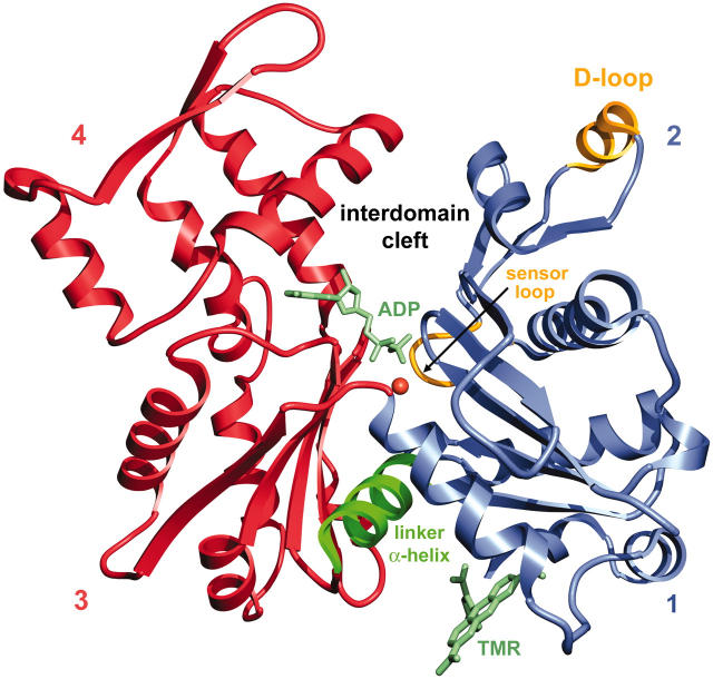
The evidence to date suggests that the transition from ATP- to ADP-actin is accompanied by a conformational change in actin subdomain 2 and, more specifically, in the DNase I binding loop (D-loop) at the top of this subdomain (Fig. 1). Indeed, the susceptibility of the D-loop to certain proteases (Strzelecka-Golaszewska et al., 1993) and the fluorescence emission of probes attached to this loop (Kim et al., 1995) depend on the state of the nucleotide. Differences in subdomain 2 also have been observed between electron microscopic reconstructions of F-actin in the ATP and ADP states (Belmont et al., 1999). More recently, a direct visualization of a conformational change in subdomain 2 was obtained from comparison of the crystal structure of monomeric actin in the ADP state (Otterbein et al., 2001) with those of ATP-actin that had been determined previously from complexes with DNase I, profilin, and gelsolin. In the ADP structure the release of the nucleotide γ-phosphate appeared to trigger a sequence of events culminating in a loop-to-helix transition in the D-loop (Fig. 1). Such a change would be expected to affect the monomer-monomer interface in F-actin and could potentially mark ADP-actin for recognition by depolymerizing proteins such as ADF/cofilin. Thus, the ADP-actin structure seemed to provide a plausible explanation for how Pi release enhances monomer dissociation. However, to crystallize ADP-actin in a monomeric state a fluorescence probe, tetramethylrhodamine-5-maleimide (TMR), was attached to Cys-374 in subdomain 1 to block polymerization (Fig. 1). This circumstance led some to suggest that the TMR modification, rather than the state of the nucleotide, produced the conformational change observed in subdomain 2 of the ADP-actin structure (Egelman, 2001; Sablin et al., 2002). Determining whether the modification at Cys-374 could have had such a profound effect on the structure of ADP-actin is important, not only because of the questions raised about this structure, but also because Cys-374 has been the single most popular site derivatized in the study of actin (Sheterline et al., 1995).
FIGURE 1.
Ribbon representation of the structure of TMR-actin in the ADP state. The two major domains on each side of the nucleotide cleft are shown in blue (subdomains 1 and 2) and red (subdomains 3 and 4). In F-actin subdomains 1 and 3 point toward the barbed end of the filament and subdomains 2 and 4 are directed toward the pointed end. Shown in yellow are the loop containing the methylated His-73 (sensor loop), which plays a key role in transmitting conformational changes from the nucleotide site to subdomain 2, and the D-loop, which changes conformation between ATP and ADP-actin. In green is the α-helix from Ile-136 to Gly-146, which serves as a hinge for rotation between the two major domains.
In the current issue of Biophysical Journal, Kudryashov and Reisler (2003) address this question by studying, side-by-side, the solution properties of TMR-modified and unmodified actin in the ATP and ADP states. They found that the susceptibility of the D-loop to subtilisin cleavage is similar for TMR-modified and unmodified actin, being severalfold slower in the ADP than in the ATP state. Both forms of actin are also similar in that the subtilisin digestion of the D-loop is insensitive to whether there is Ca2+ or Mg2+ complexed with ATP at the catalytic site. Further evidence against a long-range allosteric effect of the TMR probe on subdomain 2 is provided by the fact that neither the binding of DNase I nor the fast phase of tryptic cleavage in subdomain 2 are affected by the presence of the probe. Taken together these facts allow the authors to draw two main conclusions: the TMR modification does not, by itself, change the conformation of the D-loop, and the TMR probe does not interfere with the conformational change of the D-loop that results from Pi release.
The reduced susceptibility of the D-loop to subtilisin cleavage in the ADP state would be consistent with a more stable and less-exposed structure, which is what the crystal structure of TMR-actin in the ADP state reveals, a stable α-helix in the D-loop (Fig. 1). In the existing ATP-actin structures, on the contrary, the D-loop is either disordered or folded as a flexible β-hairpin loop, which would help explain the increased subtilisin cleavage.
Another criticism of the ADP structure of TMR-actin has been that it does not reveal an open interdomain cleft, as expected by some (Sablin et al., 2002). The closed cleft in ADP actin was suggested to be an artifact resulting from steric hindrance between the TMR probe and actin subdomains 1 and 3. Indirect evidence, including faster nucleotide exchange, increased nucleotide accessibility to collisional quenchers, increased susceptibility to trypsin cleavage at Arg-62 and Lys-68, and decreased DNase I binding have been frequently associated with an open cleft in ADP-actin. Kudryashov and Reisler show here that although both the TMR probe and gelsolin fragment 1 inhibit nucleotide exchange (albeit to different degrees), there is no effect on any of the other indicators of cleft opening (nucleotide quenching, tryptic cleavage of subdomain 2, and DNase I binding). They, therefore, conclude that the available solution methods cannot discriminate between a closed and an open cleft. As the authors also point out, further evidence of the unsuitability of these solution methods to distinguish between a closed and an open cleft comes from the fact that profilin that accelerates nucleotide exchange, supposedly an open cleft indicator, does not affect DNase I binding, which is assumed to be a closed cleft marker (Schuler et al., 2000).
Another way to establish whether the conformational change in subdomain 2 of ADP-actin is due to the TMR probe or the state of the nucleotide is to determine the structure of TMR-actin in the ATP state, which has recently been accomplished (Graceffa and Dominguez, 2003). In this work, the crystal structure of TMR-actin with bound AMPPNP, a nonhydrolyzable ATP analog, was determined to 1.85-Å resolution. In the new structure, a reversal of the conformational change previously observed in ADP-actin takes place. Because the symmetry and crystal parameters are identical for the two TMR-actin structures (ATP and ADP state), both containing the TMR modification, allosteric effects (or crystal packing) can be ruled out as the cause for the conformational change observed in subdomain 2 of ADP-actin. In addition, this work compiles all the existing structural information for members of the actin superfamily and proposes a three-state nucleotide-dependent mechanism of actin dynamics, where the open cleft state would correspond to nucleotide-free actin.
References
- Belmont, L. D., A. Orlova, D. G. Drubin, and E. H. Egelman. 1999. A change in actin conformation associated with filament instability after Pi release. Proc. Natl. Acad. Sci. USA. 96:29–34. [DOI] [PMC free article] [PubMed] [Google Scholar]
- Egelman, E. H. 2001. Actin allostery again? Nat. Struct. Biol. 8:735–736. [DOI] [PubMed] [Google Scholar]
- Graceffa, P., and R. Dominguez. 2003. Crystal structure of monomeric actin in the ATP state. Structural basis of nucleotide-dependent actin dynamics. J. Biol. Chem. 278:34172–34180. [DOI] [PubMed] [Google Scholar]
- Kim, E., M. Motoki, K. Seguro, A. Muhlrad, and E. Reisler. 1995. Conformational changes in subdomain 2 of G-actin: fluorescence probing by dansyl ethylenediamine attached to Gln-41. Biophys. J. 69:2024–2032. [DOI] [PMC free article] [PubMed] [Google Scholar]
- Kudryashov, D. S., and E. Reisler. 2003. Solution properties of tetramethylrhodamine modified G-actin. Biophys. J. 85:2466–2475. [DOI] [PMC free article] [PubMed] [Google Scholar]
- Otterbein, L. R., P. Graceffa, and R. Dominguez. 2001. The crystal structure of uncomplexed actin in the ADP state. Science. 293:708–711. [DOI] [PubMed] [Google Scholar]
- Pollard, T. D., L. Blanchoin, and R. D. Mullins. 2000. Molecular mechanisms controlling actin filament dynamics in nonmuscle cells. Annu. Rev. Biophys. Biomol. Struct. 29:545–576. [DOI] [PubMed] [Google Scholar]
- Sablin, E. P., J. F. Dawson, M. S. VanLoock, J. A. Spudich, E. H. Egelman, and R. J. Fletterick. 2002. How does ATP hydrolysis control actin's associations? Proc. Natl. Acad. Sci. USA. 99:10945–10947. [DOI] [PMC free article] [PubMed] [Google Scholar]
- Schuler, H., U. Lindberg, C. E. Schutt, and R. Karlsson. 2000. Thermal unfolding of G-actin monitored with the DNase I-inhibition assay stabilities of actin isoforms. Eur. J. Biochem. 267:476–486. [DOI] [PubMed] [Google Scholar]
- Sheterline, P., J. Clayton, and J. Sparrow. 1995. Actin. Protein Profile. 2:1–103. [PubMed] [Google Scholar]
- Strzelecka-Golaszewska, H., J. Moraczewska, S. Y. Khaitlina, and M. Mossakowska. 1993. Localization of the tightly bound divalent-cation-dependent and nucleotide-dependent conformation changes in G-actin using limited proteolytic digestion. Eur. J. Biochem. 211:731–742. [DOI] [PubMed] [Google Scholar]


