Abstract
Mammalian 5-lipoxygenase (5-LO) catalyzes conversion of arachidonic acid to leukotrienes, potent mediators of inflammation and allergy. Upon cell stimulation, 5-LO selectively binds to nuclear membranes and becomes activated, yet the mechanism of recruitment of 5-LO to nuclear membranes and the mode of 5-LO-membrane interactions are poorly understood. Here we show that membrane fluidity is an important determinant of membrane binding strength of 5-LO, penetration into the membrane hydrophobic core, and activity of the enzyme. The membrane binding strength and activity of 5-LO increase with the degree of lipid acyl chain cis-unsaturation and reach a plateau with 1-palmitoyl-2-arachidonolyl-sn-glycero-3-phosphocholine (PAPC). A fraction of tryptophans of 5-LO penetrate into the hydrocarbon region of fluid PAPC membranes, but not into solid 1,2-dipalmitoyl-sn-glycero-3-phosphocholine membranes. Our data lead to a novel concept of membrane binding and activation of 5-LO, suggesting that arachidonic-acid-containing lipids, which are present in nuclear membranes at higher fractions than in other cellular membranes, may facilitate preferential membrane binding and insertion of 5-LO through increased membrane fluidity and may thereby modulate the activity of the enzyme. The data presented in this article and earlier data allow construction of a model for membrane-bound 5-LO, including the angular orientation and membrane insertion of the protein.
INTRODUCTION
Lipoxygenase (LO) is a nonheme, iron-containing enzyme that catalyzes oxygenation of polyunsaturated fatty acids (Kühn, 1999; Peters-Golden and Brock, 2003). Both plant and mammalian LOs have been shown to undergo functionally important, Ca2+-regulated binding to membranes, followed by production of lipid-derived bioactive mediators (Rouzer and Kargman, 1988; Brock et al., 1995; Tatulian et al., 1998; Hammarberg et al., 2000; Walther et al., 2004). Mammalian 5-lipoxygenase (5-LO) is of exceptional importance because it converts arachidonic acid (AA) to 5-hydroperoxyeicosatetraenoic acid (5-HPETE) and then to leukotriene A4, a key intermediate in biosynthesis of all leukotrienes that act as potent mediators of allergy, inflammation, apoptosis, and tumorigenesis (Ghosh and Myers, 1999; Rådmark, 2000; Chen et al., 2003; Goodarzi et al., 2003; Fan et al., 2004). Leukotriene production in stimulated myeloid cells is preceded by a Ca2+-mediated binding of 5-LO to nuclear membranes (Rouzer and Kargman, 1988; Wong et al., 1991; Malaviya et al., 1993; Woods et al., 1993; Brock et al., 1995; Pouliot et al., 1996). A Ca2+-independent, phosphorylation-mediated 5-LO translocation to the nuclear membrane and activation has also been documented (Werz et al., 2000, 2002). Although the role of membrane binding in 5-LO function is well established, the mode of membrane binding of 5-LO and the reason for localization to the nuclear membrane remain poorly understood.
Preferential binding of 5-LO to nuclear membranes might be due to specific interactions between 5-LO and intrinsic proteins of nuclear membranes, such as 5-lipoxygenase activating protein (FLAP), an integral protein in nuclear membranes that significantly increases 5-LO activity (Reid et al., 1990; Abramovitz et al., 1993; Pouliot et al., 1996). However, 5-LO binding to nuclear membranes turned out to be FLAP-independent (Woods et al., 1995; Peters-Golden and Brock, 2003). In addition, artificial lipid membranes or cell plasma membranes were able to activate 5-LO in the absence of FLAP (Puustinen et al., 1988; Noguchi et al., 1994; Skorey and Gresser, 1998; Reddy et al., 2000; Pande et al., 2004), implying that FLAP is not a molecular determinant for nuclear membrane localization of 5-LO.
Among a large number of lipids examined, phosphatidylcholine (PC) turned out to be the most efficient naturally occurring lipid in terms of supporting 5-LO activity (Puustinen et al., 1988; Noguchi et al., 1994; Pande et al., 2004). The increased affinity of the N-terminal putative β-barrel domain of 5-LO for PC membranes compared to membranes containing phosphatidylglycerol (PG) or phosphatidylserine (PS) led Kulkarni et al. (2002) to conclude that high affinity of 5-LO for PC determines its selective translocation to PC-rich nuclear membranes. However, the facts that the β-barrel domain of 5-LO binds to PC more strongly than to anionic lipids and that PC supports 5-LO activity better than the anionic lipids cannot account for nuclear membrane targeting of 5-LO for two reasons. First, comparative analysis of the lipid composition of mammalian cell membranes indicated similar fractions of PC (42–48%) in the nuclear and other organellar membranes, including plasma membranes (Surette and Chilton, 1998; D'Antuono et al., 2000). Second, nuclear membranes contain large fractions of anionic lipids, including PS, PG, cardiolipin, phosphatidic acid, phosphatidylinositol and its mono- and bis-phosphates (Khandwala and Kasper, 1971; Neitcheva and Peeva, 1995; Surette and Chilton, 1998; D'Santos et al., 1999, 2000; D'Antuono et al., 2000). In fact, in human leukemia cells the (phosphatidylinositol + PS) fraction was higher in the nuclear membrane (25%) than in the whole-cell lipid extract (18%) (Surette and Chilton, 1998).
The effects of the lipid hydrocarbon chain composition and membrane fluidity on 5-LO binding and activity have not been studied. In this work, we demonstrate a significant stimulatory effect of membrane fluidity on membrane binding and activity of recombinant human 5-LO. Experiments with PCs in which the number of cis-unsaturated bonds in the sn-2 acyl chain increases from zero to six indicate that both membrane binding strength and 5-LO activity reach a maximum level with membranes composed of 1-palmitoyl-2-arachidonoyl-sn-glycero-3-phosphocholine (PAPC). Moreover, membrane depth-dependent tryptophan (Trp) fluorescence quenching by brominated lipids shows that side chains of certain tryptophans of 5-LO significantly penetrate into the interior of fluid PAPC membranes, whereas no penetration occurs into solid 1,2-dipalmitoyl-sn-glycero-3-phosphocholine (DPPC) membranes. Our results lead to a novel concept of membrane-mediated activation of 5-LO, implying that membrane fluidity supports 5-LO activity by facilitating insertion of the enzyme into the hydrocarbon region of the membrane. Considering that the nuclear membranes of various mammalian cells, including leukocytes, are enriched in AA-containing lipids more than other subcellular membranes (Neufeld et al., 1985; Capriotti et al., 1988; Albi et al., 1997; Surette and Chilton, 1998; D'Antuono et al., 2000), which results in increased fluidity of nuclear membranes (Yu et al., 1996; Albi et al., 1997; D'Antuono et al., 2000), our findings suggest that membrane fluidity may play a central role in nuclear membrane targeting and activation of 5-LO.
MATERIALS AND METHODS
Materials
Arachidonic acid was supplied by Cayman Chemical (Ann Arbor, MI). All lipids were synthetic, and cholesterol was purified from egg yolk; they all were purchased from Avanti Polar Lipids (Alabaster, AL). Most of the chemicals were purchased from Sigma (St. Louis, MO), unless specified otherwise, and the sources of other supplies are indicated below. The 5-LO plasmid, pT3-5LO, was kindly provided by Prof. Ying Yi Zhang (Boston University School of Medicine), and has been described by Zhang et al. (1992).
Expression and purification of 5-LO
Subcloning of the 5-LO gene into the expression vector pET-21a(+), as well as protein expression in E. coli BL21(DE3) cells and purification, were conducted as described previously (Pande et al., 2004). The protocol allows production of highly pure and active recombinant 5-LO that contains a 1:1 stoichiometric amount of iron cofactor. The pure protein eluted from the final size-exclusion Superdex-75 column was pooled, placed into a buffer containing 150 mM NaCl, 0.1 mM EGTA, 50 mM Tris-HCl (pH 7.5) using a G-25 desalting column, and used immediately for activity experiments. Alternatively, the protein was placed in pure deionized water by the same method, lyophilized, and stored at −80°C for biophysical experiments, which were carried out during the next 2 days. The activity of the lyophilized 5-LO was sustained at the level of ∼80% over a period of several days at −80°C.
5-LO activity assay
5-LO activity was measured in an assay buffer containing 31.64 mM Na2HPO4, 5.4 mM KH2PO4, 0.2 mM ATP, 0.1 mM dithiothreitol, 0.1 mM EGTA, and 0.3 mM CaCl2 (pH 7.5), in the presence of large (100 nm in diameter) unilamellar vesicles, at 22°C, essentially as described previously (Pande et al., 2004). Vesicles were prepared using a Liposofast extruder (Avestin, Ottawa, Canada), as described (Pande et al., 2004). 5-LO-catalyzed conversion of AA to 5-HPETE was monitored by recording time dependence of absorption at 238 nm, using a Cary 100 double-beam spectrophotometer (Varian, Palo Alto, CA). The specific activity of 5-LO was measured using an extinction coefficient of ɛ238 = 23 mM−1 cm−1 (Percival, 1991; Skorey and Gresser, 1998). Protein concentration was measured by Bradford assay (Bradford, 1976).
Measurements of membrane fluidity
Membrane fluidity was evaluated by generalized polarization (GP) of 6-lauroyl-2-(N,N-dimethylamino)-naphthalene (Laurdan) incorporated in vesicle membranes at 1 mol %. Laurdan can be excited at 350–360 nm, and its emission spectra shift with increasing membrane fluidity, resulting in a decrease in GP = (F435 − F500)/(F435 + F500), where F435 and F500 are the fluorescence emission intensities at respective wavelengths (Parasassi et al., 1994; Harris et al., 2002; Nyholm et al., 2003). Steady-state fluorescence spectra were recorded on a Jasco-810 spectrofluoropolarimeter (Jasco, Tokyo, Japan). This is a spectropolarimeter with an additional photomultiplier tube mounted at 90° with respect to the incident light beam for fluorescence measurements. Fluorescence measurements were conducted using a 0.4-cm path-length quartz cuvette at 22°C, maintained by a Peltier temperature controller. The excitation and emission slits were 4 nm and 10 nm, respectively. The emission spectra of samples containing vesicles in a buffer of 0.1 mM EGTA, 0.3 mM CaCl2, 50 mM Tris-HCl (pH 7.5) were measured between 390 and 560 nm, using excitation at 360 nm. The total lipid concentration was 0.1 mM. The emission spectra were used to calculate the Laurdan GP, which was used as a measure of membrane fluidity.
Membrane binding measurements by fluorescence spectroscopy
Binding of 5-LO to vesicle membranes was measured by resonance energy transfer (RET) from Trp residues of 5-LO to 1,2-dioleoyl-sn-glycero-3-phosphoethanolamine-N-(1-pyrenesulfonyl) (Py-PE), which was incorporated in vesicle membranes at 2 mol %. Large unilamellar vesicles were titrated into the protein solution, with continuous stirring, to yield a total lipid concentration from 4 to 760 μM. The concentration of 5-LO was 0.2 μM. After each addition of vesicles, the sample was equilibrated for 2 min and fluorescence emission spectra were recorded using a J-810 spectrofluoropolarimeter, described above. Temperature was maintained at 22°C. The excitation wavelength was 290 nm. Parallel control experiments were conducted in which vesicles without Py-PE were used to titrate protein solutions. In these control experiments, addition of lipid vesicles to the protein solution resulted in a decrease in the measured Trp fluorescence intensity due to sample dilution. In RET experiments with Py-PE-labeled vesicles, the Trp emission intensity significantly decreased upon addition of vesicles due to protein-membrane interactions and energy transfer from Trp to Py-PE. Analysis of RET data and determination of membrane binding parameters of 5-LO were conducted as described (Qin et al., 2004). Briefly, changes in Trp emission intensity at 330 nm (ΔF) were measured at each lipid concentration, corrected for the changes in the fluorescence emission as measured in control experiments, and plotted against ΔF/[L], where [L] is the lipid concentration. Each of these Scatchard plots was extrapolated with a straight line, which was used to determine the saturating value of ΔF at high lipid concentrations (ΔFmax) and the lipid concentration corresponding to ΔF = ½ΔFmax ([L]1/2), respectively from the ΔF-axis intercept and the slope of the plot. The experimental binding data were presented as the dependence of ΔF/ΔFmax on [L]. We have shown previously (Qin et al., 2004) that a simple binding model, suggesting that the membrane surface contains a finite number of binding sites that can be either free or occupied by a protein molecule, leads to the following binding isotherm:
 |
(1) |
In Eq. 1, ΔFrel ≡ ΔF/ΔFmax, KD is the dissociation constant, N is the number of lipid molecules corresponding to a protein binding site, [P] is the total protein concentration, and δ is the fraction of protein-accessible lipid. It has also been shown in the same source that
 |
(2) |
In Eq. 2, the total protein concentration, [P], is known, [L]1/2 can be determined experimentally, as described above, and the fraction of protein-accessible lipid in the external leaflet of 100-nm (in diameter) vesicles with membrane thickness of ∼4 nm (Vogel et al., 2000) is δ ≈ 0.52. Insertion of the expression for KD from Eq. 2 into Eq. 1 yields an equation with only one unknown, i.e., N. The parameter N was varied within physically reasonable limits and theoretical binding isotherms were simulated through Eq. 1 until a best fit was achieved between the experimental and theoretical isotherms. This was followed by calculation of KD through Eq. 2, using the best-fit value of N.
Tryptophan fluorescence quenching by brominated or nitroxide-labeled lipids
The degree of insertion of tryptophans into membranes was determined by measuring the quenching of Trp fluorescence of 5-LO by 1-palmitoyl-2-stearoyl(dibromo)-sn-glycero-3-phosphocholines (Br2PCs) brominated at 6,7, or 9,10, or 11,12 positions of the acyl chains, or by 1-palmitoyl-2-oleoyl-sn-glycero-3-phosphotempocholine (POPTC) nitroxide spin-labeled at the polar headgroup. First, the spectra of free 5-LO (0.24 μM in a buffer containing 0.1 mM EGTA, 0.3 mM CaCl2, and 50 mM Tris-HCl, pH 7.5) were measured between 300 and 400 nm, using a 0.4-cm optical path length quartz cuvette and an excitation wavelength of 290 nm. Then, large unilamellar vesicles were combined with 5-LO solutions to achieve the same 5-LO concentration and a final lipid concentration of 750 μM, and fluorescence emission spectra were measured. Experiments were conducted using a fluid lipid, PAPC, and a solid lipid, DPPC. In each case, vesicles of five distinct lipid compositions were prepared, i.e., plain lipid without any quencher, vesicles containing Br2PC brominated at 6,7, or 9,10, or 11,12 positions, and vesicles containing POPTC. Quenching efficiencies were calculated as F0/F, where F0 and F are peak fluorescence intensities in the absence and presence of the quencher, respectively. Brominated lipids were present at 25 mol %, and POPTC at 15 mol %. Spectra measured in the presence of 5-LO were corrected by subtracting the spectra measured under identical conditions but without 5-LO.
RESULTS
Dependence of 5-LO activity on membrane fluidity
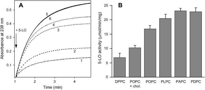
Recently we have analyzed the effect of lipid polar headgroups on 5-LO-membrane interactions and 5-LO activity (Pande et al., 2004). The results suggested that the effects of different lipids on the membrane binding and activity of 5-LO might be exerted through modulation of both membrane surface properties and overall membrane structure, including lipid packing order. In this current work, we have tested the hypothesis that membrane fluidity and increased fractions of AA-containing lipids in membranes may promote 5-LO binding and activity. The activity of 5-LO was measured in the presence of PCs containing a palmitoyl residue at the sn-1 position and fatty acid residues with zero to six cis-unsaturated bonds (nΔ) at the sn-2 position, i.e., DPPC, 1-palmitoyl-2-oleoyl-sn-glycero-3-phosphocholine (POPC), 1-palmitoyl-2-linoleoyl-sn-glycero-3-phosphocholine (PLPC), PAPC, and 1-palmitoyl-2-docosahexaenoyl-sn-glycero-3-phosphocholine (PDPC). Membranes containing lipids with unsaturated hydrocarbon chains had a significant stimulatory effect on 5-LO activity. The activity of 5-LO, as measured using the initial slope of the time dependence of absorbance at 238 nm, due to 5-HPETE production (Percival, 1991; Skorey and Gresser, 1998), increased ∼2.5 times just by incorporation of a single cis-unsaturated bond in the sn-2 acyl chain of the lipid, i.e., by replacing DPPC with POPC (Fig. 1 A). Phosphatidylcholines with 2, 4, and 6 cis-double bonds in their sn-2 acyl chains, i.e., PLPC, PAPC, and PDPC, resulted in further increase in 5-LO activity, indicating a correlation between the degree of lipid acyl chain unsaturation and 5-LO activity. It is known that cis-unsaturation of lipid acyl chains decreases lipid packing order in membranes and increases membrane fluidity (Stubbs et al., 1981; Keough et al., 1987; Cevc, 1991), and cholesterol is able to partially reverse this effect (de Almeida et al., 2003). Incorporation of 20 mol % cholesterol into POPC membranes significantly reduced 5-LO activity compared to that measured in the presence of pure POPC vesicles (Fig. 1 A), suggesting that the effect of lipid acyl chain cis-unsaturation on 5-LO activity is probably exerted through membrane fluidity. The initial slopes of kinetic curves of 5-LO activity were used to calculate the amount of 5-HPETE production per mg of 5-LO per minute (Fig. 1 B). Data of Fig. 1, A and B, imply that 5-LO activity is significantly promoted in the presence of vesicles composed of lipids with an increasing degree of unsaturation.
FIGURE 1.
Activity of 5-LO increases with an increasing degree of lipid cis-unsaturation and decreases in the presence of cholesterol in vesicle membranes. (A) Time dependence of conversion of AA to 5-HPETE, as measured by absorption at 238 nm. The buffer (specified in Materials and Methods) contains 100 μM AA and large unilamellar vesicles composed of 350 μM DPPC (1), POPC + 20 mol % cholesterol (2), POPC (3), PLPC (4), PAPC (5), and PDPC (6). The reaction is initiated by adding 2.4 μg/ml 5-LO, as indicated by the arrow. Measurements are conducted at 22°C. (B) A bar graph showing the mean values and standard deviations of 5-LO activity in the presence of vesicles of different lipid compositions, as indicated. Experimental conditions are as in A. Data shown in B are averaged based on three independent experiments.
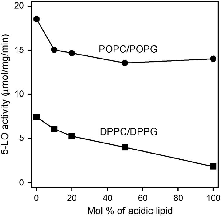
It has been shown previously that membranes composed of anionic lipids failed to support 5-LO activity as efficiently as PC (Puustinen et al., 1988; Noguchi et al., 1994; Pande et al., 2004). Here we show that the effect of the anionic charge of membrane lipids on 5-LO activity depends on the degree of lipid acyl chain unsaturation. Data of Fig. 2 demonstrate that an increase in the fraction of anionic 1-palmitoyl-2-oleoyl-sn-glycero-3-phosphoglycerol (POPG) in zwitterionic POPC membranes from 0 to 1 causes less than a twofold impairment in 5-LO activity, whereas similar fractions of 1,2-dipalmitoyl-sn-glycero-3-phosphoglycerol (DPPG) in DPPC membranes exerts a fourfold inhibition of 5-LO. In effect, 5-LO activity in the presence of fully saturated DPPG is ∼8-fold lower than in the presence of monounsaturated POPG. These data indicate that lipid acyl chain unsaturation is a key modulator of 5-LO activity both for zwitterionic and anionic lipid membranes.
FIGURE 2.
Dependence of the specific activity of 5-LO on the content of anionic lipids in fluid (POPC/POPG) and solid (DPPC/DPPG) membranes. Total lipid concentration was 350 μM in all cases, AA concentration was 100 μM, and 5-LO was added to a final concentration of 2.4 μg/ml. The buffer, method of 5-LO activity measurement, and other experimental conditions are described in Materials and Methods.
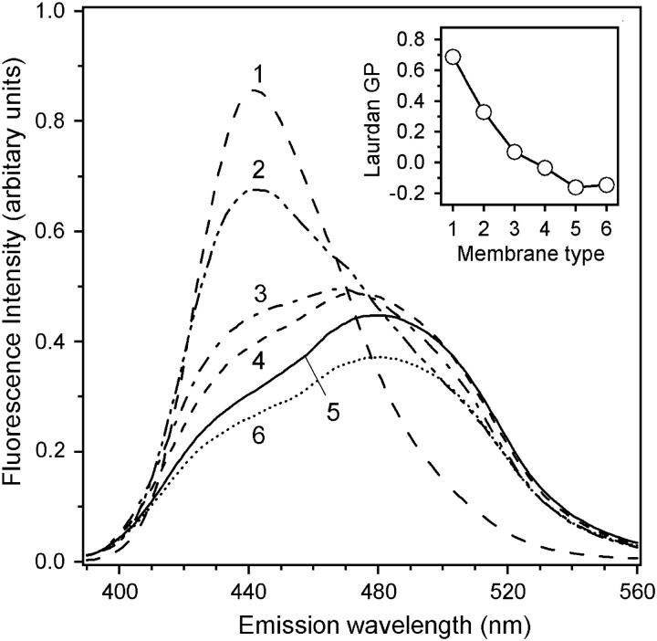
To physically interpret the effects of lipids with varying degrees of cis-unsaturation and the presence of cholesterol in membranes on 5-LO activity, the fluidity of membranes was measured by GP of Laurdan incorporated in membranes at 1 mol %. Introduction of a single double bond in the sn-2 acyl chain of the lipid, i.e., replacement of DPPC with POPC, results in a sharp decrease in GP, indicating a significant increase in membrane fluidity (Fig. 3). Membrane fluidity further increases with increasing degrees of lipid acyl chain unsaturation, and reaches a plateau at four double bonds per sn-2 chain of the lipid, corresponding to PAPC. The GP value markedly increases upon introduction of 20 mol % cholesterol in POPC membranes, indicating a decrease in membrane fluidity. Data of Figs. 1–3 provide strong evidence for modulation of 5-LO activity by membrane fluidity.
FIGURE 3.
Fluorescence emission spectra of 1 mol % Laurdan in vesicles composed of DPPC (1), POPC + 20 mol % cholesterol (2), POPC (3), PLPC (4), PAPC (5), and PDPC (6). The buffer contained 0.1 mM EGTA, 0.3 mM CaCl2, and 50 mM Tris-HCl (pH 7.5). Total lipid concentration was 100 μM in 0.4-cm path-length quartz cuvettes. The excitation wavelength was 360 nm, and the temperature was 22°C. The inset shows the dependence of GP on the lipid composition, as specified in the main figure. The values of GP were calculated as GP = (F435 − F500)/(F435 + F500), where F435 and F500 are the Laurdan fluorescence emission intensities at respective wavelengths.
Dependence of membrane binding strength of 5-LO on membrane fluidity
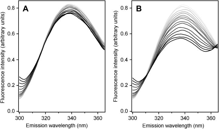
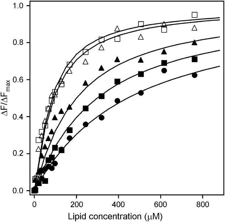
Our data indicate that 5-LO activity increases in the presence of membranes with increasing fluidity and reaches a saturating level with PAPC. To test the hypothesis that the increase in 5-LO activity results from stronger binding of the enzyme to more fluid membranes, we measured the binding of 5-LO to large unilamellar vesicles composed of DPPC, POPC, PLPC, PAPC, and PDPC. Membrane binding of 5-LO was studied using RET from tryptophans of 5-LO to 2 mol % Py-PE in the membranes. When 5-LO was titrated with phospholipid vesicles in the absence of an energy acceptor, Trp fluorescence decreased because of sample dilution (Fig. 4 A). Titration of 5-LO with Py-PE-containing vesicles resulted in a strong, lipid dose-dependent decrease in Trp fluorescence with concomitant increase in the pyrene emission, due to RET (Fig. 4 B). Since RET is based on short-range (28 Å for the Trp-pyrene pair) dipole-dipole interactions between energy donors and acceptors (Lakowicz, 1999), the observed effect indicates binding of 5-LO to vesicle membranes. Analysis of binding isotherms (Fig. 5) yielded the dissociation constants and the numbers of lipid molecules per protein binding site, as summarized in Table 1. These data indicate that membrane binding affinity of 5-LO increases with increasing degrees of lipid acyl chain cis-unsaturation, reaches the highest value for PAPC membranes (nΔ = 4), and then slightly decreases upon further increase in the degree of lipid unsaturation, i.e., with PDPC membranes (nΔ = 6). Altogether, our findings delineate a correlation between membrane fluidity, membrane binding strength, and activity of 5-LO.
FIGURE 4.
Fluorescence emission spectra of tryptophans of 5-LO in the absence and presence of large unilamellar PAPC vesicles without (A) and with (B) 2 mol % Py-PE. In both panels, increasing darkness of the lines corresponds to an increase in total lipid concentration from zero to 760 μM (see Fig. 5). Decrease in Trp emission intensity in A is due to dilution upon addition of stock vesicle suspension. In B, Trp fluorescence significantly decreases upon addition of Py-PE-containing vesicles due to energy transfer from Trp of 5-LO to Py-PE. The excitation wavelength was 290 nm, and buffer and temperature were as in Fig. 3.
FIGURE 5.
Isotherms of 5-LO binding to vesicles composed of DPPC (•), POPC (▪), PLPC (▴), PAPC (□), and PDPC (▵). The data points are obtained on the basis of the decrease in Trp fluorescence emission intensity due to energy transfer from Trp to Py-PE (2 mol % in vesicle membranes), as measured in RET experiments (e.g., Fig. 4). The theoretical curves are simulated through Eq. 1 using binding parameters summarized in Table 1.
TABLE 1.
Parameters characterizing the interaction of 5-LO with large unilamellar vesicles composed of PCs of varying fluidity
| Lipid | L1/2 (μM) | KD (μM) | N |
|---|---|---|---|
| DPPC | 443.5 | 1.15 | 184 |
| POPC | 297.8 | 0.78 | 176 |
| PLPC | 199.2 | 0.56 | 157 |
| PAPC | 83.2 | 0.24 | 127 |
| PDPC | 92.0 | 0.32 | 114 |
Parameters include lipid concentrations corresponding to binding of 50% of 5-LO (L1/2), the dissociation constants (KD), and the numbers of lipids per 5-LO binding site (N). Lipids were composed of phosphatidylcholines with varying degrees of sn-2 acyl chain unsaturation.
The data of Table 1 indicate that the number of lipid molecules per 5-LO binding site, N, decreases from 184 to 114 with increasing membrane fluidity, which likely results from larger cross-sectional area of lipids (AL) with higher degree of unsaturation (Stillwell and Wassall, 2003). Using the values of AL for all five lipids summarized in Table 1 (Stillwell and Wassall, 2003 and references therein), we estimated that the membrane surface area per 5-LO binding site is N × AL = 9600 ± 1250 Å2.
Dependence of membrane insertion of 5-LO on membrane fluidity
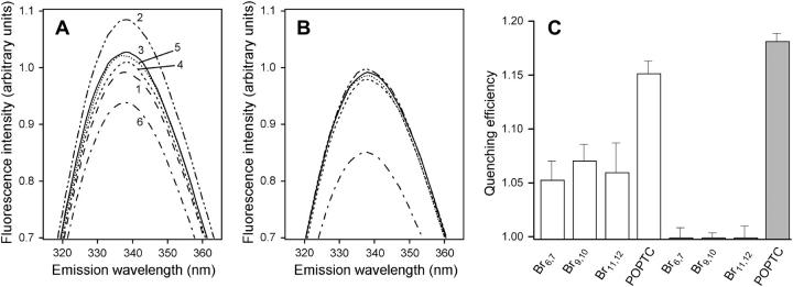
The correlation between membrane fluidity, membrane binding affinity, and activity of 5-LO suggests that the enhancement of 5-LO activity by membrane fluidity may at least partly result from stronger binding of 5-LO to membranes of higher fluidity. However, other parameters of 5-LO-membrane interaction, such as the depth of insertion of 5-LO into the hydrophobic core of membranes, may also contribute to 5-LO function. We have estimated the depth of membrane insertion of the side chains of Trp residues of 5-LO by employing the quenching of Trp fluorescence by Br2PCs brominated at 6,7, or 9,10, or 11,12 positions of the acyl chains, or by POPTC nitroxide spin-labeled at the polar headgroup. In the presence of fluid PAPC vesicles without quenchers, Trp fluorescence of 5-LO significantly increased (Fig. 6 A), indicating that some tryptophans of 5-LO experience less polar environment, apparently due to membrane binding of 5-LO. (Unlike the experiments presented in Fig. 4, in these experiments there was no dilution effect and the increase in Trp emission intensity in the presence of membranes could be seen directly.) Incorporation of Br2PCs in PAPC vesicles resulted in quenching of 5-LO fluorescence by all three brominated lipids; the maximum quenching occurred with vesicles containing 9,10-Br2PC (Fig. 6, A and C). The distances of bromine atoms in Br2PCs brominated at 6,7, or 9,10, or 11,12 positions from the bilayer center have been estimated to be 11.0, 8.3, and 6.5 Å, respectively (McIntosh and Holloway, 1987). Therefore, maximum quenching of Trp fluorescence by 9,10-Br2PC indicates that the side chains of certain Trp residues of 5-LO bound to PAPC membranes penetrate into the membrane hydrophobic core nearly halfway to the membrane center. When PAPC vesicles contained headgroup spin-labeled POPTC, Trp fluorescence was quenched to a larger extent than by any of the brominated lipids (Fig. 6, A and C). This result indicates that a larger fraction of tryptophans of 5-LO bound to PAPC membranes are located at the membrane-water interface compared to the fraction embedded into the lipid hydrocarbon region.
FIGURE 6.
Fluorescence emission spectra of tryptophans of 5-LO in the absence and presence of large unilamellar vesicles composed of a fluid lipid, PAPC (A), or a solid lipid, DPPC (B). Only the top portions of spectra are shown to make differences between spectra more discernable. The spectra are numbered as follows: 1, free 5-LO in buffer; 2, 5-LO with large unilamellar vesicles of PAPC (A) or DPPC (B); 3, 6,7-Br2PC-containing vesicles; 4, 9,10-Br2PC-containing vesicles; 5, 11,12-Br2PC-containing vesicles; and 6, POPTC-containing vesicles. Numbering in A also applies to the same line types in B. (C) Summary of the data obtained in three experiments (mean ± SD). Open and solid bars apply to PAPC and DPPC membranes, respectively. When vesicles were present, the total lipid concentration was 750 μM. Brominated lipids were present at 25 mol %, and POPTC at 15 mol %. Protein concentration was 0.24 μM. Excitation was at 290 nm. The buffer and temperature were as in Fig. 3.
In the presence of solid DPPC vesicles containing brominated lipids, none of the Br2PCs quenched the Trp fluorescence of 5-LO (Fig. 6, B and C). However, Trp fluorescence was quenched by POPTC even more efficiently than in the case of fluid PAPC membranes, presumably because tryptophans that embed into fluid PAPC membranes are located at the membrane-water interface in the case of solid DPPC membranes, thus increasing the total population of interfacially located tryptophans. Altogether, data of Fig. 6 suggest that 5-LO is able to partially intrude into the hydrocarbon region of fluid membranes, but in the case of solid membranes it interacts with the membrane surface without penetration into its hydrophobic core. In conjunction with our data that 5-LO has stronger membrane binding affinity and higher activity in the presence of fluid membranes, these data suggest that partial insertion of 5-LO into the membrane is likely to be important for the enzyme function.
A model for membrane-bound 5-LO
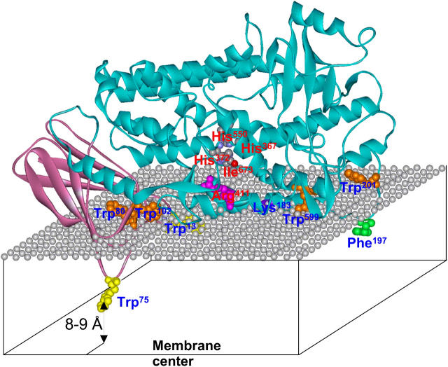
The available data on membrane binding of human 5-LO, its N-terminal putative β-barrel domain, and rabbit 15-LO allow construction of a model of 5-LO bound to a phospholipid membrane in the fluid phase (Fig. 7). We have modeled the structure of 5-LO using the structure of rabbit reticulocyte 15-LO as a template (Protein Data Bank entry, 1lox), which shares 37% sequence identity and 57% similarity with 5-LO. Homology modeling was achieved using the internet-based SWISS-MODEL server (Schwede et al., 2003). The modeled structure of 5-LO was positioned at the membrane surface using the following criteria: 1), the symmetry axis of the N-terminal β-barrel is oriented at ∼45° with respect to the membrane normal (Pande et al., 2004); 2), tryptophans 13, 75, and 102 likely interact with the membrane (Kulkarni et al., 2002); 3), Trp181 of rabbit 15-LO, which corresponds to Lys183 in the aligned 5-LO sequence, is likely to interact with the membrane (Walther et al., 2002, 2004); and 4), at least one Trp penetrates into the membrane to a depth of 8–9 Å from the membrane center (this work). Positioning of the modeled 5-LO structure on the membrane surface according to these constraints indicates that Trp75 likely penetrates into the membrane deeper than any other residue, Trp13 inserts into the hydrophobic part of the membrane, but not as deep as Trp75, and Trp102 is closer to the membrane-water interface. Note that Kulkarni et al. (2002) have shown that Trp102 was more important for the interaction of the β-barrel domain of 5-LO with membranes than Trp13 or Trp75. This is not necessarily in conflict with our model because tryptophans located at the polar headgroup/hydrocarbon boundary of membranes can facilitate protein-membrane interactions by means of both H-bonding and nonpolar interactions (Yau et al., 1998) and thus may contribute to the interaction more than totally embedded tryptophans that are not involved in H-bonding with lipid carbonyl oxygens. Also, the truncated β-barrel domain may not be oriented at the membrane surface as the full-length protein. Lys183 is properly located to make ionic and/or H-bonding interaction with the phosphate group of membrane phospholipids. In Fig. 7, the residue Arg411 is highlighted to indicate the putative site of entrance of AA into the substrate binding cleft, because Gillmor et al. (1997) have suggested that the corresponding residue in the rabbit 15-LO sequence (Arg403) forms an ionic contact with the carboxyl group of arachidonate during the initial step of enzyme-substrate interaction. Also shown are the iron-coordinating residues (histidines 367, 372, 550, and Ile673) to identify the catalytic site of the enzyme (Zhang et al., 1992). The model indicates that most surface-exposed tryptophans of 5-LO are at the polar headgroup region of the membrane, whereas Trp75 and Trp13 insert into the hydrophobic part of the membrane. This agrees with the data indicating stronger quenching of Trp fluorescence by headgroup nitroxide-labeled POPTC than by acyl chain-brominated lipids (Fig. 6). In the case of solid membranes, all tryptophans are presumably located at the membrane-water interface without membrane insertion, resulting in negligible quenching by brominated lipids and stronger quenching by POPTC (Fig. 6). The more “peripheral” binding of 5-LO to solid membranes probably contributes to the decreased membrane affinity and lower activity of 5-LO (Figs. 1 and 5, Table 1).
FIGURE 7.
A model for 5-LO bound to a phospholipid membrane in the fluid phase, constructed on the basis of homology modeling and data on membrane insertion and angular orientation of 5-LO. The protein structure is modeled using SWISS-MODEL on the basis of rabbit 15-LO crystal structure as a template, and is presented in a ribbon format. The N-terminal β-barrel and the catalytic domains are shown in plum and aqua, respectively. The mesh of gray spheres represents the plane of the phosphate groups of phospholipid molecules. Membrane-interacting and catalytically important residues are shown in the ball-and-stick format and are labeled by blue and red labels, respectively. Tryptophans that insert into the membrane hydrocarbon region are shown in yellow, and those located at the membrane-water interface are shown in orange. Phe197 and Lys183, which interact with the membrane by nonpolar and ionic interactions, are shown in green and purple, respectively. Arg411, which presumably forms an ionic contact with the carboxyl group of arachidonate during the initial step of enzyme-substrate interaction, is shown in magenta. Residues involved in coordination of the iron cofactor are colored according to the atom type. Trp13, and partially Trp599 and Lys183, are underneath the polar groups of lipids and can hardly be seen. His367 is hidden behind the helical ribbon. According to our earlier results (Pande et al., 2004), the symmetry axis of the N-terminal β-barrel domain is tilted at ∼45° with respect to the membrane normal, and the current data suggest that at least one Trp, most likely the Trp75, inserts into the membrane as deep as 8–9 Å from the membrane center.
DISCUSSION
Binding to intracellular membranes in vivo or to artificial lipid membranes in vitro is an essential step in activation of 5-LO. Despite this, little is known about the mechanism of interaction of 5-LO with membranes. No quantitative data on 5-LO-membrane interactions, such as binding constants or 5-LO-lipid binding stoichiometries, have been reported so far, and the effects of physical properties of membranes, such as membrane fluidity, on membrane binding and activity of 5-LO are poorly understood. Previously we have conducted a detailed analysis of the effects of lipid polar headgroups, as well as of Ca2+ and lipid concentrations, on 5-LO-membrane interactions and 5-LO activity (Pande et al., 2004). In this work, we concentrate on the effect of lipid acyl chain unsaturation and membrane fluidity on membrane binding strength, membrane insertion, and activity of 5-LO. Our results provide unprecedented evidence that membrane fluidity is an important determinant for the membrane-binding mode and activity of 5-LO.
To study the effect of lipid acyl chain unsaturation on the membrane-binding mode and activity of 5-LO, we have used PCs that contain a saturated palmitoyl chain at the sn-1 position, whereas the acyl chain at the sn-2 position was either a palmitoyl (16:0), oleoyl (18:1), linoleoyl (18:2), arachidonoyl (20:4), or docosahexaenoyl (22:6) residue (shown in parentheses are the ratios of carbon atoms to double bonds per chain). The choice of these lipids was based on the fact that in major lipids of mammalian cell membranes the sn-1 position is usually occupied by a saturated chain, such as a palmitoyl residue, and the sn-2 position is most frequently occupied by one of the unsaturated fatty acyl chains specified above (Albi et al., 1997; Surette and Chilton, 1998; D'Antuono et al., 2000). Replacement of fully saturated DPPC membranes by membranes containing lipids with cis-unsaturated acyl chains results in an ∼3-fold increase in 5-LO activity. It is known that membranes composed of DPPC are in the solid gel phase at temperatures below 41°C, whereas membranes composed of POPC, PLPC, PAPC, or PDPC are in the fluid, liquid crystalline phase above 0°C (Stubbs et al., 1981; Litman et al., 1991; Stillwell and Wassall, 2003). Therefore, our data indicate that the membrane-binding mode and activity of 5-LO are controlled by membrane fluidity. Remarkably, the maximum stimulatory effect on 5-LO activity was reached with vesicles composed of PAPC. The fact that both the membrane-binding strength and activity of 5-LO correlate with Laurdan GP (Figs. 1, 3, and 5) indicates that the effect of PAPC on 5-LO function is exerted through variation of membrane fluidity rather than specific 5-LO-PAPC interactions. The activity of 5-LO in the presence of POPC membranes containing 20 mol % cholesterol, which consist of coexisting disordered and ordered phases (de Almeida et al., 2003), is between activities measured in the presence of solid (DPPC) and fluid membranes (e.g., POPC). This is consistent with the concept of modulation of 5-LO activity by membrane fluidity.
Our data indicate that the values of dissociation constants (KD) of 5-LO for phospholipid membranes decrease from 1.15 to 0.24 μM upon an increase of the degree of lipid sn-2 acyl chain cis-unsaturation from zero to four, and undergoes little change upon further increase in nΔ (Fig. 5 and Table 1). This resembles the increase in 5-LO activity as a function of membrane fluidity (Fig. 1). Although quantitative data on 5-LO binding to membranes are not available, surface plasmon resonance experiments yielded a KD value of 0.6 nM for the binding of the putative β-barrel domain of 5-LO to POPC vesicles in the presence of 0.1 mM CaCl2 (Kulkarni et al., 2002), which indicates much stronger binding of the β-barrel domain compared to that of the full-length 5-LO determined in this work (0.78 μM). Differences in methods used by Kulkarni et al. (2002) and in this work might partially account for discrepancies in measured dissociation constants. On the other hand, this divergence is congruent with earlier data showing that intracellular translocation of the N-terminal β-barrel domain of 5-LO to the nuclear membrane was much faster than that of the full-length protein (Chen and Funk, 2001). Furthermore, stronger membrane binding by itself may not necessarily favor the enzyme function. It is possible that the catalytic domain, while reducing the binding strength, facilitates a defined, productive mode binding of the enzyme to the membrane surface.
The average area per 5-LO binding site at the membrane surface is estimated to be Abs = 9600 ± 1250 Å2. Using the model of 5-LO (Fig. 7), we have determined that the protein molecule can be roughly presented as a prolate ellipsoid of revolution with semiaxes of 48 and 23 Å. This would correspond to a maximum cross-sectional area of π × 48 × 23 ≈ 3500 Å2, implying that Abs is ∼2.7 times larger than the maximum cross-sectional area of the molecule. Because of the rotational freedom of membrane-bound protein molecules around the membrane normal, the effective area per membrane-bound 5-LO would be π × 482 ≈ 7200 Å2. On the other hand, even at the saturation of adsorption, there will be “empty” surface area between membrane-bound protein molecules. This means that upon binding of each protein molecule, an area of (96 Å)2 ≈ 9200 Å2 will become inaccessible to other protein molecules, which is in good agreement with the experimentally determined effective area per protein binding site of 9600 ± 1250 Å2.
Previous studies indicated that the N-terminal putative β-barrel domain of 5-LO had ∼20 times stronger affinity (in terms of KD) for PC membranes than for membranes containing 50 mol % of PG or PS (Kulkarni et al., 2002). This led to the conclusion that increased affinity of the β-barrel domain of 5-LO for PC versus anionic lipid membranes dictates the localization of 5-LO to PC-rich nuclear membranes (Kulkarni et al., 2002). However, the content of anionic lipids in nuclear membranes is comparable to or even higher than that of other cellular membranes (Khandwala and Kasper, 1971; Neitcheva and Peeva, 1995; Surette and Chilton, 1998; D'Santos et al., 1999, 2000; D'Antuono et al., 2000). Also, PC is abundant not only in nuclear but in many other membranes of eukaryotic cells (Hanahan, 1997; Surette and Chilton, 1998; Mathews et al., 1999; D'Antuono et al., 2000). For example, analysis of phospholipid content in the plasma membrane, endoplasmic reticulum, and the nuclear membrane of rat papillary cells indicated that PC was the most abundant lipid (42–48 mol %) in all three cases (D'Antuono et al., 2000). No significant difference was found in the PC content in nuclear membranes and whole-cell lipid extract for human monocytes (Surette and Chilton, 1998). Therefore, preference of 5-LO for PC versus anionic lipids cannot account for selective recruitment of 5-LO to nuclear membranes.
Nuclear membranes are enriched in AA-containing lipids more than other cellular membranes (Neufeld et al., 1985; Capriotti et al., 1988; Albi et al., 1997; Surette and Chilton, 1998; D'Antuono et al., 2000). Thus, up to 48% of the total cellular AA-containing lipids were found in the nuclear membranes of various mammalian cell lines, including leukemia cells (Surette and Chilton, 1998). In rat renal papillary cells, the fraction of AA-containing lipids was 1.5 times higher, whereas the fraction of lipids with saturated chains was 1.6 times lower in nuclear than in endoplasmic reticulum membranes (D'Antuono et al., 2000). Large fractions of AA-containing lipids result in elevated fluidity of nuclear membranes (Yu et al., 1996; Albi et al., 1997; D'Antuono et al., 2000). This leads to a mechanism suggesting that increased membrane fluidity via high levels of AA-containing lipids may be an important factor for localization of 5-LO to nuclear membranes. Our data indeed provide support for this mechanism by indicating that among PCs with a palmitoyl residue in the sn-1 position and various acyl chains in the sn-2 position, PAPC was the most efficient lipid in terms of facilitating membrane binding, membrane insertion, and activity of 5-LO. Lipids with lower or higher degrees of acyl chain unsaturation were not as efficient as PAPC. Taking into account that cytosolic phospholipase A2 (PLA2), as well as FLAP, are also colocalized to nuclear membranes (Woods et al., 1993; Pouliot et al., 1996), this mechanism would lead to a congregation of all enzymes, regulators, and substrates for efficient metabolism of AA-containing lipids to leukotrienes.
Our model of 5-LO binding to a membrane in the fluid phase (Fig. 7) shows that partial insertion of 5-LO into the hydrophobic core of the membrane may be required for membrane-proximal location of the orifice of the substrate-binding pocket, marked by Arg411, and better access of 5-LO to membrane-residing AA. Partial membrane insertion of 5-LO may also be necessary for positioning 5-LO at the membrane surface in a productive mode, because the Ca2+-mediated electrostatic mechanism alone is apparently not enough to do this.
Although details of membrane binding of 5-LO have not been described before, membrane-binding modes of several C2 domain-containing proteins or truncated C2 domains have been assessed in the past. Cytosolic PLA2α, which colocalizes with 5-LO at the nuclear membrane and produces AA for eicosanoid production, was shown to considerably penetrate into the hydrocarbon region of phospholipid monolayers (Lichtenbergova et al., 1998). Certain residues of the truncated C2-domain of cytosolic PLA2α penetrate into the hydrocarbon region of membranes as deep as 5–10 Å below the plane of the lipid phosphate groups (Ball et al., 1999; Malmberg et al., 2003). C2 domains of other membrane-binding proteins, e.g., protein kinase Cα and synaptotagmin I, penetrate into the polar headgroup region of membranes but not into the hydrocarbon region as deep as cytosolic PLA2α (Frazier et al., 2003; Kohout et al., 2003; Rufener et al., 2005). The hydrophobic residues Phe70, Leu71, Trp181, and Leu195of rabbit 15-LO, which are clustered at the membrane-binding surface of 15-LO that contains the substrate-binding pocket, were found to play a major role in membrane anchoring of the protein, probably involving partial membrane insertion (Walther et al., 2002, 2004). It seems likely that hydrophobic protein-membrane interactions and partial membrane penetration constitute a common feature of C2-domain-containing proteins. This may be necessary to stabilize the membrane docking of these proteins. It should be noted that all experiments with cytosolic PLA2α, protein kinase Cα, and synaptotagmin I were conducted using fluid membranes composed of lipids containing an oleic acid moiety at the sn-2 or at both positions, and in experiments with 15-LO submitochondrial particles were used (Walther et al., 2002, 2004). Thus, it was not possible to assess the dependence of membrane insertion on membrane fluidity based on these studies. Our data, however, clearly demonstrate that membrane insertion of 5-LO only occurs with fluid, but not solid membranes, indicating an important role of membrane fluidity in modulating the function of 5-LO and probably other enzymes that undergo interfacial activation via membrane binding.
Finally, partial membrane insertion of 5-LO indicates a resemblance between the membrane binding modes of lipoxygenases and the other major class of enzymes involved in eicosanoid biosynthesis, cyclooxygenases. The latter enzymes are embedded in nuclear and perinuclear endoplasmic reticulum membranes as monotopic proteins (Spencer et al., 1998; Marvin et al., 2000; Picot and Garavito, 1994; Picot et al., 1997). Enrichment of these membranes with AA-containing lipids probably facilitates membrane binding and insertion of both cyclooxygenases and 5-LO through increased membrane fluidity and thus plays a regulatory role in the biosynthesis of prostanoids and leukotrienes.
Acknowledgments
We thank David Moe for expert technical assistance at the initial stages of this work and Dr. Jeffrey L. Urbauer for useful comments during preparation of the manuscript.
This work was supported in part by National Heart, Lung, and Blood Institute grant HL65524.
References
- Abramovitz, M., E. Wong, M. E. Cox, C. D. Richardson, C. Li, and P. J. Vickers. 1993. 5-lipoxygenase-activating protein stimulates the utilization of arachidonic acid by 5-lipoxygenase. Eur. J. Biochem. 215:105–111. [DOI] [PubMed] [Google Scholar]
- Albi, E., M. L. Tomassoni, and M. Viola-Magni. 1997. Effect of lipid composition on rat liver nuclear membrane fluidity. Cell Biochem. Funct. 3:181–190. [DOI] [PubMed] [Google Scholar]
- Ball, A., R. Nielsen, M. H. Gelb, and B. H. Robinson. 1999. Interfacial membrane docking of cytosolic phospholipase A2 C2 domain using electrostatic potential-modulated spin relaxation magnetic resonance. Proc. Natl. Acad. Sci. USA. 96:6637–6642. [DOI] [PMC free article] [PubMed] [Google Scholar]
- Bradford, M. M. 1976. A rapid and sensitive method for the quantitation of microgram quantities of protein utilizing the principle of protein-dye binding. Anal. Biochem. 72:248–254. [DOI] [PubMed] [Google Scholar]
- Brock, T. G., R. W. McNish, and M. Peters-Golden. 1995. Translocation and leukotriene synthetic capacity of nuclear 5-lipoxygenase in rat basophilic leukemia cells and alveolar macrophages. J. Biol. Chem. 270:21652–21658. [DOI] [PubMed] [Google Scholar]
- Capriotti, A. M., E. E. Furth, M. E. Arrasmith, and M. Laposata. 1988. Arachidonate released upon agonist stimulation preferentially originates from arachidonate most recently incorporated into nuclear membrane phospholipids. J. Biol. Chem. 263:10029–10034. [PubMed] [Google Scholar]
- Cevc, G. 1991. How membrane chain-melting phase-transition temperature is affected by the lipid chain asymmetry and degree of unsaturation: an effective chain-length model. Biochemistry. 30:7186–7193. [DOI] [PubMed] [Google Scholar]
- Chen, X.-S., and C. D. Funk. 2001. The N-terminal “β-barrel” domain of 5-lipoxygenase is essential for nuclear membrane translocation. J. Biol. Chem. 276:811–818. [DOI] [PubMed] [Google Scholar]
- Chen, X., N. Li, S. Wang, N. Wu, J. Hong, X. Jiao, M. J. Krasna, D. G. Beer, and C. S. Yang. 2003. Leukotriene A4 hydrolase in rat and human esophageal adenocarcinomas and inhibitory effects of bestatin. J. Natl. Cancer Inst. 95:1053–1061. [DOI] [PubMed] [Google Scholar]
- D'Antuono, C., M. C. Fernandez-Tome, N. Sterin-Speziale, and D. L. Bernik. 2000. Lipid-protein interactions in rat renal subcellular membranes: a biophysical and biochemical study. Arch. Biochem. Biophys. 382:39–47. [DOI] [PubMed] [Google Scholar]
- de Almeida, R. F., A. Fedorov, and M. Prieto. 2003. Sphingomyelin/phosphatidylcholine/cholesterol phase diagram: boundaries and composition of lipid rafts. Biophys. J. 85:2406–2416. [DOI] [PMC free article] [PubMed] [Google Scholar]
- D'Santos, C. S., J. H. Clarke, R. F. Irvine, and N. Divecha. 1999. Nuclei contain two differentially regulated pools of diacylglycerol. Curr. Biol. 9:437–440. [DOI] [PubMed] [Google Scholar]
- D'Santos, C., J. H. Clarke, M. Roefs, J. R. Halstead, and N. Divecha. 2000. Nuclear inositides. Eur. J. Histochem. 44:51–60. [PubMed] [Google Scholar]
- Fan, X. M., S. P. Tu, S. K. Lam, W. P. Wang, J. Wu, W. M. Wong, M. F. Yuen, M. C. Lin, H. F. Kung, and B. C. Wong. 2004. Five-lipoxygenase-activating protein inhibitor MK-886 induces apoptosis in gastric cancer through upregulation of p27kip1 and bax. J. Gastroenterol. Hepatol. 19:31–37. [DOI] [PubMed] [Google Scholar]
- Frazier, A. A., C. R. Roller, J. J. Havelka, A. Hinderliter, and D. S. Cafiso. 2003. Membrane-bound orientation and position of the synaptotagmin I C2A domain by site-directed spin labeling. Biochemistry. 42:96–105. [DOI] [PubMed] [Google Scholar]
- Ghosh, J., and C. E. Myers. 1999. Central role of arachidonate 5-lipoxygenase in the regulation of cell growth and apoptosis in human prostate cancer cells. Adv. Exp. Med. Biol. 469:577–582. [DOI] [PubMed] [Google Scholar]
- Gillmor, S. A., A. Villaseñor, R. Fletterick, E. Sigal, and M. F. Browner. 1997. The structure of mammalian 15-lipoxygenase reveals similarity to the lipases and the determinants of substrate specificity. Nat. Struct. Biol. 4:1003–1009. [DOI] [PubMed] [Google Scholar]
- Goodarzi, K., M. Goodarzi, A. M. Tager, A. D. Luster, and U. H. von Andrian. 2003. Leukotriene B4 and BLT1 control cytotoxic effector T cell recruitment to inflamed tissues. Nat. Immunol. 4:965–973. [DOI] [PubMed] [Google Scholar]
- Hammarberg, T., P. Provost, B. Persson, and O. Rådmark. 2000. The N-terminal domain of 5-lipoxygenase binds calcium and mediates calcium stimulation of enzyme activity. J. Biol. Chem. 275:38787–38793. [DOI] [PubMed] [Google Scholar]
- Hanahan, D. J. 1997. A Guide to Phospholipid Chemistry. Oxford University Press, New York.
- Harris, F. M., K. B. Best, and J. D. Bell. 2002. Use of laurdan fluorescence intensity and polarization to distinguish between changes in membrane fluidity and phospholipid order. Biochim. Biophys. Acta. 1565:123–128. [DOI] [PubMed] [Google Scholar]
- Keough, K. M., B. Giffin, and N. Kariel. 1987. The influence of unsaturation on the phase transition temperatures of a series of heteroacid phosphatidylcholines containing twenty-carbon chains. Biochim. Biophys. Acta. 902:1–10. [DOI] [PubMed] [Google Scholar]
- Khandwala, A. S., and C. B. Kasper. 1971. The fatty acid composition of individual phospholipids from rat liver nuclear membrane and nuclei. J. Biol. Chem. 246:6242–6246. [PubMed] [Google Scholar]
- Kohout, S. C., S. Corbalan-Garcia, J. C. Gomez-Fernandez, and J. J. Falke. 2003. C2 domain of protein kinase Cα: elucidation of the membrane docking surface by site-directed fluorescence and spin labeling. Biochemistry. 42:1254–1265. [DOI] [PMC free article] [PubMed] [Google Scholar]
- Kühn, H. 1999. Lipoxygenases. In Prostaglandins, Leukotrienes, and Other Eicosanoids. From Biogenesis to Clinical Applications. F. Marks and G. Fürstenberger, editors. Wiley-VCH, Weinheim, Germany. 109–141.
- Kulkarni, S., S. Das, C. D. Funk, D. Murray, and W. Cho. 2002. Molecular basis of the specific subcellular localization of the C2-like domain of 5-lipoxygenase. J. Biol. Chem. 277:13167–13174. [DOI] [PubMed] [Google Scholar]
- Lakowicz, J. R. 1999. Principles of Fluorescence Spectroscopy, 2nd ed.. Kluwer Academic/Plenum Publishers, New York.
- Lichtenbergova, L., E. T. Yoon, and W. Cho. 1998. Membrane penetration of cytosolic phospholipase A2 is necessary for its interfacial catalysis and arachidonate specificity. Biochemistry. 37:14128–14136. [DOI] [PubMed] [Google Scholar]
- Litman, B. J., E. N. Lewis, and I. W. Levin. 1991. Packing characteristics of highly unsaturated bilayer lipids: Raman spectroscopic studies of multilamellar phosphatidylcholine dispersions. Biochemistry. 30:313–319. [DOI] [PubMed] [Google Scholar]
- Malaviya, R., R. Malaviya, and B. A. Jakschik. 1993. Reversible translocation of 5-lipoxygenase in mast cells upon IgE/antigen stimulation. J. Biol. Chem. 268:4939–4944. [PubMed] [Google Scholar]
- Malmberg, N. J., D. R. Van Buskirk, and J. J. Falke. 2003. Membrane-docking loops of the cPLA2 C2 domain: detailed structural analysis of the protein-membrane interface via site-directed spin-labeling. Biochemistry. 42:13227–13240. [DOI] [PMC free article] [PubMed] [Google Scholar]
- Marvin, K. W., R. L. Eykholt, and M. D. Mitchell. 2000. Subcellular localization of prostaglandin H synthase-2 in a human amnion cell line: implications for nuclear localized prostaglandin signaling pathways. Prostaglandins Leukot. Essent. Fatty Acids. 62:7–11. [DOI] [PubMed] [Google Scholar]
- Mathews, C. K., K. E. van Holde, and K. G. Ahern. 1999. Biochemistry, 3rd ed. Benjamin/Cummings, San Francisco, CA.
- McIntosh, T. J., and P. W. Holloway. 1987. Determination of the depth of bromine atoms in bilayers formed from bromolipid probes. Biochemistry. 26:1783–1788. [DOI] [PubMed] [Google Scholar]
- Neitcheva, T., and D. Peeva. 1995. Phospholipid composition, phospholipase A2 and sphingomyelinase activities in rat liver nuclear membrane and matrix. Int. J. Biochem. Cell Biol. 27:995–1001. [DOI] [PubMed] [Google Scholar]
- Neufeld, E. J., P. W. Majerus, C. M. Krueger, and J. E. Saffitz. 1985. Uptake and subcellular distribution of [3H]arachidonic acid in murine fibrosarcoma cells measured by electron microscope autoradiography. J. Cell Biol. 101:573–581. [DOI] [PMC free article] [PubMed] [Google Scholar]
- Noguchi, M., M. Miyano, T. Matsumoto, and M. Noma. 1994. Human 5-lipoxygenase associates with phosphatidylcholine liposomes and modulates LTA4 synthetase activity. Biochim. Biophys. Acta. 1215:300–306. [DOI] [PubMed] [Google Scholar]
- Nyholm, T., M. Nylund, A. Soderholm, and J. P. Slotte. 2003. Properties of palmitoyl phosphatidylcholine, sphingomyelin, and dihydrosphingomyelin bilayer membranes as reported by different fluorescent reporter molecules. Biophys. J. 84:987–997. [DOI] [PMC free article] [PubMed] [Google Scholar]
- Pande, A. H., D. Moe, K. N. Nemec, S. Qin, S. Tan, and S. A. Tatulian. 2004. Modulation of human 5-lipoxygenase activity by membrane lipids. Biochemistry. 43:14653–14666. [DOI] [PubMed] [Google Scholar]
- Parasassi, T., M. Di Stefano, M. Loiero, G. Ravagnan, and E. Gratton. 1994. Influence of cholesterol on phospholipid bilayers phase domains as detected by Laurdan fluorescence. Biophys. J. 66:120–132. [DOI] [PMC free article] [PubMed] [Google Scholar]
- Percival, M. D. 1991. Human 5-lipoxygenase contains an essential iron. J. Biol. Chem. 266:10058–10061. [PubMed] [Google Scholar]
- Peters-Golden, M., and T. G. Brock. 2003. 5-lipoxygenase and FLAP. Prostaglandins Leukot. Essent. Fatty Acids. 69:99–109. [DOI] [PubMed] [Google Scholar]
- Picot, D., and R. M. Garavito. 1994. Prostaglandin H synthase: implications for membrane structure. FEBS Lett. 346:21–25. [DOI] [PubMed] [Google Scholar]
- Picot, D., P. J. Loll, and R. M. Garavito. 1997. X-ray crystallographic study of the structure of prostaglandin H synthase. Adv. Exp. Med. Biol. 400A:107–111. [DOI] [PubMed] [Google Scholar]
- Pouliot, M., P. P. McDonald, E. Krump, J. A. Mancini, S. R. McColl, P. K. Weech, and P. Borgeat. 1996. Colocalization of cytosolic phospholipase A2, 5-lipoxygenase, and 5-lipoxygenase-activating protein at the nuclear membrane of A23187-stimulated human neutrophils. Eur. J. Biochem. 238:250–258. [DOI] [PubMed] [Google Scholar]
- Puustinen, T., M. M. Scheffer, and B. Samuelsson. 1988. Regulation of the human leukocyte 5-lipoxygenase: stimulation by micromolar Ca2+ levels and phosphatidylcholine vesicles. Biochim. Biophys. Acta. 960:61–66. [DOI] [PubMed] [Google Scholar]
- Qin, S., A. H. Pande, K. N. Nemec, and S. A. Tatulian. 2004. The N-terminal α-helix of pancreatic phospholipase A2 determines productive-mode orientation of the enzyme at the membrane surface. J. Mol. Biol. 344:71–89. [DOI] [PubMed] [Google Scholar]
- Rådmark, O. P. 2000. The molecular biology and regulation of 5-lipoxygenase. Am. J. Respir. Crit. Care Med. 161:S11–S15. [DOI] [PubMed] [Google Scholar]
- Reddy, K. V., T. Hammarberg, and O. Rådmark. 2000. Mg2+ activates 5-lipoxygenase in vitro: dependency on concentrations of phosphatidylcholine and arachidonic acid. Biochemistry. 39:1840–1848. [DOI] [PubMed] [Google Scholar]
- Reid, G. K., S. Kargman, P. J. Vickers, J. A. Mancini, C. Leveille, D. Ethier, D. K. Miller, J. W. Gillard, R. A. Dixon, and J. F. Evans. 1990. Correlation between expression of 5-lipoxygenase-activating protein, 5-lipoxygenase, and cellular leukotriene synthesis. J. Biol. Chem. 265:19818–19823. [PubMed] [Google Scholar]
- Rouzer, C. A., and S. Kargman. 1988. Translocation of 5-lipoxygenase to the membrane in human leukocytes challenged with ionophore A23187. J. Biol. Chem. 263:10980–10988. [PubMed] [Google Scholar]
- Rufener, E., A. A. Frazier, C. M. Wieser, A. Hinderliter, and D. S. Cafiso. 2005. Membrane-bound orientation and position of the synaptotagmin C2B domain determined by site-directed spin labeling. Biochemistry. 44:18–28. [DOI] [PubMed] [Google Scholar]
- Schwede, T., J. Kopp, N. Guex, and M. C. Peitsch. 2003. SWISS-MODEL: an automated protein homology-modeling server. Nucleic Acids Res. 31:3381–3385. [DOI] [PMC free article] [PubMed] [Google Scholar]
- Skorey, K. I., and M. J. Gresser. 1998. Calcium is not required for 5-lipoxygenase activity at high phosphatidylcholine vesicle concentrations. Biochemistry. 37:8027–8034. [DOI] [PubMed] [Google Scholar]
- Spencer, A. G., J. W. Woods, T. Arakawa, I. I. Singer, and W. L. Smith. 1998. Subcellular localization of prostaglandin endoperoxide H synthases-1 and -2 by immunoelectron microscopy. J. Biol. Chem. 273:9886–9893. [DOI] [PubMed] [Google Scholar]
- Stillwell, W., and S. R. Wassall. 2003. Docosahexaenoic acid: membrane properties of a unique fatty acid. Chem. Phys. Lipids. 126:1–27. [DOI] [PubMed] [Google Scholar]
- Stubbs, C. D., T. Kouyama, K. Kinosita, and A. Ikegami. 1981. Effect of double bonds on the dynamic properties of the hydrocarbon region of lecithin bilayers. Biochemistry. 20:4257–4262. [DOI] [PubMed] [Google Scholar]
- Surette, M. E., and F. H. Chilton. 1998. The distribution and metabolism of arachidonate-containing phospholipids in cellular nuclei. Biochem. J. 330:915–921. [DOI] [PMC free article] [PubMed] [Google Scholar]
- Tatulian, S. A., J. Steczko, and W. Minor. 1998. Uncovering a calcium-regulated membrane-binding mechanism for soybean lipoxygenase-1. Biochemistry. 37:15481–15490. [DOI] [PubMed] [Google Scholar]
- Vogel, M., C. Münster, W. Fenzl, and T. Salditt. 2000. Thermal unbinding of highly oriented phospholipid membranes. Phys. Rev. Lett. 84:390–393. [DOI] [PubMed] [Google Scholar]
- Walther, M., M. Anton, M. Wiedmann, R. Fletterick, and H. Kühn. 2002. The N-terminal domain of the reticulocyte-type 15-lipoxygenase is not essential for enzymatic activity but contains determinants for membrane binding. J. Biol. Chem. 277:27360–27366. [DOI] [PubMed] [Google Scholar]
- Walther, M., R. Wiesner, and H. Kühn. 2004. Investigations into calcium-dependent membrane association of 15-lipoxygenase-1. Mechanistic role of surface-exposed hydrophobic amino acids and calcium. J. Biol. Chem. 279:3717–3725. [DOI] [PubMed] [Google Scholar]
- Werz, O., E. Bürkert, B. Samuelsson, O. Rådmark, and D. Steinhilber. 2002. Activation of 5-lipoxygenase by cell stress is calcium independent in human polymorphonuclear leukocytes. Blood. 99:1044–1052. [DOI] [PubMed] [Google Scholar]
- Werz, O., J. Klemm, B. Samuelsson, and O. Rådmark. 2000. 5-lipoxygenase is phosphorylated by p38 kinase-dependent MAPKAP kinases. Proc. Natl. Acad. Sci. USA. 97:5261–5266. [DOI] [PMC free article] [PubMed] [Google Scholar]
- Wong, A., M. N. Cook, J. J. Foley, H. M. Sarau, P. Marshall, and S. M. Hwang. 1991. Influx of extracellular calcium is required for the membrane translocation of 5-lipoxygenase and leukotriene synthesis. Biochemistry. 30:9346–9354. [DOI] [PubMed] [Google Scholar]
- Woods, J. W., J. F. Evans, D. Ethier, S. Scott, P. J. Vickers, L. Hearn, J. A. Heibein, S. Charleson, and I. I. Singer. 1993. 5-lipoxygenase and 5-lipoxygenase-activating protein are localized in the nuclear envelope of activated human leukocytes. J. Exp. Med. 178:1935–1946. [DOI] [PMC free article] [PubMed] [Google Scholar]
- Woods, J. W., M. J. Coffey, T. G. Brock, I. I. Singer, and M. Peters-Golden. 1995. 5-Lipoxygenase is located in the euchromatin of the nucleus in resting human alveolar macrophages and translocates to the nuclear envelope upon cell activation. J. Clin. Invest. 95:2035–2046. [DOI] [PMC free article] [PubMed] [Google Scholar]
- Yau, W.-M., W. C. Wimley, K. Gawrisch, and S. H. White. 1998. The preference of tryptophan for membrane interfaces. Biochemistry. 37:14713–14718. [DOI] [PubMed] [Google Scholar]
- Yu, W., P. T. So, T. French, and E. Gratton. 1996. Fluorescence generalized polarization of cell membranes: a two-photon scanning microscopy approach. Biophys. J. 70:626–636. [DOI] [PMC free article] [PubMed] [Google Scholar]
- Zhang, Y. Y., O. Rådmark, and B. Samuelsson. 1992. Mutagenesis of some conserved residues in human 5-lipoxygenase: effects on enzyme activity. Proc. Natl. Acad. Sci. USA. 89:485–489. [DOI] [PMC free article] [PubMed] [Google Scholar]


