

Abstract
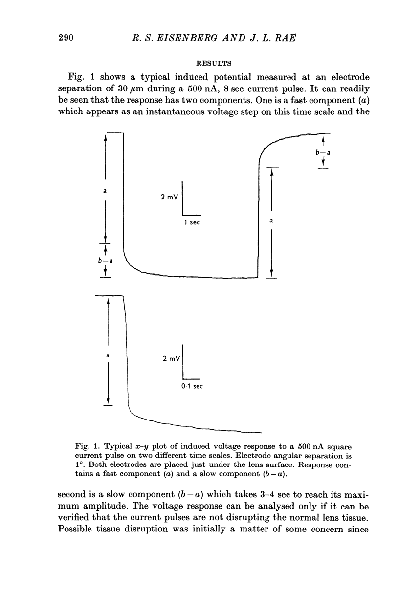
1. Electrical coupling between the cells of the crystalline lens of the frog eye was studied using two intralenticular micro-electrodes, one to pass current and one to record potential. In most experiments, both electrodes were placed just inside the posterior surface of the lens at a depth of approximately 200 mum from the surface. Step functions of current were applied and the time course of the resulting change in voltage was measured at many different electrode separations. 2. The voltage change has both a fast component, which occurs only locally in the region close to the current passing micro-electrode, and a slow component, which is spatially uniform, independent of distance from the current micro-electrode. 3. This behaviour is predicted by an electrical model of a single large spherical cell, and so that model can be used to analyse our data. 4. The resistivity of the lens 'interior' (both cytoplasm and coupling resistivity) is 625 omega cm; the resistance of the lens 'membrane' is 2751 omega cm2. 5. The data and analysis help to reconcile discrepancies between previous measurements of the electrical properties of the lens and show clearly that there is substantial electrical coupling from cell to cell. The method should allow investigation of the role of electrical coupling in cataract formation in the crystalline lens.
Full text
PDF


Selected References
These references are in PubMed. This may not be the complete list of references from this article.
- ANDREE G. Uber die Natur des transkapsularen Potentials der Linse. Pflugers Arch. 1958;267(2):109–116. doi: 10.1007/BF00362977. [DOI] [PubMed] [Google Scholar]
- BRINDLEY G. S. Resting potential of the lens. Br J Ophthalmol. 1956 Jul;40(7):385–391. doi: 10.1136/bjo.40.7.385. [DOI] [PMC free article] [PubMed] [Google Scholar]
- Duncan G., Bushell A. R. Ion analyses of human cataractous lenses. Exp Eye Res. 1975 Mar;20(3):223–230. doi: 10.1016/0014-4835(75)90136-0. [DOI] [PubMed] [Google Scholar]
- Duncan G. The site of the ion restricting membranes in the toad lens. Exp Eye Res. 1969 Oct;8(4):406–412. doi: 10.1016/s0014-4835(69)80006-0. [DOI] [PubMed] [Google Scholar]
- Eisenberg R. S., Engel E. The spatial variation of membrane potential near a small source of current in a spherical cell. J Gen Physiol. 1970 Jun;55(6):736–757. doi: 10.1085/jgp.55.6.736. [DOI] [PMC free article] [PubMed] [Google Scholar]
- Loewenstein W. R. Permeability of membrane junctions. Ann N Y Acad Sci. 1966 Jul 14;137(2):441–472. doi: 10.1111/j.1749-6632.1966.tb50175.x. [DOI] [PubMed] [Google Scholar]
- Peskoff A., Eisenberg R. S. Interpretation of some microelectrode measurements of electrical properties of cells. Annu Rev Biophys Bioeng. 1973;2:65–79. doi: 10.1146/annurev.bb.02.060173.000433. [DOI] [PubMed] [Google Scholar]
- Rae J. L., Blankenship J. E. Bioelectric measurements in the frog lens. Exp Eye Res. 1973 Feb;15(2):209–217. doi: 10.1016/0014-4835(73)90121-8. [DOI] [PubMed] [Google Scholar]
- Rae J. L. Potential profiles in the crystalline lens of the frog. Exp Eye Res. 1974 Sep;19(3):227–234. doi: 10.1016/0014-4835(74)90141-9. [DOI] [PubMed] [Google Scholar]
- Rae J. L. The movement of procion dye in the crystalline lens. Invest Ophthalmol. 1974 Feb;13(2):147–150. [PubMed] [Google Scholar]
- Rafferty N. S., Esson E. A. An electron-microscope study of adult mouse lens: some ultrastructural specializations. J Ultrastruct Res. 1974 Feb;46(2):239–253. doi: 10.1016/s0022-5320(74)80059-6. [DOI] [PubMed] [Google Scholar]
- Rafferty N. S., Goossens W. Ultrastructural studies of traumatic cataractogenesis: observations of a repair process in mouse lens. Am J Anat. 1975 Feb;142(2):177–199. doi: 10.1002/aja.1001420204. [DOI] [PubMed] [Google Scholar]