Abstract
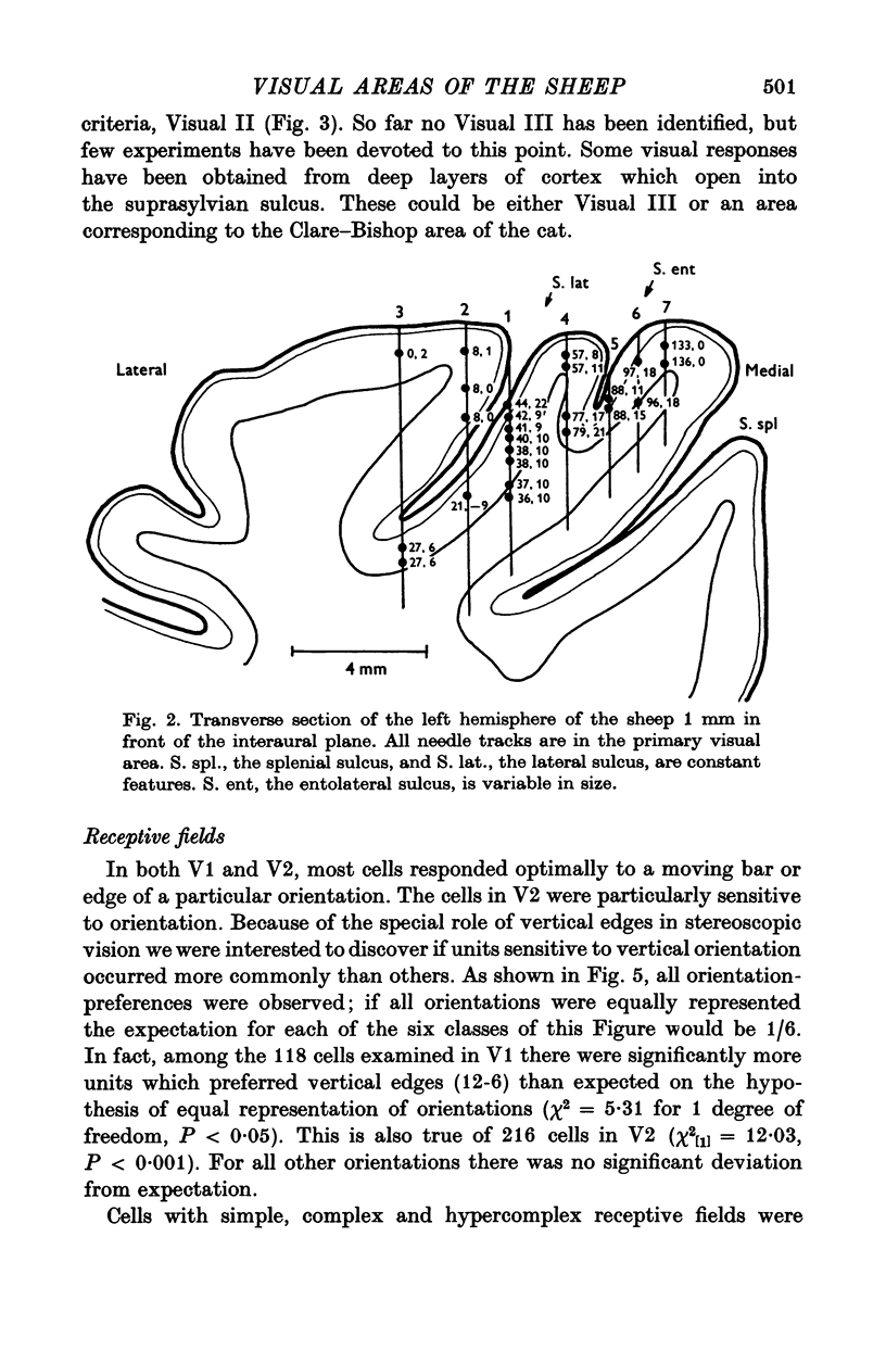
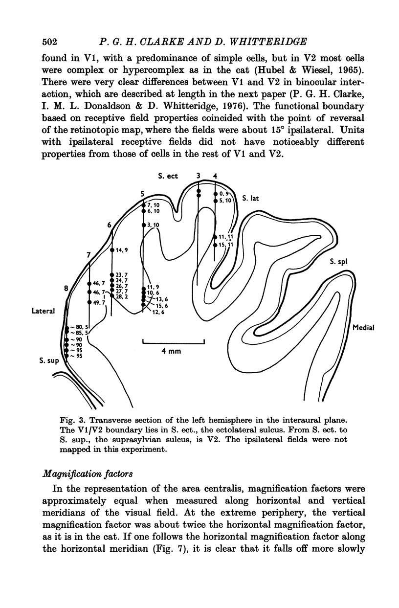
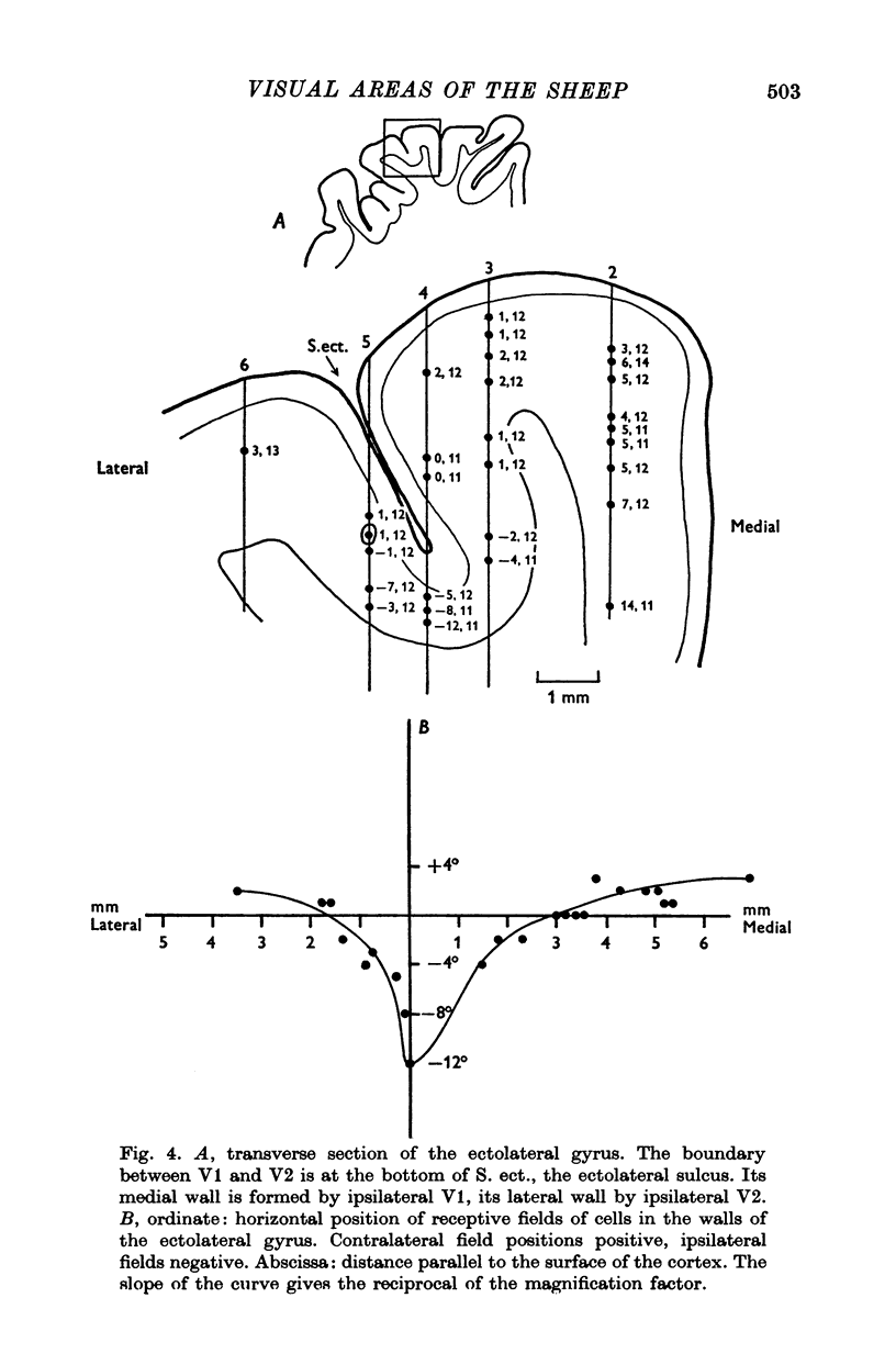
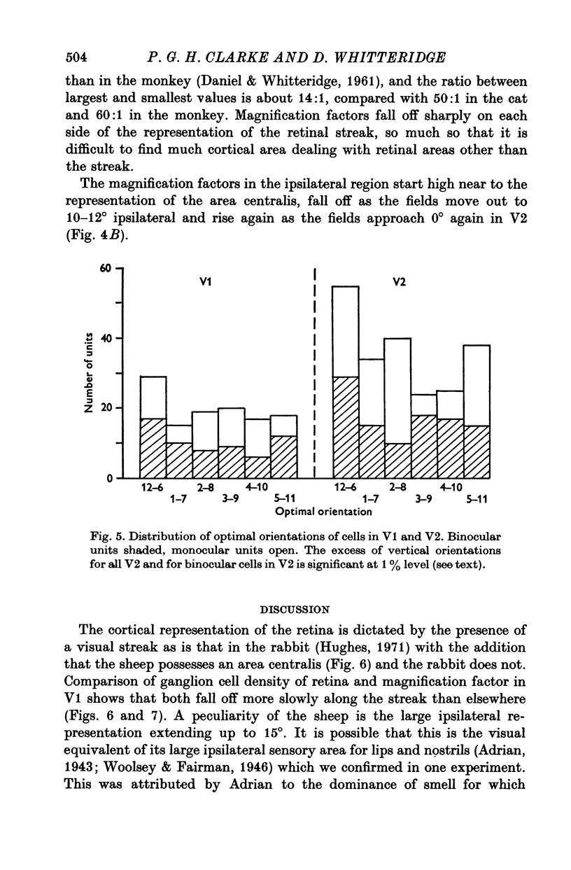
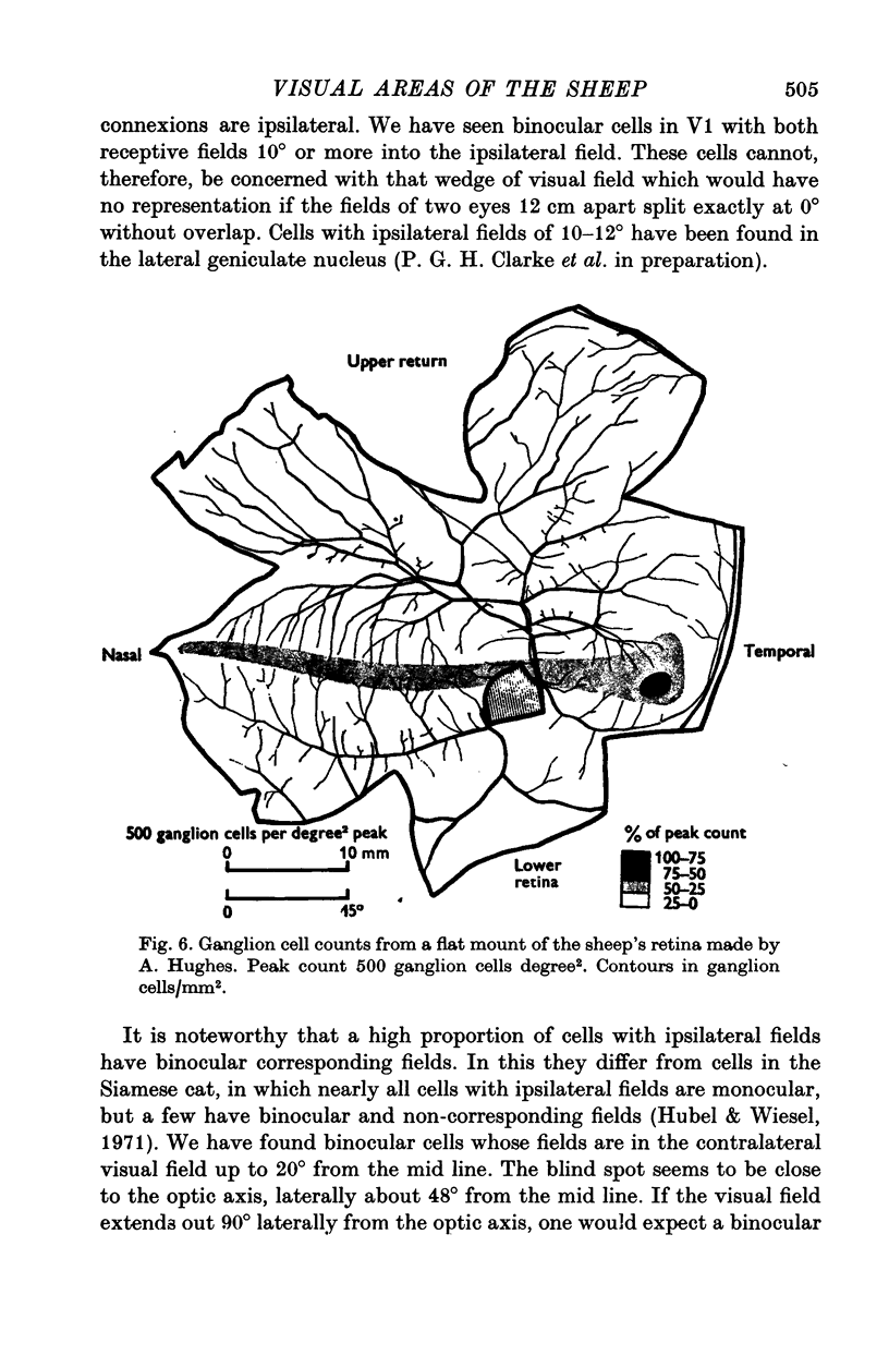
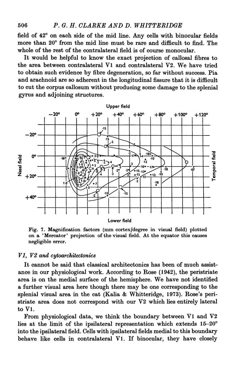
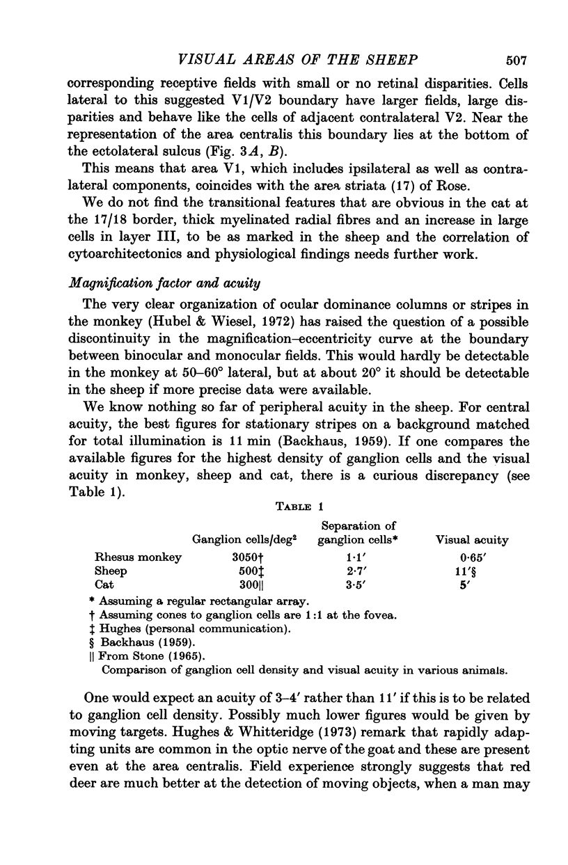
1. A stereotaxic method for the sheep brain is described. 2. At its widest part the primary visual area (Visual I) of each hemisphere extends approximately 20 mm anteroposteriorly and, when unfolded, approximately 35 mm from side to side. It occupies both walls of the lateral sulcus, and extends medially to the medial wall of the hemisphere and to the depth of the ectolateral sulcus laterally. 3. The most lateral part of the primary visual area includes 10-15 degrees of the ipsilateral field; the contralateral field is represented to 135 degrees from the mid line. 4. Visual II also includes a strip of ipsilateral representation on its medial edge and extends to the supra-sylvian sulcus on the lateral surface of the brain. The furthest lateral representation recorded was 130 degrees lateral. 5. Most of both visual areas is concerned with the area centralis and the visual streak. The remainder of the retina has very little cortical representation. 6. Most cells in Visual I are simple with orientational and sometimes directional sensitivity. Some complex and hypercomplex cells have been seen in Visual I, and these predominate in Visual II. Receptive field sizes from 0-25 to 10 degree were found. Within 15 degrees of the vertical meridian, binocular cells are common in both Visual I and II.
Full text
PDF
Images in this article
Selected References
These references are in PubMed. This may not be the complete list of references from this article.
- BISHOP P. O., KOZAK W., VAKKUR G. J. Some quantitative aspects of the cat's eye: axis and plane of reference, visual field co-ordinates and optics. J Physiol. 1962 Oct;163:466–502. doi: 10.1113/jphysiol.1962.sp006990. [DOI] [PMC free article] [PubMed] [Google Scholar]
- COOPER S., DANIEL P. M., WHITTERIDGE D. Muscle spindles and other sensory endings in the extrinsic eye muscles; the physiology and anatomy of these receptors and of their connexions with the brain-stem. Brain. 1955;78(4):564–583. doi: 10.1093/brain/78.4.564. [DOI] [PubMed] [Google Scholar]
- Clarke P. G., Donaldson I. M., Whitteridge D. Binocular visual mechanisms in cortical areas I and II of the sheep. J Physiol. 1976 Apr;256(3):509–526. doi: 10.1113/jphysiol.1976.sp011336. [DOI] [PMC free article] [PubMed] [Google Scholar]
- DANIEL P. M., WHITTERIDGE D. The representation of the visual field on the cerebral cortex in monkeys. J Physiol. 1961 Dec;159:203–221. doi: 10.1113/jphysiol.1961.sp006803. [DOI] [PMC free article] [PubMed] [Google Scholar]
- Guillery RW THE LAMINAR D. a new interpretation. J Comp Neurol. 1970 Mar;138(3):339–366. doi: 10.1002/cne.901380307. [DOI] [PubMed] [Google Scholar]
- HUBEL D. H., WIESEL T. N. RECEPTIVE FIELDS AND FUNCTIONAL ARCHITECTURE IN TWO NONSTRIATE VISUAL AREAS (18 AND 19) OF THE CAT. J Neurophysiol. 1965 Mar;28:229–289. doi: 10.1152/jn.1965.28.2.229. [DOI] [PubMed] [Google Scholar]
- Harker D. W. The structure and innervation of sheep superior rectus and levator palpebrae extraocular muscles. II. Muscle spindles. Invest Ophthalmol. 1972 Dec;11(12):970–979. [PubMed] [Google Scholar]
- Hubel D. H., Wiesel T. N. Aberrant visual projections in the Siamese cat. J Physiol. 1971 Oct;218(1):33–62. doi: 10.1113/jphysiol.1971.sp009603. [DOI] [PMC free article] [PubMed] [Google Scholar]
- Hubel D. H., Wiesel T. N. Laminar and columnar distribution of geniculo-cortical fibers in the macaque monkey. J Comp Neurol. 1972 Dec;146(4):421–450. doi: 10.1002/cne.901460402. [DOI] [PubMed] [Google Scholar]
- Hughes A. Topographical relationships between the anatomy and physiology of the rabbit visual system. Doc Ophthalmol. 1971 Sep 12;30:33–159. doi: 10.1007/BF00142518. [DOI] [PubMed] [Google Scholar]
- Hughes A., Whitteridge D. The receptive fields and topographical organization of goat retinal ganglion cells. Vision Res. 1973 Jun;13(6):1101–1114. doi: 10.1016/0042-6989(73)90147-8. [DOI] [PubMed] [Google Scholar]
- Joshua D. E., Bishop P. O. Binocular single vision and depth discrimination. Receptive field disparities for central and peripheral vision and binocular interaction on peripheral single units in cat striate cortex. Exp Brain Res. 1970;10(4):389–416. doi: 10.1007/BF02324766. [DOI] [PubMed] [Google Scholar]
- Kalia M., Whitteridge D. The visual areas in the splenial sulcus of the cat. J Physiol. 1973 Jul;232(2):275–283. doi: 10.1113/jphysiol.1973.sp010269. [DOI] [PMC free article] [PubMed] [Google Scholar]
- Stone J. A quantitative analysis of the distribution of ganglion cells in the cat's retina. J Comp Neurol. 1965 Jun;124(3):337–352. doi: 10.1002/cne.901240305. [DOI] [PubMed] [Google Scholar]


