

Abstract
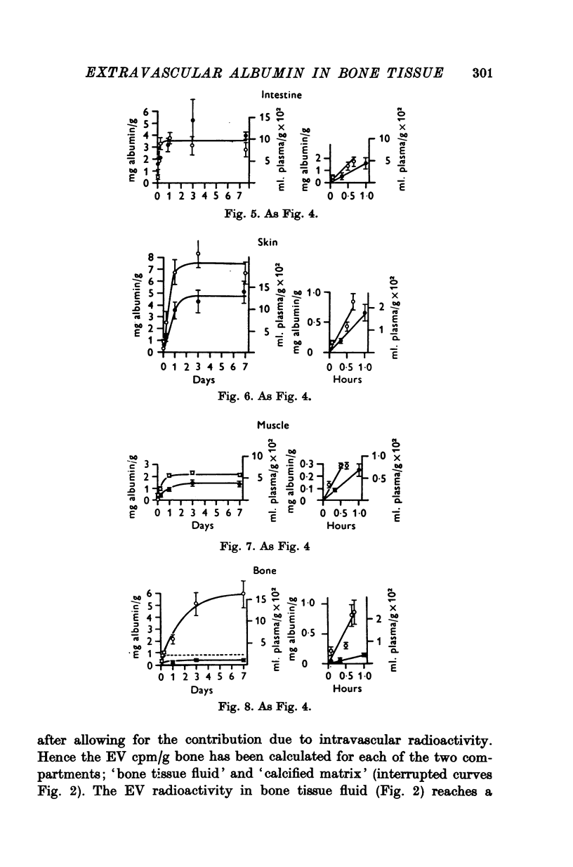
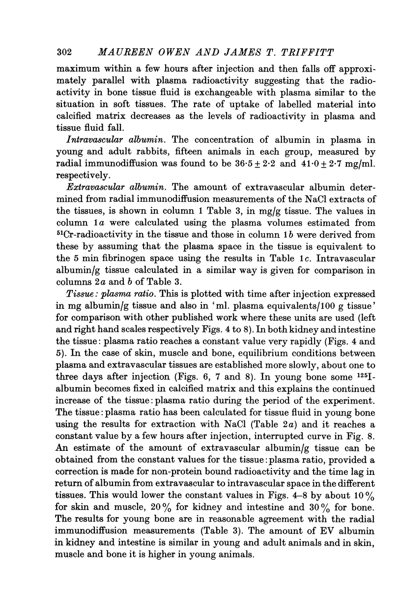
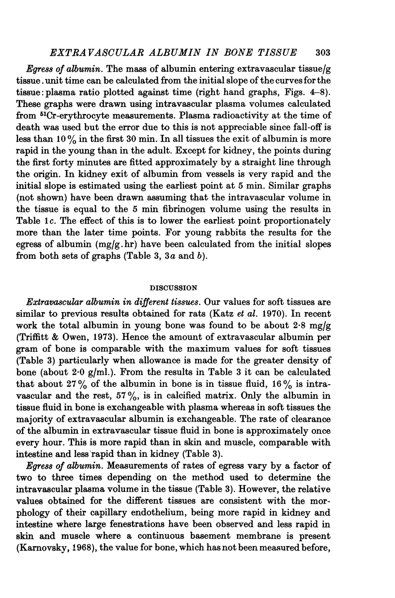
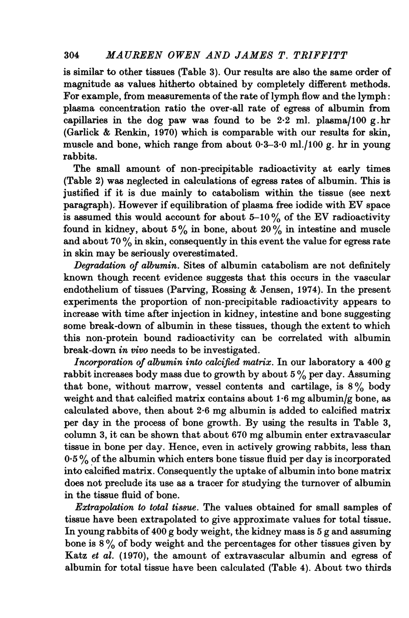
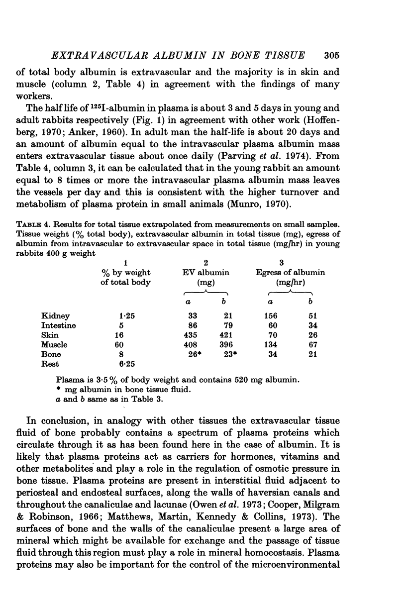
1. The amount of albumin in extravascular tissue fluid in bone, kidney, intestine, skin and muscle and in plasma of young rabbits has been measured by radial immunodiffusion. 2. The majority of extravascular albumin in kidney, intestine, skin and muscle is exchangeable with plasma albumin, whereas in bone, only the proportion which is in tissue fluid is readily exchangeable; the remaining fraction in calcified matrix is more permanently fixed. 3. About 27% of the albumin in young bone is in tissue fluid, about 57% in calcified matrix and about 16% is intravascular. The total amount of extravascular albumin per unit mass of bone is similar to that found in soft tissues. 4. The volume of intravascular plasma in tissues was determined in two ways: from 51Cr-erythrocyte radioactivity and the venous haematocrit and from the '5 min 125I-fibrinogen space'. 5. The rate of egress of albumin from blood vessels has been estimated from the initial slope of the ratio of extravascular radioactivity in the tissue to plasma radioactivity plotted against time after injection of 125I-albumin. 6. The rate of clearance of the albumin in extravascular tissue fluid in bone is approximately once every hour. This is more rapid than in skin and muscle, comparable with intestine and less rapid than in kidney. 7. The amount of albumin incorporated into calcified matrix of bone per day is calculated to be less than 0-5% of the total albumin passing through the tissue fluid of bone per day.
Full text
PDF
Selected References
These references are in PubMed. This may not be the complete list of references from this article.
- Ashton B. A., Triffitt J. T., Herring G. M. Isolation and partial characterization of a glycoprotein from bovine cortical bone. Eur J Biochem. 1974 Jun 15;45(2):525–533. doi: 10.1111/j.1432-1033.1974.tb03577.x. [DOI] [PubMed] [Google Scholar]
- Cooper R. R., Milgram J. W., Robinson R. A. Morphology of the osteon. An electron microscopic study. J Bone Joint Surg Am. 1966 Oct;48(7):1239–1271. [PubMed] [Google Scholar]
- Garlick D. G., Renkin E. M. Transport of large molecules from plasma to interstitial fluid and lymph in dogs. Am J Physiol. 1970 Dec;219(6):1595–1605. doi: 10.1152/ajplegacy.1970.219.6.1595. [DOI] [PubMed] [Google Scholar]
- Mancini G., Carbonara A. O., Heremans J. F. Immunochemical quantitation of antigens by single radial immunodiffusion. Immunochemistry. 1965 Sep;2(3):235–254. doi: 10.1016/0019-2791(65)90004-2. [DOI] [PubMed] [Google Scholar]
- Neuman W. F., Bareham B. J. Evidence for the presence of secondary calcium phosphate in bone and its stabilization by acid production. Calcif Tissue Res. 1975 Sep 5;18(3):161–172. doi: 10.1007/BF02546238. [DOI] [PubMed] [Google Scholar]
- Neuman W. F. The milieu interieur of bone: Claude Bernard revisited. Fed Proc. 1969 Nov-Dec;28(6):1846–1850. [PubMed] [Google Scholar]
- Parving H. H., Rossing N., Jensen H. A. Increased metabolic turnover rate and transcapillary escape rate of albumin in essential hypertension. Circ Res. 1974 Oct;35(4):544–552. doi: 10.1161/01.res.35.4.544. [DOI] [PubMed] [Google Scholar]
- Studer R., Potchen J. The radioisotopic assessment of regional microvascular permeability to macromolecules. Microvasc Res. 1971 Jan;3(1):35–48. doi: 10.1016/0026-2862(71)90005-7. [DOI] [PubMed] [Google Scholar]
- Swan H., Nelson A. W. Blood volume measurement: concepts and technology. J Cardiovasc Surg (Torino) 1971 Sep-Oct;12(5):389–401. [PubMed] [Google Scholar]
- Triffitt J. T., Owen M. Studies on bone matrix glycoproteins. Incorporation of (1-14C)glucosamine and plasma (14C)glycoprotein into rabbit cortical bone. Biochem J. 1973 Sep;136(1):125–134. doi: 10.1042/bj1360125. [DOI] [PMC free article] [PubMed] [Google Scholar]