

Abstract
1. The directional selectivity of individual cones was examined by intracellular recording in the eye of the turtle. Sensitivites were determined from linear responses to dim flashes of monochromatic light incident on a cell over a range of angles to its long axis. 2. With light near the optimum wave-length, some red- and green-sensitive cones showed a high sensitivity for light entering axially and lower sensitivities for light entering obliquely. In contrast, other cells had lower peak sensitivities and less pronounced directional selectivities. The highest axial sensitivities observed in red receptors were about 320 muV photon(-1) mu2; in these cells, the sensitivity declined to half for rays 6-9 degrees off the axis as measured in the retina. Green receptors had lower axial sensitivities and broader angular profiles. 3. On the assumption that rays at all angles contribute independently to the over-all sensitivity, the sensitivity of a cell to large cones of rays was successfully predicted from the angular selectivity determined with a narrow pencil of rays. The shape of small responses to dim stimuli delivered on and off the axis of the cell was invariant, implying that a cone signals the number of photons absorbed but not their angle of incidence. 4. Short wave-lengths have previously been shown to be filtered out by the oil droplets present in turtle cones. At short wave-lengths, the angular profiles showed a depression in axial sensitivity consistent with this filtering action. 5. Diameters of inner segments, oil droplets, and outer segments were measured in red-, green-, and blue-sensitive cones, since these dimensions are expected to influence the cones' angular acceptances and ability to collect light. The diameters of the structure were in approximately the same proportions for each type of receptor, but the absolute values of the diameters were found to be scaled in relation to the wave-length of maximum sensitivity. 6. Optical determinations of the efficiency with which axial rays are concentrated by red receptors gave a mean value of 55%. 7. Receptors in histological sections of the whole eye were found to be oriented with their long axes directed approximately toward the pupil. 8. The observed directional selectivities and collecting efficiencies agree well with the behaviour of a model retinal cone developed by Winston & Enoch (1971) on a geometrical optical treatment. 9. Effective collecting areas are derived for red-, green- and blue-sensitive cones; these permit conversion of observed flash sensitivities into the mean peak hyperpolarization produced by isomerization of a visual pigment molecule. The figure obtained is about 25 muV for red-sensitive cones and 21muV for green-sensitive cones.
Full text
PDF


Images in this article
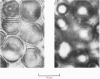
Selected References
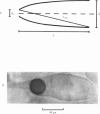
These references are in PubMed. This may not be the complete list of references from this article.
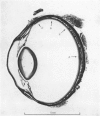
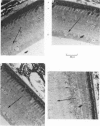
- BARER R. Refractometry and interferometry of living cells. J Opt Soc Am. 1957 Jun;47(6):545–556. doi: 10.1364/josa.47.000545. [DOI] [PubMed] [Google Scholar]
- Baylor D. A., Fuortes M. G. Electrical responses of single cones in the retina of the turtle. J Physiol. 1970 Mar;207(1):77–92. doi: 10.1113/jphysiol.1970.sp009049. [DOI] [PMC free article] [PubMed] [Google Scholar]
- Baylor D. A., Fuortes M. G., O'Bryan P. M. Receptive fields of cones in the retina of the turtle. J Physiol. 1971 Apr;214(2):265–294. doi: 10.1113/jphysiol.1971.sp009432. [DOI] [PMC free article] [PubMed] [Google Scholar]
- Baylor D. A., Hodgkin A. L. Detection and resolution of visual stimuli by turtle photoreceptors. J Physiol. 1973 Oct;234(1):163–198. doi: 10.1113/jphysiol.1973.sp010340. [DOI] [PMC free article] [PubMed] [Google Scholar]
- DENTON E. J. The contributions of the orientated photosensitive and other molecules to the absorption of whole retina. Proc R Soc Lond B Biol Sci. 1959 Jan 27;150(938):78–94. doi: 10.1098/rspb.1959.0009. [DOI] [PubMed] [Google Scholar]
- Granda A. M., Haden K. W. Retinal oil globule counts and distributions in two species of turtles: Pseudemys scripta elegans (Wied) and Chelonia mydas mydas (Linnaeus). Vision Res. 1970 Jan;10(1):79–84. doi: 10.1016/0042-6989(70)90064-7. [DOI] [PubMed] [Google Scholar]
- JOHNSON B. K., TANSLEY K. The cones of the grass snake's eye. Nature. 1956 Dec 8;178(4545):1285–1286. doi: 10.1038/1781285a0. [DOI] [PubMed] [Google Scholar]
- LIEBMAN P. A. In situ microspectrophotometric studies on the pigments of single retinal rods. Biophys J. 1962 Mar;2:161–178. doi: 10.1016/s0006-3495(62)86847-7. [DOI] [PMC free article] [PubMed] [Google Scholar]
- Laties A. M., Liebman P. A., Campbell C. E. Photoreceptor orientation in the primate eye. Nature. 1968 Apr 13;218(5137):172–173. doi: 10.1038/218172a0. [DOI] [PubMed] [Google Scholar]
- Liebman P. A., Granda A. M. Microspectrophotometric measurements of visual pigments in two species of turtle, Pseudemys scripta and Chelonia mydas. Vision Res. 1971 Feb;11(2):105–114. doi: 10.1016/0042-6989(71)90227-6. [DOI] [PubMed] [Google Scholar]
- O'BRIEN B. Vision and resolution in the central retina. J Opt Soc Am. 1951 Dec;41(12):882–894. doi: 10.1364/josa.41.000882. [DOI] [PubMed] [Google Scholar]
- Richter A., Simon E. J. Electrical responses of double cones in the turtle retina. J Physiol. 1974 Nov;242(3):673–683. doi: 10.1113/jphysiol.1974.sp010730. [DOI] [PMC free article] [PubMed] [Google Scholar]
- SIDMAN R. L. The structure and concentration of solids in photoreceptor cells studied by refractometry and interference microscopy. J Biophys Biochem Cytol. 1957 Jan 25;3(1):15–30. doi: 10.1083/jcb.3.1.15. [DOI] [PMC free article] [PubMed] [Google Scholar]
- STILES W. S. The directional sensitivity of the retina. Ann R Coll Surg Engl. 1962 Feb;30:73–101. [PMC free article] [PubMed] [Google Scholar]
- STROTHER G. K. Absorption spectra of retinal oil globules in turkey, turtle and pigeon. Exp Cell Res. 1963 Jan;29:349–355. doi: 10.1016/0014-4827(63)90389-6. [DOI] [PubMed] [Google Scholar]
- Safir A., Hyams L., Philpot J. The retinal directional effect: a model based on the gaussian distribution of cone orientations. Vision Res. 1971 Aug;11(8):819–831. doi: 10.1016/0042-6989(71)90004-6. [DOI] [PubMed] [Google Scholar]
- Snyder A. W., Pask C. The Stiles-Crawford effect--explanation and consequences. Vision Res. 1973 Jun;13(6):1115–1137. doi: 10.1016/0042-6989(73)90148-x. [DOI] [PubMed] [Google Scholar]
- Winston R., Enoch J. M. Retinal cone receptor as an ideal light collector. J Opt Soc Am. 1971 Aug;61(8):1120–1122. doi: 10.1364/josa.61.001120. [DOI] [PubMed] [Google Scholar]