Abstract
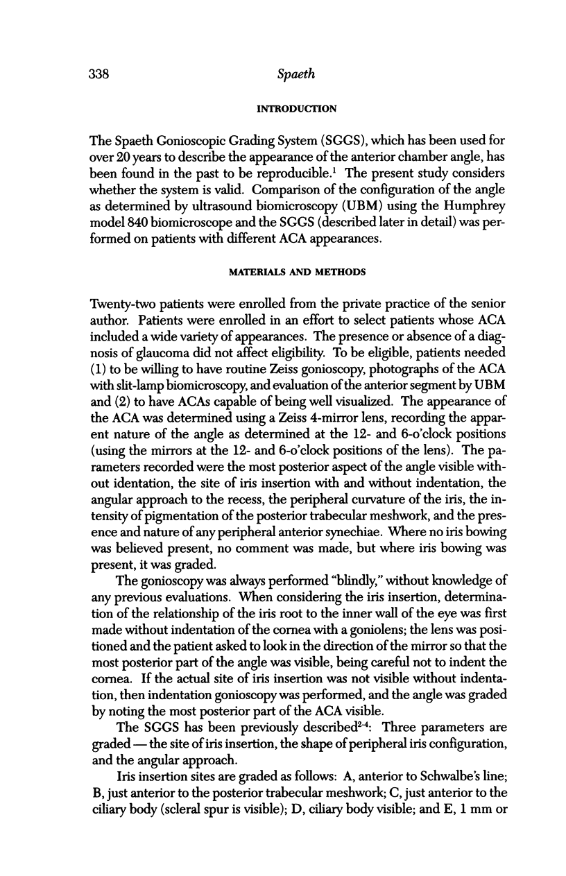
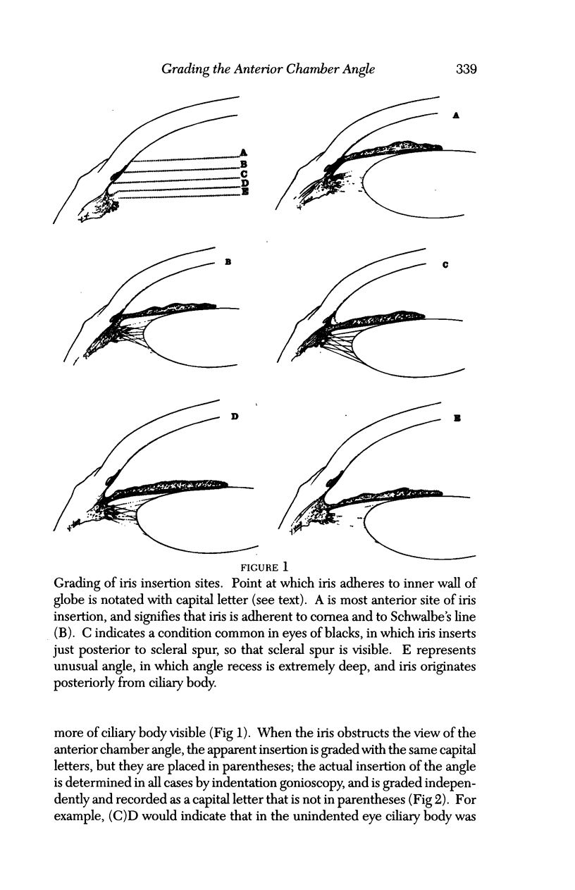
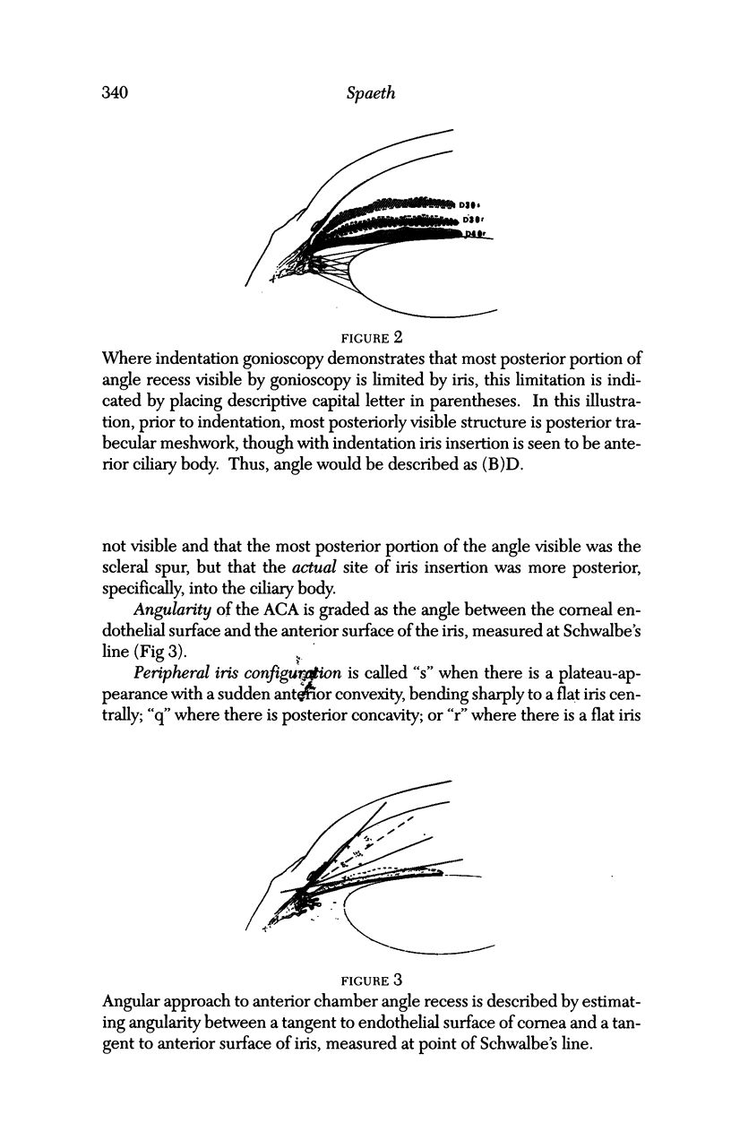
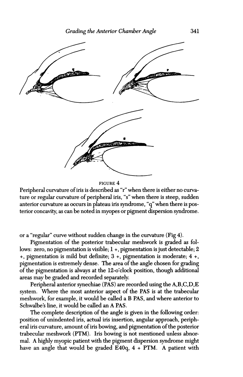
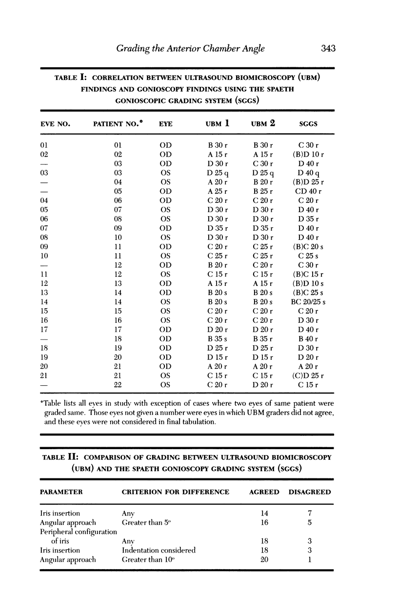
PURPOSE: The primary purpose of this investigation was to validate anatomically, if possible, the Spaeth gonioscopic grading system. METHODS: The gonioscopic appearance of the anterior chamber angle of 22 patients was described, using a Zeiss 4-mirror gonioscopic lens; the angle was graded according to the Spaeth gonioscopic grading system (SGGS) previously described elsewhere. This system provides a more comprehensive characterization of the angle than other methods, including the site of iris insertion, the angular approach to the recess and the curvature of the peripheral iris. The same patients were then evaluated with biomicroscopic ultrasound examination of the anterior segment; the nature of the anterior chamber angle was described independently by two observers. The two methods of characterizing the anterior chamber angle were then compared. RESULTS: There was a high correlation between the two different methods of characterizing the anterior chamber angle. Ultrasound biomicroscopy had limitations relating to inability to distinguish between iris-cornea apposition and iris-cornea adhesion, and to exact location of the posterior trabecular meshwork. The angularity of the approach to the anterior chamber angle using the SGGS tended to be underestimated by about 5 degrees. Other aspects showed high correlation. CONCLUSION: The SGGS appears to be an accurate method of characterizing the anatomic appearance of the anterior chamber angle in patients. It also provides a more comprehensive description of the angle that other grading systems.
Full text
PDF


Images in this article
Selected References
These references are in PubMed. This may not be the complete list of references from this article.
- Forbes M. Gonioscopy with corneal indentation. A method for distinguishing between appositional closure and synechial closure. Arch Ophthalmol. 1966 Oct;76(4):488–492. doi: 10.1001/archopht.1966.03850010490005. [DOI] [PubMed] [Google Scholar]
- Oh Y. G., Minelli S., Spaeth G. L., Steinman W. C. The anterior chamber angle is different in different racial groups: a gonioscopic study. Eye (Lond) 1994;8(Pt 1):104–108. doi: 10.1038/eye.1994.20. [DOI] [PubMed] [Google Scholar]
- Pavlin C. J., Ritch R., Foster F. S. Ultrasound biomicroscopy in plateau iris syndrome. Am J Ophthalmol. 1992 Apr 15;113(4):390–395. doi: 10.1016/s0002-9394(14)76160-4. [DOI] [PubMed] [Google Scholar]
- Pavlin C. J., Sherar M. D., Foster F. S. Subsurface ultrasound microscopic imaging of the intact eye. Ophthalmology. 1990 Feb;97(2):244–250. doi: 10.1016/s0161-6420(90)32598-8. [DOI] [PubMed] [Google Scholar]
- Potash S. D., Tello C., Liebmann J., Ritch R. Ultrasound biomicroscopy in pigment dispersion syndrome. Ophthalmology. 1994 Feb;101(2):332–339. doi: 10.1016/s0161-6420(94)31331-5. [DOI] [PubMed] [Google Scholar]
- SCHEIE H. G. Width and pigmentation of the angle of the anterior chamber; a system of grading by gonioscopy. AMA Arch Ophthalmol. 1957 Oct;58(4):510–512. doi: 10.1001/archopht.1957.00940010526005. [DOI] [PubMed] [Google Scholar]
- SHAFFER R. N. Primary glaucomas. Gonioscopy, ophthalmoscopy and perimetry. Trans Am Acad Ophthalmol Otolaryngol. 1960 Mar-Apr;64:112–127. [PubMed] [Google Scholar]
- Spaeth G. L. Gonioscopy: uses old and new. The inheritance of occludable angles. Ophthalmology. 1978 Mar;85(3):222–232. doi: 10.1016/s0161-6420(78)35675-x. [DOI] [PubMed] [Google Scholar]
- Spaeth G. L. The normal development of the human anterior chamber angle: a new system of descriptive grading. Trans Ophthalmol Soc U K. 1971;91:709–739. [PubMed] [Google Scholar]
- Van Herick W., Shaffer R. N., Schwartz A. Estimation of width of angle of anterior chamber. Incidence and significance of the narrow angle. Am J Ophthalmol. 1969 Oct;68(4):626–629. doi: 10.1016/0002-9394(69)91241-0. [DOI] [PubMed] [Google Scholar]


