

Abstract
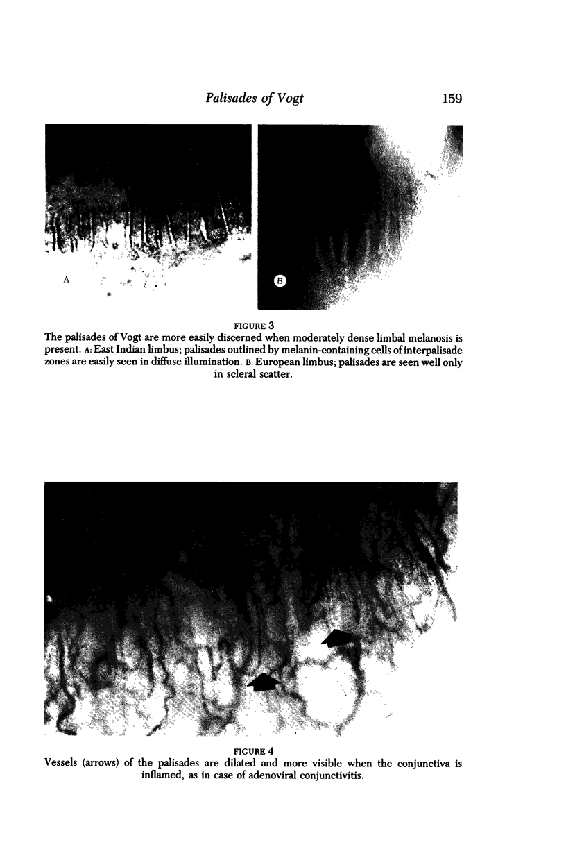
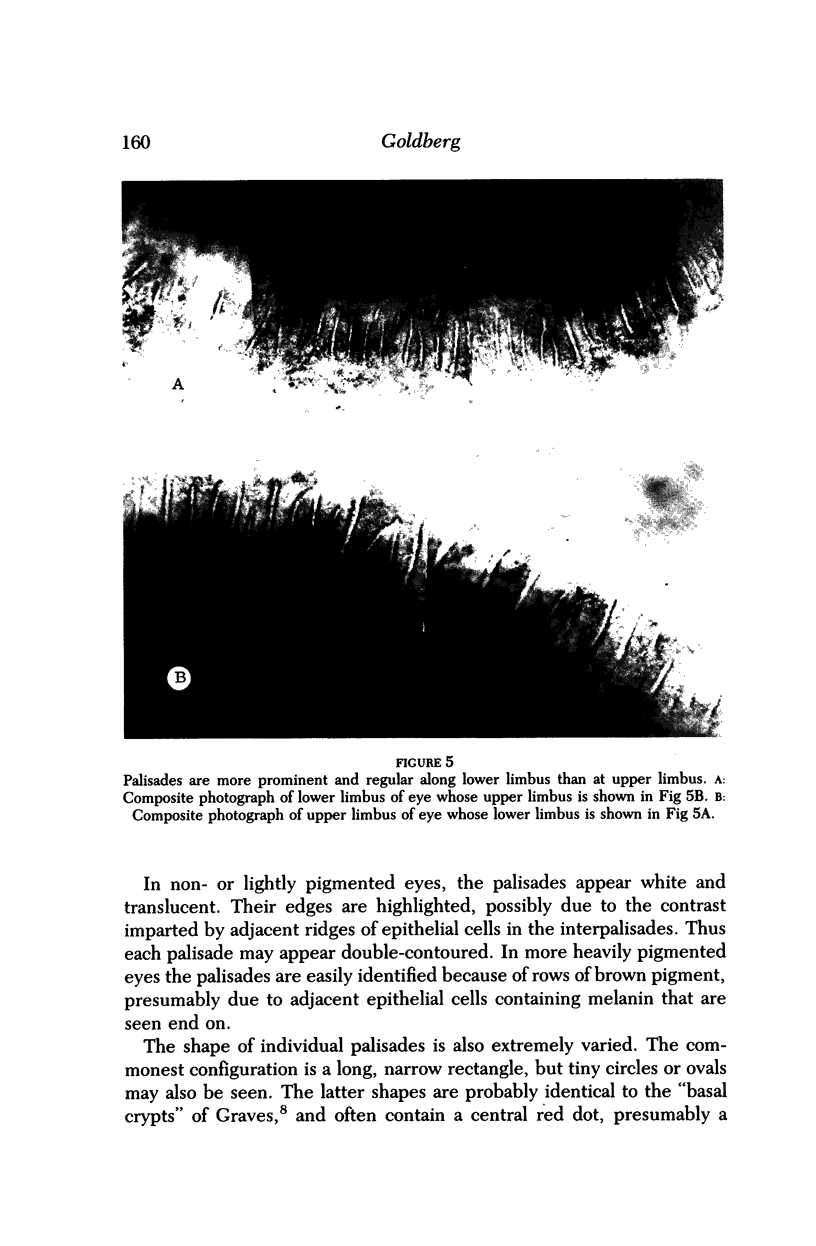
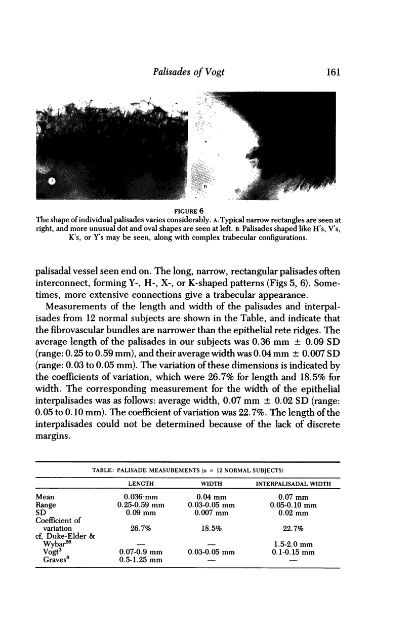
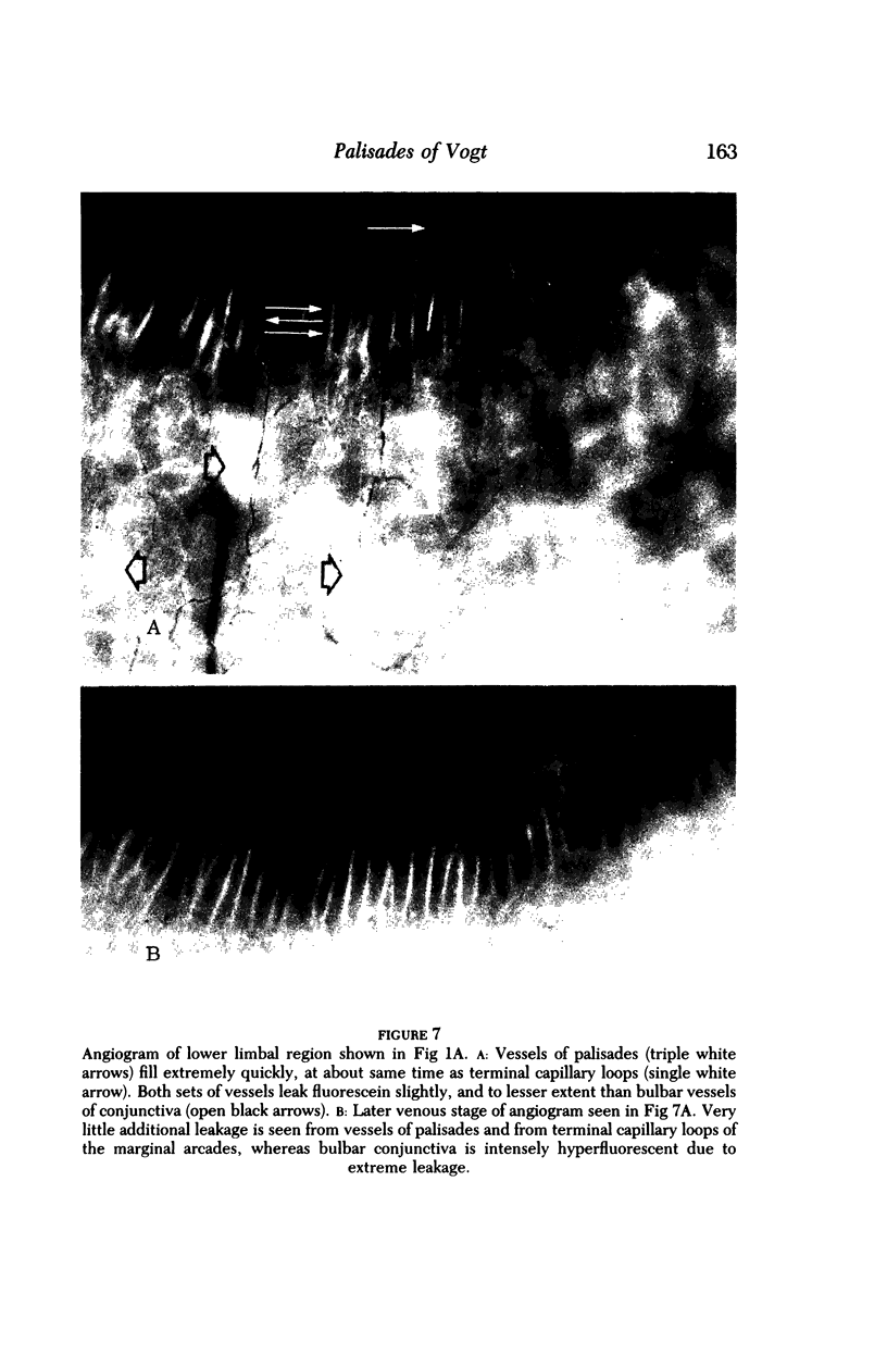
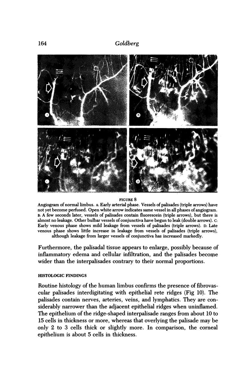
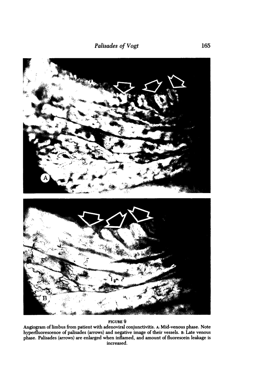
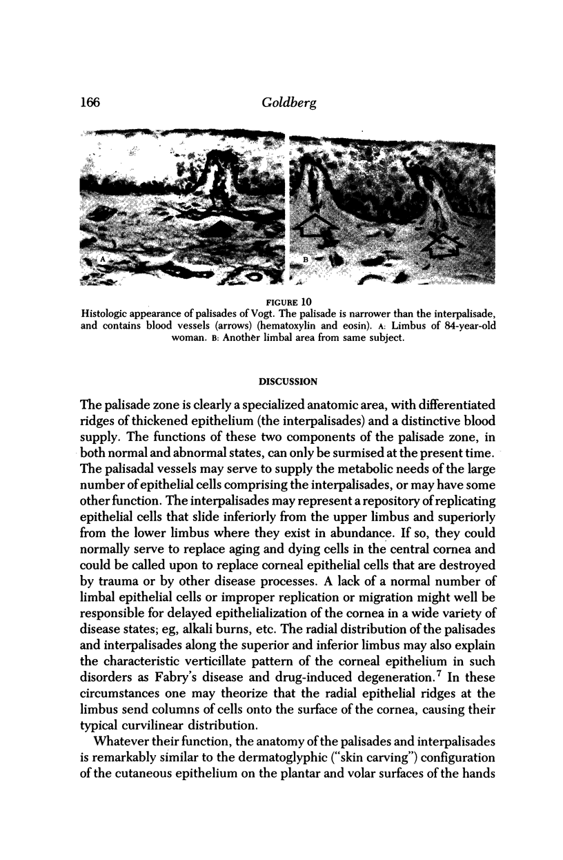
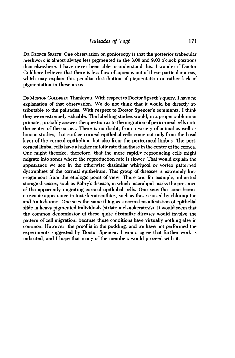
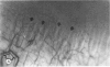
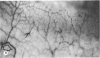
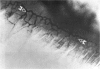
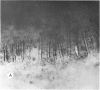
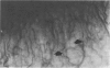
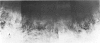
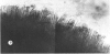
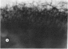
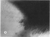
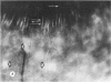
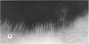
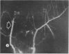
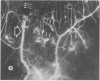
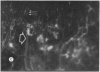
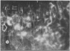
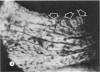
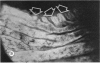
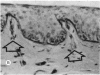
The palisades of Vogt are distinctive normal features of the human corneoscleral limbus. Our clinical studies indicate that they are more discrete in younger and in more heavily pigmented individuals, and that they appear more regular and prominent at the lower limbus than at the upper limbus. They are seen only infrequently in the horizontal meridian. There is some symmetry (though it is not exact) from one eye to the other in the same person. The anatomy of the palisades appears to be unique for a given individual. In this respect, as well as in their microscopic anatomy, the palisades of Vogt appear comparable to fingerprints, and the term "conjunctivoglyphics" ("conjunctival carvings") or "limboglyphics" is suggested in analogy with "dermatoglyphics." The palisades of Vogt have a distinct vasculature with narrow, barely visible, arterial and venous components of radially oriented hairpin loops. Angiography reveals that these vessels leak fluorescein relatively late and only to a moderate extent. They respond to inflammation by dilatation and gross breakdown of their physiologic barrier properties. The functions of the palisades of Vogt are not known with certainty, but their interpalisadal epithelial rete ridges may serve as a repository for corneal epithelial cells. They may thus be important in both aging and diseases of the cornea.
Full text
PDF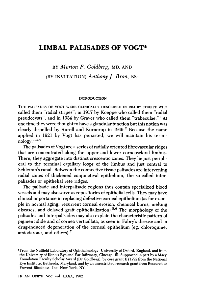
Images in this article

Selected References
These references are in PubMed. This may not be the complete list of references from this article.
- Amalric P., Rebiére P. Nouvelles indications de l'angiographie fluoresceinique du segment anterieur de l'oeil. IV. Quelques exemples de pathologie sclerale et corneene. Ann Ocul (Paris) 1971 Jul;204(7):731–concl. [PubMed] [Google Scholar]
- Bron A. J., Easty D. L. Fluorescein angiography of the globe and anterior segment. Trans Ophthalmol Soc U K. 1970;90:339–367. [PubMed] [Google Scholar]
- Bron A. J. Vortex patterns of the corneal epithelium. Trans Ophthalmol Soc U K. 1973;93(0):455–472. [PubMed] [Google Scholar]
- Bruun-Jensen J. Fluorescein angiography of the anterior segment. Am J Ophthalmol. 1969 Jun;67(6):842–845. doi: 10.1016/0002-9394(69)90076-2. [DOI] [PubMed] [Google Scholar]
- DOBREE J. H. Superficial perilimbal vessels in the normal and congested eye. Br J Ophthalmol. 1950 Dec;34(12):720–726. doi: 10.1136/bjo.34.12.720. [DOI] [PMC free article] [PubMed] [Google Scholar]
- Easty D. L., Bron A. J. Fluorescein angiography of the anterior segment. Its value in corneal disease. Br J Ophthalmol. 1971 Oct;55(10):671–682. doi: 10.1136/bjo.55.10.671. [DOI] [PMC free article] [PubMed] [Google Scholar]
- Fetkenhour C. L., Chromokos E. Anterior segment fluorescein angiography with a retinal fundus camera. Arch Ophthalmol. 1978 Apr;96(4):711–713. doi: 10.1001/archopht.1978.03910050401020. [DOI] [PubMed] [Google Scholar]
- Graves B. CERTAIN CLINICAL FEATURES OF THE NORMAL LIMBUS. Br J Ophthalmol. 1934 Jun;18(6):305–341. doi: 10.1136/bjo.18.6.305. [DOI] [PMC free article] [PubMed] [Google Scholar]
- Holt S. B. The significance of dermatoglyphics in medicine. A short survey and summary. Clin Pediatr (Phila) 1973 Aug;12(8):471–484. doi: 10.1177/000992287301200904. [DOI] [PubMed] [Google Scholar]
- Marsh R. J., Ford S. M. Blood flow in the anterior segment of the eye. Trans Ophthalmol Soc U K. 1980 Sep;100(3):388–397. [PubMed] [Google Scholar]
- Miller J. R. Dermatoglyphics. J Invest Dermatol. 1973 Jun;60(6):435–442. doi: 10.1111/1523-1747.ep12702906. [DOI] [PubMed] [Google Scholar]
- Minatoya H., Acacio I., Goldberg M. Fluorescein angiography of the bulbar conjunctiva in sickle cell disease. Ann Ophthalmol. 1973 Sep;5(9):980–992. [PubMed] [Google Scholar]
- Mitsui Y., Matsubara M., Kanagawa M. Fluorescence irido-corneal photography. Br J Ophthalmol. 1969 Aug;53(8):505–512. doi: 10.1136/bjo.53.8.505. [DOI] [PMC free article] [PubMed] [Google Scholar]
- Preus M., Fraser F. C. Dermatoglyphics and syndromes. Am J Dis Child. 1972 Dec;124(6):933–943. doi: 10.1001/archpedi.1972.02110180135022. [DOI] [PubMed] [Google Scholar]
- Talusan E. D., Schwartz B. Fluorescein angiography. Demonstration of flow pattern of anterior ciliary arteries. Arch Ophthalmol. 1981 Jun;99(6):1074–1080. doi: 10.1001/archopht.1981.03930011074018. [DOI] [PubMed] [Google Scholar]
- Verbov J. Clinical significance and genetics of epidermal ridges--a review of dermatoglyphics. J Invest Dermatol. 1970 Apr;54(4):261–271. doi: 10.1111/1523-1747.ep12258550. [DOI] [PubMed] [Google Scholar]