Abstract
Peroxins are genetically defined as proteins necessary for peroxisome biogenesis. By screening for reduced response to indole-3-butyric acid, which is metabolized to active auxin in peroxisomes, we isolated an Arabidopsis thaliana peroxin4 (pex4) mutant. This mutant displays sucrose-dependent seedling development and reduced lateral root production, characteristics of plant peroxisome malfunction. We used yeast two-hybrid analysis to determine that PEX4, an apparent ubiquitin-conjugating enzyme, interacts with a previously unidentified Arabidopsis protein, PEX22. A pex4 pex22 double mutant enhanced pex4 defects, confirming that PEX22 is a peroxin. Expression of both Arabidopsis genes together complemented yeast pex4 or pex22 mutant defects, whereas expression of either gene individually failed to rescue the corresponding yeast mutant. Therefore, it is likely that the Arabidopsis proteins can function similarly to the yeast PEX4–PEX22 complex, with PEX4 ubiquitinating substrates and PEX22 tethering PEX4 to the peroxisome. However, the severe sucrose dependence of the pex4 pex22 mutant is not accompanied by correspondingly strong defects in peroxisomal matrix protein import, suggesting that this peroxin pair may have novel plant targets in addition to those important in fungi. Isocitrate lyase is stabilized in pex4 pex22, indicating that PEX4 and PEX22 may be important during the remodeling of peroxisome matrix contents as glyoxysomes transition to leaf peroxisomes.
INTRODUCTION
During early development before photosynthetic growth begins, plants break down seed storage compounds for energy; this process is required for seedling establishment after germination. Because plants metabolize storage fatty acids primarily, or perhaps exclusively, by peroxisomal β-oxidation (Gerhardt, 1992; Kindl, 1993; Graham and Eastmond, 2002), peroxisomal function is essential for early seedling development in oilseed plants. Although peroxisomes are best known for their roles in fatty acid β-oxidation and hydrogen peroxide metabolism, peroxisomes are ubiquitous organelles implicated in diverse cellular and developmental processes (Hayashi and Nishimura, 2003; Titorenko and Rachubinski, 2004). Approximately 280 Arabidopsis thaliana proteins are predicted to be targeted to the peroxisome matrix (Reumann et al., 2004); some of these act in photorespiration (Reumann, 2002), embryonic development (Lin et al., 1999; Hu et al., 2002; Schumann et al., 2003; Sparkes et al., 2003; Woodward and Bartel, 2005a), photomorphogenesis (Hu et al., 2002), and hormone metabolism, including jasmonic acid biosynthesis (Weber, 2002) and conversion of the protoauxin indole-3-butyric acid (IBA) into active indole-3-acetic acid (IAA) (Bartel et al., 2001; Woodward and Bartel, 2005b).
Proteins acting in the peroxisome matrix are imported posttranslationally via a peroxisome-targeting signal (PTS1 or PTS2). More than 30 peroxin proteins required for peroxisomal function have been identified, many in yeast mutant screens, and defects in the corresponding human proteins cause a number of diseases, collectively known as peroxisomal biogenesis disorders (Gould and Valle, 2000; Wanders and Waterham, 2005). Forward genetic screens have identified several Arabidopsis peroxins, including PEROXIN2 (PEX2) (Hu et al., 2002), PEX5 (Zolman et al., 2000), PEX6 (Zolman and Bartel, 2004), PEX14 (Hayashi et al., 2000a), and PEX16 (Lin et al., 1999). In addition, Arabidopsis PEX7 (Woodward and Bartel, 2005a), PEX10 (Schumann et al., 2003; Sparkes et al., 2003), and PEX12 (Fan et al., 2005), originally identified by sequence similarity, have been verified as peroxins using reverse genetic analyses. However, several yeast and mammalian peroxins lack obvious plant homologs, suggesting that overlapping but distinct groups of proteins are required for peroxisomal function in different organisms. Alternatively, sequence divergence may preclude homology-based identification of some peroxins, even when functions are conserved.
Based on studies in yeast and other organisms, a cycling mechanism for peroxisomal matrix protein import has been established. The matrix protein receptors PEX5 and PEX7 bind peroxisome-targeted proteins in the cytoplasm, dock with membrane peroxins, and translocate with the cargo into the matrix. The receptors dissociate after cargo release and return to the cytoplasm for another round of import (Collins et al., 2000; Dammai and Subramani, 2001; Costa-Rodrigues et al., 2004; Nair et al., 2004). Although the mechanism by which PEX5 recycles to the cytosol after each round of import remains incompletely understood, epistasis experiments in Pichia pastoris implicate four proteins: two ATPases, Pex1p and Pex6p, the ubiquitin-conjugating (UBC) enzyme Pex4p, and the Pex4p-docking protein Pex22p (Collins et al., 2000). In addition to the PEX4 UBC enzyme, the PEX2, PEX10, and PEX12 RING finger peroxins are candidate ubiquitin protein ligases that further implicate ubiquitination in peroxisome function (Lazarow, 2003).
Here, we describe an Arabidopsis pex4 mutant that displayed characteristic peroxisome-defective phenotypes: sucrose dependence during early seedling development and resistance to the endogenous auxin IBA in both root elongation inhibition and lateral root promotion. Yeast two-hybrid analysis revealed that PEX4 interacts with a previously unidentified Arabidopsis peroxin, PEX22. A pex4 pex22 double mutant displayed enhanced defects compared with either parent. In addition, expression of both Arabidopsis genes in yeast pex4 and pex22 mutants complemented the mutant defects, indicating that the Arabidopsis proteins can function similarly to the PEX4–PEX22 complex in other species.
RESULTS
pex4 Is an IBA-Response Mutant Defective in a Putative UBC Enzyme
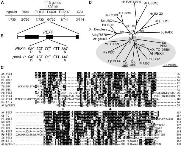
Response to the hormone IBA is a facile assay of plant peroxisomal function, as the peroxisomal conversion of IBA to IAA inhibits root elongation. Therefore, certain mutants defective in peroxisomal processes are IBA-resistant, with longer roots on IBA than in wild-type controls (Zolman et al., 2000, 2001a, 2001b; Zolman and Bartel, 2004; Adham et al., 2005; Woodward and Bartel, 2005a). Using positional information, we localized the defect in a previously isolated IBA-response mutant (Zolman et al., 2000) to a region north of the chromosome 5 centromere (Figure 1A). We identified a gene in this interval (At5g25760) encoding a protein ∼35% identical to Saccharomyces cerevisiae Pex4p, a UBC enzyme (Wiebel and Kunau, 1992; Crane et al., 1994; van der Klei et al., 1998; Koller et al., 1999). Because yeast pex4 mutants have peroxisome-defective phenotypes, we hypothesized that disruption of the Arabidopsis protein could reduce IBA responses. We sequenced mutant DNA from this region and identified a C-to-T mutation at position 532 (where 1 is the A of the initiator ATG), changing a conserved Pro residue to Leu (Figure 1B).
Figure 1.
pex4-1 Is Defective in a UBC Enzyme.
(A) Recombination mapping was used to localize pex4-1 to the middle of chromosome 5, represented by the gray bar. IBA-resistant plants were scored using PCR-based markers (above the bar), and the number of recombination events/total number of chromosomes are shown below the bar.
(B) Sequence analysis of a candidate gene using mutant DNA revealed a C-to-T mutation in the third exon (black rectangle) of PEX4, altering a conserved Pro to a Leu in the encoded protein.
(C) Sequence alignment of Arabidopsis (At) PEX4 with its closest Arabidopsis homolog (At1g78870) and similar proteins from the monocot plant rice (Oryza sativa; Os); the algae Pavlova lutheri (Pl); the protists Dictyostelium discoideum (Dd), Trypanosoma brucei (Tb), and Plasmodium falciparum (Pf); the yeast Pichia angusta (Pa), P. pastoris (Pp), and S. cerevisiae (Sc); and the metazoan Homo sapiens (Hs). Sequences were aligned with the MEGALIGN program (DNASTAR) using the ClustalW method with Gonnet series protein weight matrix. Residues identical in a majority of sequences are indicated by black boxes. The active site Cys is marked with an asterisk, and the pex4-1 mutation is indicated by a circle.
(D) Phylogenetic tree of PEX4 relatives from (C) with additional UBC proteins, including representatives from Drosophila melanogaster (Dm) and Yarrowia lipolytica (Yl). The unrooted phylogram was generated with PAUP 4.0b5 (Swofford, 2001) using the alignment shown in Supplemental Figure 1 online. The bootstrap method was performed for 100 replicates with a distance optimality criterion, and all characters were weighted equally. Bootstrap values are at the tree nodes, and characterized and predicted PEX4 proteins are highlighted in the gray oval.
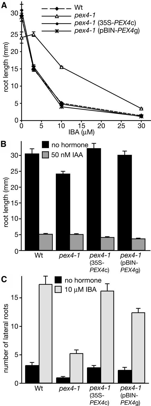
As shown in Figure 2A, pex4-1 is resistant to a range of IBA concentrations. As the mutant retains wild-type sensitivity to IAA (Figure 2B), the mutation appears to specifically disrupt IBA responses rather than general auxin responses. To confirm that the mutation in PEX4 is responsible for the IBA-response defect, we transformed pex4-1 plants with genomic PEX4 driven by its endogenous promoter (pBIN-PEX4g) or a PEX4 cDNA driven by the constitutive 35S promoter (35S-PEX4c). Lines homozygous for either construct displayed restored IBA responsiveness (Figure 2), indicating that we had identified the defective gene in the mutant.
Figure 2.
pex4-1 IBA-Response Defects.
(A) and (B) Root elongation of the wild type, the pex4-1 mutant, and homozygous lines of pex4-1 (35S-PEX4c) and pex4-1 (pBIN-PEX4g) grown for 8 d on medium containing 0.5% sucrose plus the indicated IBA concentrations (A) or 50 nM IAA (B).
(C) pex4-1 has a lateral root initiation defect. Seeds were sown on medium without hormone and grown for 4 d, then transferred to medium with or without 10 μM IBA and grown for 4 d.
Error bars indicate se (n ≥ 11).
Like other auxins, exogenous IBA stimulates lateral root formation (Hartmann et al., 1990; Zolman et al., 2000). Endogenous IBA also promotes root branching, as suggested by the decreased lateral root initiation of mutants with IBA-response defects grown on unsupplemented medium (Zolman et al., 2001b; Zolman and Bartel, 2004; Woodward and Bartel, 2005a). pex4-1 is resistant to the stimulatory effects of applied IBA on lateral root initiation and has fewer lateral roots in the absence of hormone (Figure 2C), indicating that endogenous pathways are disrupted. Both defects were restored by introducing wild-type PEX4 into the mutant (Figure 2C).
pex4 Has a Peroxisome-Defective Phenotype
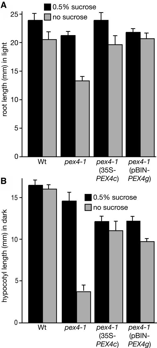
In addition to IBA resistance, mutants defective in peroxisomal processes often display sucrose dependence during seedling establishment (Hayashi et al., 1998, 2000a, 2002; Zolman et al., 2000, 2001a, 2001b; Footitt et al., 2002; Fulda et al., 2004; Zolman and Bartel, 2004; Adham et al., 2005; Woodward and Bartel, 2005a), because peroxisomal β-oxidation of fatty acids during germination provides energy before photosynthetic energy production. We found that pex4-1 seedlings have substantial growth defects in the absence of exogenous sucrose, reflected in reduced root elongation in the light and hypocotyl elongation in the dark (Figure 3). These defects are largely restored by sucrose supplementation (Figure 3). The pex4-1 sucrose dependence was accompanied by slowed utilization of the primary seed storage fatty acid (C20:1) in mutant seedlings (Zolman et al., 2000). Unlike some peroxisomal mutants (Hayashi et al., 2000a, 2005; Zolman and Bartel, 2004; Fan et al., 2005), adult pex4-1 plants have wild-type growth, fertility, and pigmentation, indicating no obvious defects in peroxisomal photorespiration.
Figure 3.
pex4-1 Sucrose Dependence during Early Seedling Growth.
The wild type, the pex4-1 mutant, and homozygous rescue lines pex4-1 (35S-PEX4c) and pex4-1 (pBIN-PEX4g) were germinated and grown with or without sucrose for 6 d, and root length in the light (A) or hypocotyl length in the dark (B) was measured. Error bars indicate se (n ≥ 11).
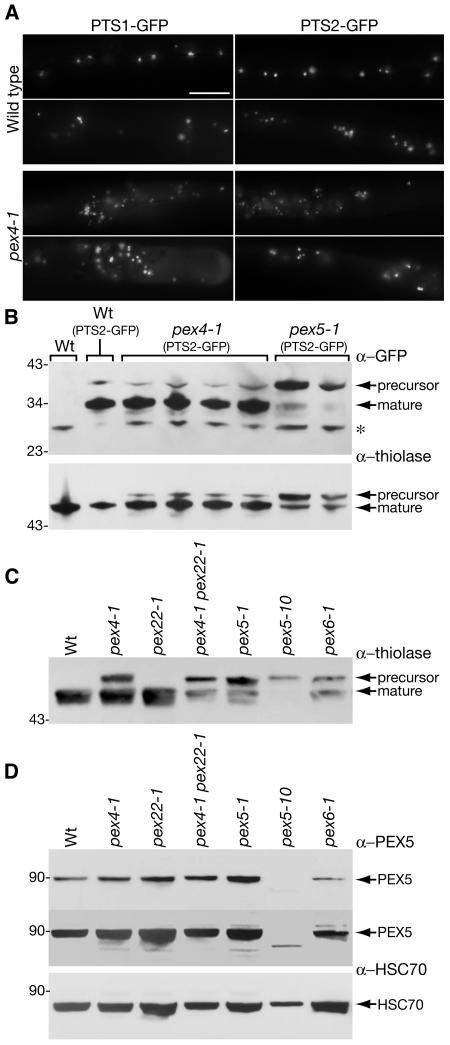
To visualize peroxisomal matrix protein import in pex4-1, we examined fluorescence localization in mutant plants expressing green fluorescent protein (GFP) targeted to peroxisomes by either PTS1 or PTS2 sequences. In the wild type, fluorescence from lines expressing either GFP construct is punctate (Figure 4A), consistent with peroxisomal localization (Zolman and Bartel, 2004; Woodward and Bartel, 2005a). The pex4-1 mutant displayed a wild-type fluorescence pattern with both PTS1- and PTS2-tagged GFP (Figure 4A), suggesting that at least some peroxisome matrix import occurs and that peroxisomes are of approximately normal size and numbers in the pex4-1 mutant.
Figure 4.
Peroxisomal Matrix Protein Import in pex4.
(A) Analysis of matrix protein import using peroxisome-targeted GFPs. Five-day-old wild-type or pex4-1 seedlings expressing GFP modified to contain a PTS1 (Zolman and Bartel, 2004) or PTS2 (Woodward and Bartel, 2005a) signal were examined using fluorescence microscopy. Two root hairs are shown for each line. The punctate pattern indicates peroxisomal protein import. All images were taken at ×100 magnification. Bar = 50 μm.
(B) Analysis of PTS2-GFP and thiolase processing in the wild type, pex4-1, and pex5-1. Immunoblotting using α-GFP (top) or α-thiolase (bottom) was performed on protein samples from 2-d-old untransformed Col-0 (wild type) and wild type, pex4-1, and pex5-1 expressing the PTS2-GFP transgene. The top bands (precursor) represent unprocessed PTS2-GFP or thiolase, the bottom bands represent the mature moiety, and the asterisk marks a cross-reacting protein in the α-GFP panel. pex5-1 (missense mutation in the matrix protein receptor PEX5; Zolman et al., 2000) is shown as a control for PTS2 processing; PEX5 interaction with the PTS2 receptor PEX7 is required for PTS2 matrix protein import (Hayashi et al., 2005; Woodward and Bartel, 2005a). The positions of molecular mass markers (kD) are shown at left.
(C) Analysis of thiolase processing in peroxisome-defective mutants. Immunoblotting using α-thiolase was performed on protein from 3-d-old light-grown seedlings. pex5-1, pex5-10 (T-DNA insertion in PEX5), and pex6-1 (missense mutation in the ATPase PEX6; Zolman and Bartel, 2004) are shown as controls.
(D) Analysis of PEX5 levels in peroxisome-defective mutants. Immunoblotting using α-PEX5 (Zolman and Bartel, 2004) was performed on protein from 3-d-old light-grown seedlings. The top panel shows different exposures of the same membrane to demonstrate similar PEX5 levels in the pex4-1, pex22-1, pex4-1 pex22-1, and pex5-1 mutants; the second exposure shows a lack of intact PEX5 in the pex5-10 mutant. The bottom panel shows the PEX5 membrane reprobed with α-HSC70 as a loading control.
A second method of assessing peroxisomal matrix protein import is direct analysis of PTS2 proteins, because the N-terminal PTS2 is cleaved after peroxisomal import. Examining the ratio of full-length to processed protein can reveal defects in PTS2 protein import. For example, in the peroxisome defective2 mutant, which is defective in the peroxisomal membrane protein PEX14 required for matrix protein import (Hayashi et al., 2000a), both processed and unprocessed thiolase are visible on immunoblots (Hayashi et al., 1998). To directly observe the extent of PTS2-GFP cleavage in pex4-1, we examined protein extracts using immunoblot analysis with a GFP antibody. We found that the PTS2 was removed from PTS2-GFP similarly in the wild type and pex4-1 (Figure 4B), consistent with the apparent peroxisomal localization of this protein in both lines (Figure 4A). We also used an anti-thiolase antibody (Kato et al., 1996) to examine the targeting of endogenous thiolase. In the pex4-1 mutant, we found most thiolase processed into the smaller form, as in the wild type, but we also detected some unprocessed thiolase not visible in the wild-type sample (Figures 4B and 4C). These results suggest that the pex4-1 mutant imports and processes thiolase nearly as efficiently as the wild type. Both PTS2-GFP and thiolase were inefficiently processed in the pex5-1 mutant (Figures 4B and 4C), which carries a missense mutation in the PTS1 receptor (Zolman et al., 2000). This result is consistent with the previously reported failure of PTS2 targeting in pex5 mutants: PEX5 and the PTS2 receptor PEX7 physically interact (Nito et al., 2002), and PTS2 import by PEX7 requires PEX5 (Hayashi et al., 2005; Woodward and Bartel, 2005a). The pex5-1 missense mutation falls within the proposed PEX7 binding domain, and PTS2 import is disrupted in the mutant, as visualized by mislocalized PTS2-GFP and incomplete thiolase processing (Woodward and Bartel, 2005a). We also found a thiolase-processing defect in pex6-1 (Zolman and Bartel, 2004) (Figure 4C). PEX6 is an ATPase that in yeast facilitates receptor recycling (Collins et al., 2000).
Several peroxisome-defective mutants, including P. pastoris pex4, have reduced levels of the matrix protein receptor PEX5 (Koller et al., 1999; Collins et al., 2000). We visualized PEX5 on immunoblots using a PEX5 peptide antibody (Zolman and Bartel, 2004) as an ∼90-kD band. To confirm the specificity of our antibody, we isolated a T-DNA insertion mutant in PEX5 from the Salk Institute collection (Alonso et al., 2003). This pex5-10 mutant has a T-DNA inserted in exon 5, probably causing a null allele. Indeed, we did not detect intact PEX5 protein in the pex5-10 mutant (Figure 4D). pex5-10 has a strong peroxisome-defective phenotype, including an absolute requirement for sucrose during early development, high seedling lethality, and delayed development (B.K. Zolman and B. Bartel, unpublished data). The pex5-10 individuals that develop into adult plants have reduced seed production (B.K. Zolman and B. Bartel, unpublished data). Consistent with a severe defect in peroxisome matrix import, thiolase was largely unprocessed in pex5-10 seedlings (Figure 4C).
We found that PEX5 levels in pex4-1 are similar to those in the wild type (Figure 4D). PEX5 levels also appear unaffected in the pex5-1 missense mutant but are reduced in an Arabidopsis pex6 mutant (Figure 4D), as reported previously (Zolman and Bartel, 2004). Thus, although pex4-1 has growth phenotypes suggesting greatly reduced peroxisome functioning, pex4-1 defects in peroxisomal matrix protein import are modest at best and do not seem to be accompanied by reduced PEX5 levels. This molecular phenotype may be limited because the pex4-1 missense allele retains partial PEX4 function and/or because Arabidopsis PEX4 has important roles in addition to matrix protein import.
PEX4 Interacts with Another Peroxisomal Protein
Arabidopsis and yeast PEX4 proteins lack peroxisome-targeting signals or apparent membrane-spanning regions. Yeast Pex4p is tethered to the peroxisome by the Pex22p membrane protein (Koller et al., 1999). Analysis of several sequenced genomes has not revealed a PEX22 candidate in any plant species. We conducted a yeast two-hybrid experiment to identify PEX4-interacting proteins, which might include the Arabidopsis PEX22, ubiquitin protein ligases, ubiquitinated substrates, or other components of the import machinery.
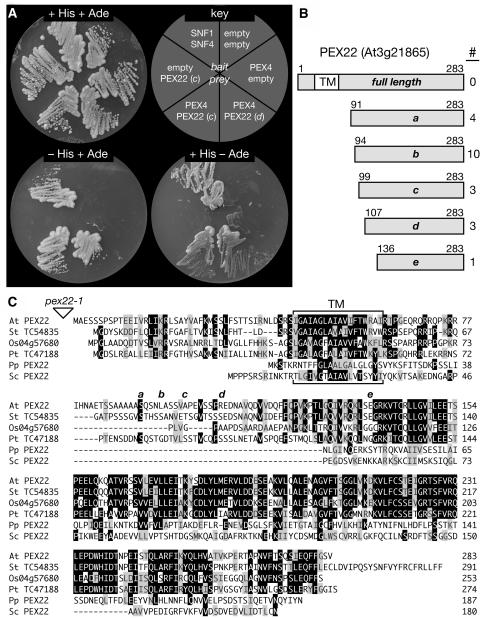
Using PEX4 as bait to probe an Arabidopsis cDNA library, we screened 2,665,000 transformants and identified 42 putative interacting proteins that conferred growth on medium lacking His. A secondary screen on medium lacking adenine reduced the number of potential PEX4 interactors to 21. Sequence analysis of the prey plasmids revealed that the clones represented five independent cDNAs from a single gene, At3g21865. The five clones were truncated at different 5′ positions, but each was in frame with the Gal4p activation domain and had the potential to encode the complete C terminus of At3g21865 (Figure 5). Analysis of the predicted At3g21865 protein using SMART (Schultz et al., 1998) revealed one potential transmembrane domain near the N terminus, from amino acids 45 to 62 (Figure 5). None of the cDNA clones obtained in the two-hybrid experiment contained this region, likely because transmembrane domains can interfere in the assay.
Figure 5.
PEX4 Interacts with Arabidopsis PEX22.
(A) A yeast two-hybrid assay with PEX4 fused to the GAL4 DNA binding domain (bait) was used to query an Arabidopsis cDNA library fused to the GAL4 activation domain (prey). The growth of the resulting yeast strains is shown on plates containing complete medium (top left) or under selective conditions indicating protein interaction (−His at bottom left, −adenine [Ade] at bottom right). Two PEX4-interactor strains are shown, SNF1 and SNF4 are positive controls (Fields and Song, 1989), and the empty vector combinations are negative controls.
(B) All 21 PEX4 interactors are PEX22 (At3g21865). The gray bar represents the PEX22 protein; a predicted transmembrane domain (TM) is indicated by the white box. The five identified clones are designated a to e, with the terminal amino acid numbers above the bar and the number of isolates indicated at right.
(C) Protein alignment of Arabidopsis (At) PEX22 with similar putative proteins from potato (Solanum tuberosum; St), rice (Os), and loblolly pine (Pinus taeda; Pt) and characterized yeast PEX22 proteins from S. cerevisiae (Sc) and P. pastoris (Pp). Potato and pine sequences are from genome projects assembled in The Institute for Genomic Research Gene Index Database (http://www.tigr.org/tdb/tgi; Quackenbush et al., 2001), and Os0457680 is from The Institute for Genomic Research Rice Genome Annotation (http://www.tigr.org/tdb/e2k1/osa1/index.shtml; Yuan et al., 2005). Sequences were aligned with the MEGALIGN program (DNASTAR) using the ClustalW method with BLOSUM series protein weight matrix. Residues identical or chemically similar in a majority of sequences are indicated by black or gray boxes, respectively. The T-DNA insertion disrupting pex22-1 upstream of the ATG is represented by the triangle. The predicted transmembrane domain (TM) is bracketed, and the starting amino acids of each of the five two-hybrid clones represented in (B) are indicated by a to e above the sequence.
Additional analysis of this PEX4-interacting protein revealed no recognized domains. Although the protein has apparent plant homologs (Figure 5C), none of these are characterized, and there were no obvious homologs outside the plant lineage. We hypothesized that At3g21865 might be the Arabidopsis PEX22 protein. Although the Arabidopsis protein is only 6 to 12% identical to characterized yeast Pex22p proteins, they have comparable sizes and predicted topologies, including similarly positioned N-terminal transmembrane domains (Figure 5C).
A Mutant Defective in PEX22
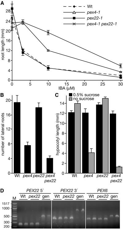
To determine whether this candidate PEX22 protein was indeed a peroxin, we sought a loss-of-function mutation in At3g21865. Although no T-DNA insertions in publicly available collections disrupt the 2.2-kb primary transcript of this gene, we isolated a mutant with a T-DNA insert near the coding region from the Salk Institute collection (Alonso et al., 2003). The T-DNA in this pex22-1 mutant is inserted at position −107 relative to the initiator ATG (the 5′ untranslated region is 103 nucleotides). Because of the PEX4 interaction, we anticipated that reduced levels of this protein might result in defective IBA responses similar to those of pex4-1. However, characterization of this mutant revealed wild-type responses to IBA in root elongation (Figure 6A) and lateral root assays (Figure 6B) and normal growth in the absence of sucrose in both the dark (Figure 6C) and light (data not shown). In addition, the mutant had normal levels of the PEX5 matrix protein receptor (Figure 4D) and no obvious defect in thiolase processing (Figure 4C).
Figure 6.
Enhanced Phenotypes in pex4-1 pex22-1 Double Mutants.
(A) to (C) Responses of the wild type, pex4-1, pex22-1, and the pex4-1 pex22-1 double mutant were measured as described in the legends to Figures 2 and 3. The double mutant has enhanced resistance to IBA inhibition of root elongation (A), more severe lateral root initiation defects (B), and increased sucrose dependence in the dark during early development (C). Error bars indicate se (n ≥ 7).
(D) pex22-1 seedling PEX22 transcript levels resemble wild-type levels. Two biological replicates of RNA from 7-d-old wild-type and pex22-1 mutant plants were reverse-transcribed. The resulting cDNAs and genomic DNA (gen) were amplified for 32 cycles with intron-spanning primers near the 5′ (left) and 3′ (middle) of PEX22. A control reaction amplifying PEX6 is shown (right), and the positions of markers (M) in bp are indicated at left. Similar results were observed after allowing the PCR to proceed for 25 or 40 cycles.
Because the pex22-1 insertion is upstream of the coding region, disruption of this gene probably is incomplete. To test whether the T-DNA insertion affects PEX22 expression, we isolated RNA from mutant and wild-type seedlings and performed RT-PCR using gene-specific primers. We found PEX22 transcript at similar levels in both samples (Figure 6D), indicating that the T-DNA insertion does not dramatically alter PEX22 transcript levels in 7-d-old seedlings.
Because pex22-1 does not eliminate PEX22 transcription and pex4-1 is a missense allele (Figure 1) that may incompletely compromise PEX4 function, we constructed a pex4-1 pex22-1 double mutant to determine whether combining both defects would enhance the mutant phenotypes. As in the single mutants, PEX5 levels remained similar to those in the wild type in the pex4-1 pex22-1 double mutant (Figure 4D). However, the pex4-1 pex22-1 double mutant displayed more severe phenotypes than either parent in both IBA responses and sucrose dependence (Figure 6). Moreover, the slight thiolase-processing defect observed in pex4-1 was enhanced in the pex4-1 pex22-1 double mutant (Figure 4C). This genetic interaction indicates that PEX22 contributes to peroxisome functions in vivo.
Arabidopsis PEX4 and PEX22 Functionally Complement Yeast Mutants
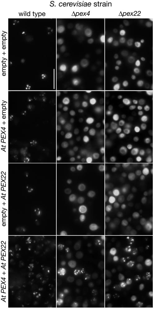
To confirm that this new protein functions as the Arabidopsis PEX22 and to determine whether the Arabidopsis PEX4 and PEX22 proteins can act similarly to their yeast counterparts, we tested whether the Arabidopsis proteins could complement the corresponding S. cerevisiae mutants. Yeast pex4 and pex22 mutants fail to import a PTS1-GFP reporter into peroxisomes (Wiebel and Kunau, 1992; Koller et al., 1999; Eckert and Johnsson, 2003) (Figure 7). We expressed the Arabidopsis PEX4 and PEX22 genes both individually and together in yeast pex4 and pex22 mutants. Neither PEX4 nor PEX22 alone rescued the corresponding yeast mutants (Figure 7). However, introducing Arabidopsis PEX4 and PEX22 together partially restored the ability of either mutant to import PTS1-GFP into peroxisomes (Figure 7). The ability of the genes to rescue when added together indicates that we have identified the Arabidopsis PEX22 protein and that the PEX4–PEX22 complex can perform a similar task in yeast and plants.
Figure 7.
Dual Expression of Arabidopsis PEX4 and PEX22 Rescues Yeast pex4 and pex22 Mutant Defects.
Wild-type, Δpex4, and Δpex22 yeast strains were transformed with the indicated combinations of empty vectors or vectors expressing Arabidopsis (At) PEX4 or PEX22 from the constitutive GPD promoter. Yeast strains were examined microscopically as described in the legend to Figure 4A. Wild-type fluorescence is punctate, indicating peroxisomal localization of GFP, whereas Δpex4 and Δpex22 display diffuse cytoplasmic fluorescence unless Arabidopsis PEX4 and PEX22 cDNAs are coexpressed, which partially restores the punctate pattern. All images were made at ×100 magnification using the same exposure time. Bar = 50 μm.
Isocitrate Lyase Is Stabilized in the pex4-1 pex22-1 Double Mutant
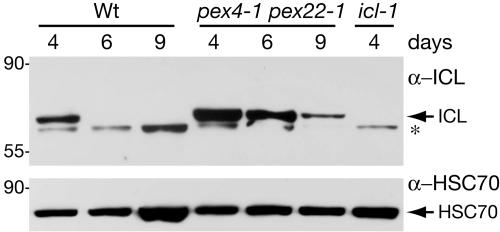
The strong sucrose dependence of the pex4-1 pex22-1 mutant is not accompanied by correspondingly strong defects in peroxisomal matrix protein import, suggesting that this peroxin pair may have novel plant targets in addition to those important in fungi. One possible plant-specific role is in the transition of seedling glyoxysomes to leaf peroxisomes. Glyoxysomes, specialized peroxisomes that function early in seedling development, convert acetyl-CoA liberated by fatty acid β-oxidation to substrates that are metabolized to sucrose via gluconeogenesis. As photosynthesis begins, glyoxysomes are converted to leaf peroxisomes, which house photorespiration enzymes (Titus and Becker, 1985). Several glyoxysome-specific enzymes are not found in leaf peroxisomes. For instance, isocitrate lyase (ICL), an essential enzyme of the glyoxylate cycle, has high mRNA levels and enzyme activity soon after imbibition, but these levels quickly decline 3 to 4 d later (Eastmond et al., 2000). To determine whether PEX4/PEX22 plays a role in protein degradation at this transition, we examined the rate of ICL disappearance during wild-type and pex4-1 pex22-1 seedling development. We used an antibody raised against castor bean (Ricinus communis) ICL (Maeshima et al., 1988) to monitor ICL levels and the icl-1 null mutant (Eastmond et al., 2000) to determine the specificity of the antibody in Arabidopsis. We found that ICL was readily detected by immunoblotting in 4-d-old wild-type seedlings and was absent in 4-d-old icl-1 seedlings (Figure 8). As expected, ICL levels declined in older wild-type seedlings; ICL was not apparent 6 or 9 d after sowing (Figure 8). By contrast, ICL was more abundant in pex4-1 pex22-1 seedlings at every time point assayed and was easily detected even in 9-d-old pex4-1 pex22-1 seedlings (Figure 8). This result indicates that the PEX4–PEX22 pair is necessary to efficiently remove ICL during seedling development.
Figure 8.
ICL Is Stabilized in the pex4-1 pex22-1 Mutant.
Extracts from wild-type and pex4-1 pex22-1 seedlings grown for 4, 6, or 9 d in the light on medium with sucrose were analyzed by immunoblotting using α-ICL (top). Four-day-old icl-1 seedlings, in which the single-copy ICL gene is disrupted by an En-1 insertion in exon 3 (Eastmond et al., 2000), were used as a control for the castor bean ICL antibody (Maeshima et al., 1988). This antibody detects a protein of the expected size of Arabidopsis ICL (66 kD), which is absent in the icl-1 mutant, and a smaller unidentified cross-reacting band (asterisk). The bottom panel shows the membrane reprobed with α-HSC70 as a loading control. The positions of molecular mass markers (kD) are shown at left.
DISCUSSION
Because the protoauxin IBA appears to be converted to active IAA in a mechanism paralleling peroxisomal fatty acid β-oxidation (Bartel et al., 2001; Woodward and Bartel, 2005b), certain Arabidopsis mutants defective in general peroxisomal or β-oxidation processes affect both fatty acid utilization and IBA responses (Zolman et al., 2000, 2001a, 2001b; Zolman and Bartel, 2004; Adham et al., 2005; Woodward and Bartel, 2005a). Related screens using 2,4-dichlorophenoxybutyric acid, which may be similarly converted to the active auxin-like derivative 2,4-D, have revealed a partially overlapping set of mutants (Hayashi et al., 1998, 2000a, 2002; Lange et al., 2004).
PEX4 Is a UBC Enzyme Important for Peroxisome Function in Plants and Fungi
We isolated the Arabidopsis pex4-1 mutant in an IBA-response screen (Zolman et al., 2000). The IBA-response phenotype is apparent in root growth, lateral root initiation on exogenous hormone, and lateral root initiation in the absence of applied hormone, indicating that endogenous pathways are disrupted (Figure 2). In addition, pex4-1 has reduced growth after germination in the absence of sucrose in the dark and light (Figure 3). These phenotypes reflect defects in peroxisomal β-oxidation of seed storage lipids and the parallel process of IBA-to-IAA conversion.
PEX4 encodes an apparent UBC enzyme, which transfers ubiquitin from ubiquitin-activating enzymes to ubiquitin protein ligase–substrate complexes. Arabidopsis contains ∼45 UBC enzymes (Bachmair et al., 2001; Vierstra, 2003); PEX4 appears to be a single-copy gene. Apparent PEX4 homologs are found in fungi, protists, and throughout the plant lineage, including algae (Figures 1C and 1D). Although all UBC proteins are related, phylogenetic analysis reveals a cluster of PEX4-like proteins distinct from other UBCs (Figure 1D). Interestingly, PEX4 has not been identified in any metazoan, and none of the human peroxisomal biogenesis disorder complementation groups are known to be defective in a UBC enzyme (Gould and Valle, 2000; Wanders and Waterham, 2005). The human protein most similar to PEX4, E2 N, functions in mitotic progression and DNA repair (Hofmann and Pickart, 1999; Bothos et al., 2003). Several Arabidopsis proteins are highly similar to this protein and cluster together, distinct from the PEX4 clade.
The apparent absence of PEX4 in metazoans may reflect protein divergence that confounds homology-based identification, redundancy that prevents the occurrence of a pex4-based peroxisome biogenesis disorder, or mechanistic differences between yeast, plants, and animals. However, peroxins in addition to PEX4 implicate ubiquitin in peroxisome function. Three candidate ubiquitin protein ligases, the RING finger peroxins PEX2, PEX10, and PEX12, are necessary for peroxisome function in mammals and yeast (Lazarow, 2003). Arabidopsis PEX2 and PEX10 are members of the RING-HCa subclass of putative and confirmed ubiquitin protein ligases (Stone et al., 2005), and all three proteins are required for embryonic development (Hu et al., 2002; Schumann et al., 2003; Sparkes et al., 2003; Fan et al., 2005).
An Arabidopsis pex22 Mutant
Although PEX4 proteins lack a PTS, P. pastoris Pex4p is recruited to the outer surface of the peroxisome (Crane et al., 1994) via interaction with the peroxisomal membrane protein Pex22p. P. pastoris pex22 mutants are defective in peroxisomal matrix protein import (Koller et al., 1999), similar to pex4. The N-terminal transmembrane domain of P. pastoris Pex22p has a membrane PTS sufficient for peroxisomal targeting (Koller et al., 1999). Pex22p interacts with Pex4p via the cytoplasmic C-terminal domain, and Pex4p is cytosolic and unstable in the P. pastoris pex22 mutant, suggesting that Pex22p tethers Pex4p to peroxisomes (Koller et al., 1999).
We identified the Arabidopsis functional equivalent of Pex22p using a yeast two-hybrid assay with the Arabidopsis PEX4 protein. Arabidopsis PEX22 is encoded by a single-copy gene not previously identified by sequence comparison; indeed, it displays limited similarity to the yeast counterparts, although there are several highly similar proteins in other plants (Figure 5C). Arabidopsis PEX22 is structurally similar to yeast PEX22 proteins, with a single predicted transmembrane domain near the N terminus. We isolated a pex22 mutant likely to confer a partial loss of function and found that it lacked notable phenotypes on its own but enhanced all pex4-1 mutant defects (Figure 6), indicating that PEX4 and PEX22 interact genetically and that PEX22 contributes to peroxisome functioning.
Remarkably, coexpression of Arabidopsis PEX4 and PEX22 partially rescues the yeast pex4 and pex22 mutant defects in matrix protein localization (Figure 7). By contrast, neither PEX4 nor PEX22 alone rescued the corresponding yeast mutant phenotype. The inability of the single genes to rescue is not surprising considering the limited similarity between the PEX22 homologs (Figure 5); the native Pex4p protein probably is unable to interact with the introduced PEX22 protein, and vice versa. In fact, S. cerevisiae Pex22p does not interact with P. pastoris Pex4p in a yeast two-hybrid assay (Koller et al., 1999), and expression of S. cerevisiae PEX4 or PEX22 does not complement the respective P. pastoris pex4 or pex22 mutant (Crane et al., 1994; Koller et al., 1999). However, expression of the interacting pair from Arabidopsis restores peroxisomal function in the yeast mutants, indicating that PEX4 and PEX22 can function similarly in plants and yeast.
PEX4 and PEX22 Affect Peroxisomal Processes
pex4-1 seedling defects in the absence of sucrose are consistent with reduced peroxisomal function. However, the molecular cause of these defects is not apparent. We did not detect localization defects in peroxisomally targeted GFP in pex4-1 (Figure 4A), in contrast with pex5-1 (Woodward and Bartel, 2005a), pex7-1 (Woodward and Bartel, 2005a), pex12 (Fan et al., 2005), and pex14-1 (Hayashi et al., 2000a). However, we did find a subtle defect in the import or processing of PTS2-containing thiolase in pex4-1 (Figures 4B and 4C). Thiolase maturation defects are found in mutants defective in peroxisomal matrix protein import, including Arabidopsis pex5, pex6, pex7, and pex14 (Hayashi et al., 1998; Zolman and Bartel, 2004; Woodward and Bartel, 2005a) (Figure 4C). These mutant defects may reflect reduced PTS2 import or may be indirect, as Arabidopsis PEX7 requires the PTS1 receptor PEX5 for function (Hayashi et al., 2005; Woodward and Bartel, 2005a) and the PTS2-cleaving protease has not been described and may contain a PTS1. The ability to detect thiolase maturation defects in pex4-1 and pex6-1, which display apparently normal peroxisomal localization of PTS-GFP, may reflect higher sensitivity in the thiolase assay for detecting partial losses of function. However, it is intriguing that pex4-1 and pex6-1 both show severe sucrose dependence (Zolman and Bartel, 2004) (Figure 3) but have only mild thiolase-processing defects (Figure 4C), whereas pex5-1 displays only slight sucrose dependence (Zolman et al., 2000) accompanied by a more severe block in thiolase processing (Woodward and Bartel, 2005a) (Figures 4B and 4C).
PEX4 mutants have been identified in several yeast species (Wiebel and Kunau, 1992; Crane et al., 1994; van der Klei et al., 1998; Koller et al., 1999). In P. pastoris, decreased Pex4p activity results in ∼10-fold decreased Pex5p levels attributable to increased protein degradation (Koller et al., 1999; Collins et al., 2000). By contrast, S. cerevisiae pex4 mutants have nearly wild-type Pex5p levels (Platta et al., 2004; Kiel et al., 2005), similar to our findings with Arabidopsis pex4-1 (Figure 4D).
Pex5p localization changes from predominantly cytosolic in wild-type P. pastoris to primarily peroxisomal in a pex4 mutant (van der Klei et al., 1998; Collins et al., 2000), suggesting that Pex4p assists Pex5p movement out of peroxisomes. In Arabidopsis, however, cyan fluorescent protein (CFP)-tagged PEX5 is peroxisomal in both the wild type (Tian et al., 2004) and in the pex4-1 mutant (data not shown). It is not known whether the C-terminal CFP tag interferes with PEX5 cycling; in yeast, addition of a C-terminal GFP tag changes PEX7 localization from cytosolic to primarily peroxisomal (Nair et al., 2004). Further studies are needed to investigate Arabidopsis PEX5 localization in the wild type and peroxin mutants.
In yeast, Pex4p is proposed to act with the ATPases Pex1p and Pex6p, recycling Pex5p after import (Collins et al., 2000). As predicted from this model, Arabidopsis pex4-1 and pex6-1 share similar phenotypes: both have strong growth defects in the absence of sucrose and IBA-resistant root growth. However, unlike Arabidopsis pex6-1 (Zolman and Bartel, 2004), pex4-1 seedlings do not have reduced PEX5 levels (Figure 4D) and are not rescued by PEX5 overexpression (data not shown), suggesting that Arabidopsis PEX4 is not limiting for PEX5 recycling. As pex4-1 is a missense allele and no insertion alleles are currently available, definitive determination of a role for PEX4 in maintaining PEX5 levels in plants awaits the isolation of a pex4 null allele.
Functional similarity to the yeast homologs suggests that the PEX4–PEX22 complex will be peroxisomal. Accumulating evidence, however, indicates that some membrane peroxins originate in the endoplasmic reticulum (Titorenko and Rachubinski, 1998; Mullen et al., 2001; Geuze et al., 2003; Hoepfner et al., 2005; Karnik and Trelease, 2005). Future subcellular localization studies may reveal whether PEX4 and PEX22 also reside, either transiently or continuously, in the endoplasmic reticulum.
Possible Roles for Ubiquitin in Peroxisome Function
Ubiquitination most commonly is associated with protein degradation, but it also can regulate protein–protein interactions, alter protein function, and facilitate the intracellular movement of tagged proteins (Schnell and Hicke, 2003). In yeast, Pex4p UBC activity is not required for Pex4p localization (Wiebel and Kunau, 1992) or Pex4p–Pex22p interaction (Koller et al., 1999), but it is required for functional peroxisomes (Wiebel and Kunau, 1992; Crane et al., 1994).
Recent reports indicate that S. cerevisiae Pex5p is ubiquitinated at the peroxisome membrane during matrix protein import (Platta et al., 2004, 2005; Kiel et al., 2005; Kragt et al., 2005). Peroxisomal import of matrix-bound proteins occurs via a receptor-recycling mechanism, in which the Pex5p and Pex7p receptors bind peroxisome-bound cargo, escort the proteins into the peroxisome, and then dissociate and return to the cytoplasm for another round of import (Dammai and Subramani, 2001; Costa-Rodrigues et al., 2004; Nair et al., 2004). In wild-type yeast, Pex5p is monoubiquitinated (Kragt et al., 2005), whereas several yeast peroxin mutants have altered ubiquitination patterns (Platta et al., 2004; Kiel et al., 2005; Kragt et al., 2005); these mutants all are proposed to act in recycling Pex5p from the peroxisome and include pex4 and pex22. These data suggest a model in which Pex5p ubiquitination normally assists receptor recycling. When Pex5p accumulates in the membrane (as in recycling mutants such as pex4), it may be targeted for degradation. Many questions remain regarding this model, including the number of ubiquitin molecules required for recycling and the mechanism of receptor release (Platta et al., 2005). We have not tested whether this model applies in plants, as our PEX5 antibody does not reliably detect higher molecular weight PEX5 species that might be ascribed to ubiquitinated PEX5.
In addition to a role for PEX4 in PEX5 recycling, we speculate that PEX4-mediated ubiquitination may function in retrotranslocation of damaged proteins from the peroxisome matrix to the cytosol, where they would be subject to ubiquitin-dependent proteasomal degradation. This model is based on the function of the yeast Ubc7p enzyme in the retrotranslocation and degradation of misfolded endoplasmic reticulum proteins (Meusser et al., 2005). Intriguingly, Ubc7p is tethered to the endoplasmic reticulum membrane by Cue1p, a small, single membrane-spanning protein, as PEX4 is tethered to peroxisomes by PEX22. Moreover, endoplasmic reticulum retrotranslocation of misfolded luminal proteins requires the Cdc48p ATPase (Meusser et al., 2005), which is similar to PEX6 and PEX1 (Zolman and Bartel, 2004), and the endoplasmic reticulum membrane RING finger ubiquitin protein ligase Der3p/Hrd1p (Meusser et al., 2005), which might function analogously to one or more of the RING finger peroxins PEX2, PEX10, and PEX12.
PEX4-mediated ubiquitination also could function in removing matrix proteins during transitions between different types of peroxisomes. During the conversion of glyoxysomes to leaf peroxisomes in seedling development, proteins acting in early developmental processes, such as the glyoxylate cycle, disappear, whereas proteins acting in leaf peroxisomes, including photorespiration enzymes, appear. These changes are correlated with changes in gene expression and protein degradation (Nishimura et al., 1996; Olsen, 1998; Hayashi et al., 2000b). The mechanism of peroxisomal protein degradation is unknown. Several uncharacterized protease-like proteins are predicted to be targeted to the Arabidopsis peroxisomal matrix (Kikuchi et al., 2004; Reumann et al., 2004). In addition to degradation within the peroxisome, peroxisomal proteins could be retrotranslocated with the assistance of PEX4-mediated ubiquitination for cytosolic proteasomal degradation. This latter hypothesis is supported by our observation that ICL, which normally functions only very early in seedling development (Eastmond et al., 2000), persists in the pex4-1 pex22-1 mutant for days after the protein has disappeared in the wild type (Figure 8). If hypothetical misfolded or accumulating peroxisomal proteins impede peroxisomal processes, this model could explain why the severe physiological phenotypes seen in the pex4-1 pex22-1 mutant are not accompanied by correspondingly severe defects in PEX5 and matrix protein import.
We have characterized an Arabidopsis pex4 mutant and identified the previously unknown Arabidopsis PEX22 protein, which genetically and physically interacts with PEX4. Our results suggest a role for ubiquitination in plant peroxisomal function that may be partially conserved in yeast and provide the groundwork for the identification of the unique and overlapping PEX4 targets and accessories in plants.
METHODS
Plant Materials and Growth Conditions
The pex4-1 mutant of Arabidopsis thaliana was described previously as B29, an ethyl methanesulfonate–induced IBA-response mutant in the Columbia (Col-0) background (Zolman et al., 2000). The pex22-1 (SALK_030743) and pex5-10 (SALK_124577) mutants, also in the Col-0 accession, were from the Salk Institute sequence indexed insertion collection (Alonso et al., 2003), and pex5-1 (Zolman et al., 2000), pex6-1 (Zolman and Bartel, 2004), and icl-1 (Eastmond et al., 2000) were described previously. T-DNA insertion positions were confirmed by sequencing amplification products of genomic DNA using a modified LBb1 T-DNA primer (5′-CAAACCAGCGTGGACCGCTTGCTGCA-3′; http://signal.salk.edu) and PEX22 (5′-AAGGAACCAGATTTCGAATCGATGTAGTGG-3′) or PEX5 (5′-GATATCAAATGCGACTCAAACACCTGATGAC-3′) gene-specific primers.
Surface-sterilized seeds were plated on plant nutrient medium (Haughn and Somerville, 1986) solidified with 0.6% (w/v) agar and supplemented with sucrose, hormones, kanamycin, or Basta (glufosinate-ammonium; Crescent Chemical) as indicated. Plates were placed at 22°C under continuous light. To slow the breakdown of indolic compounds, auxin plates were incubated under yellow light filters (Stasinopoulos and Hangarter, 1990). Seedlings were transferred to soil (Metromix 200; Scotts) and grown at 18 to 24°C under continuous illumination.
Mutant Characterization
The pex4-1 and pex22-1 mutants were backcrossed to the wild type (Col-0) at least once before analyses, and all assays were conducted at least twice with similar results. In root elongation assays, seedlings were grown for 6 or 8 d and the length of the primary root was measured. In lateral root assays, seedlings were grown for 4 d and transferred to medium containing IBA or no hormone, and lateral roots were counted with a dissecting microscope after an additional 4 d. For hypocotyl elongation assays, seeds were incubated for 24 h under white light before being transferred to the dark. Hypocotyl lengths were measured after an additional 5 d.
pex4-1 was outcrossed to Landsberg erecta tt4 and Wassilewskija for mapping, and DNA was isolated (Celenza et al., 1995) from IBA-resistant F2 plants with 100% sucrose-dependent progeny. The mutation was mapped using PCR-based molecular markers (http://www.arabidopsis.org). The At5g25760 candidate gene was amplified by PCR from mutant DNA and sequenced directly (SeqWright).
PEX4 Isolation and pex4-1 Complementation
For pex4-1 mutant complementation, a 2.7-kb EcoRI-SpeI fragment containing the PEX4 coding sequence, 0.9-kb 5′ flanking sequence, and 0.6-kb 3′ flanking sequence was subcloned from the F18A17 BAC into the EcoRI-XbaI sites of the plant transformation vector pBIN19 (Bevan, 1984) to make pBIN-PEX4g. This plasmid was electroporated into Agrobacterium tumefaciens GV3101 (Koncz and Schell, 1986) and transformed into pex4-1 (Clough and Bent, 1998). Transformants were selected on 12 μg/mL kanamycin.
A PEX4 cDNA in pBluescript SK− was obtained from the Kasuza DNA Research Institute (Asamizu et al., 2000). The cDNA was excised using HincII and XhoI and subcloned into pBluescript II KS+ cut with EcoRV and XhoI (pKS-PEX4c) or into SmaI-XhoI–cut 35SpBARN (LeClere and Bartel, 2001) behind the constitutive 35S cauliflower mosaic virus promoter (35S-PEX4c). 35S-PEX4c was transformed into pex4-1 plants, and transformants were selected on 7.5 μg/mL Basta.
Microscopy
Wild-type (Col-0) plants transformed with PTS1-GFP (Zolman and Bartel, 2004; Woodward and Bartel, 2005a) or PEX5-CFP (Tian et al., 2004) were crossed to pex4-1, and mutant plants carrying the transgene were identified by PCR-based genotyping. PTS2-GFP (Woodward and Bartel, 2005a) was transformed into pex4-1, and progeny of the transformants were compared with Col-0 and pex5-1 plants transformed with PTS2-GFP (Woodward and Bartel, 2005a). Fluorescence localization was examined in root hairs of 4- or 6-d-old light-grown seedlings using a Zeiss Axioplan 2 fluorescence microscope equipped with narrow-band GFP and cyan GFP filter sets (41020 and 31044v2; Chroma Technology).
Yeast Methods
The pBI770PEX4 bait construct for the two-hybrid assay was made by performing oligonucleotide-directed mutagenesis on pKS-PEX4c to create an in-frame SalI site immediately 5′ of the initiator ATG using the oligonucleotide 5′-CAATTCGTTCTCTGTCGACCCATGGAGATGCAGGC-3′ (altered residues are underlined). The PEX4 coding sequence was excised using SalI and NotI and subcloned into the pBI770 vector (Kohalmi et al., 1998) cut with the same enzymes. For library transformation, 200 μL of competent AH109 (MATa, trp1-901, leu2-3, 112, ura3-52, his3-200, gal4Δ, gal80Δ, LYS2::GAL1UAS-GAL1TATA-HIS3, GAL2UAS-GAL2TATA-ADE2, URA3::MEL1UAS-MEL1TATA-lacZ, MEL1; Clontech) yeast was transformed according to the manufacturer's instructions with 50 μg of pBI770PEX4 and 20 μg of Arabidopsis cDNA library in the pBI771 prey vector (Kohalmi et al., 1998). Transformations were plated on synthetic complete plates lacking Leu, Trp, and His and containing 2 mM 3-amino-1,2,4-triazole to reduce background growth. Positive transformants were assayed for growth on plates lacking Leu, Trp, and adenine.
Plasmid DNA was isolated from yeast colonies (Monroe-Augustus, 2004) and purified using the Wizard DNA clean-up system (Promega A7280). One Shot MACH1-T1 Escherichia coli cells (Invitrogen) were transformed with yeast DNA, and cDNA inserts were sequenced. Growth was retested after cotransforming AH109 cells with purified pBI771 cDNA clones and pBI770PEX4.
For yeast mutant complementation, a full-length PEX22 cDNA clone (U11677) was identified (Yamada et al., 2003). This cDNA was excised from the pUNI51 vector with EcoRI and NotI and subcloned into pBluescript II KS+ cut with the same enzymes, forming pKS-PEX22c. PEX22 was amplified by PCR with 5′-GGGATCCGCCGTCAAGGCCAGAAGG-3′ and 5′-GCGGCCGCTTAAACACTTCCAAAGAACTGTTC-3′ primers to add BamHI and NotI sites (underlined) on the 5′ and 3′ ends, respectively. The PEX22 cDNA was subcloned into the yeast transformation vector pTGPD (a pRS314 derivative containing the GPD promoter and TRP1) cut with BamHI and NotI to make pTGPD-PEX22. The PEX4 cDNA was removed from pKS-PEX4c with EcoRI and SalI and subcloned into pRS426-GPD (containing the GPD promoter and URA3) cut with EcoRI and XhoI. PCR amplification using 5′-AACTCGACAAGCTTAAAGGGAACAAAAGCTG-3′ and 5′-GTGCGGCCGCGTAATACGACTCACTATAGGG-3′ amplified the GPD promoter and PEX4, adding HindIII-NotI sites (underlined) at the ends; the resulting amplicon was ligated into pRS415 (containing LEU2) cut with the same enzymes to make pRS415-PEX4.
S. cerevisiae pex4 and pex22 deletion mutants (Eckert and Johnsson, 2003) and the wild-type parent were transformed with pEW88, which encodes a peroxisomally targeted GFP under URA3 selection (Hettema et al., 1998). The three resulting lines were transformed with combinations of empty pRS415 and pTGPD vectors, pRS415-PEX4 and pTGPD-PEX22. Transformants were selected on medium lacking Leu, Trp, and uracil. Matrix protein import was scored using GFP fluorescence of yeast grown on selective medium plus 0.3% glucose for 4 d. Images were made as described above.
Protein Analysis
Immunoblot analysis was performed as described previously (Zolman and Bartel, 2004) on proteins extracted from light-grown seedlings. Membranes were incubated with rabbit primary antibodies raised against Arabidopsis PEX5 (Zolman and Bartel, 2004; used at a 1:200 dilution), plant thiolase (Kato et al., 1996; used at a 1:2500 or 1:5000 dilution), GFP (BD Biosciences 8372-2; used at a 1:350 dilution), or castor bean (Ricinus communis) ICL (Maeshima et al., 1988; used at a 1:2000 dilution), followed by horseradish peroxidase–linked goat anti-rabbit secondary antibody (Santa Cruz Biotechnology sc-2030; used at a 1:1000 or 1:5000 dilution). As a loading control, membranes subsequently were incubated with a mouse monoclonal antibody against spinach (Spinacia oleracea) Hsc70 (Stressgen Bioreagents SPA-817; used at a 1:2000 dilution), followed by horseradish peroxidase–linked goat anti-mouse secondary antibody (Santa Cruz Biotechnology sc-2031; used at a 1:1000 or 1:5000 dilution). All antibodies were visualized using LumiGLO (Cell Signaling).
RNA Analysis
Wild-type and pex22-1 seedlings were grown for 7 d under white light on medium containing 0.5% sucrose covered with filter paper. Tissue was harvested by immersion in liquid nitrogen and ground to a fine powder, and RNA was isolated using RNeasy Mini kits (Qiagen). RNA was reverse-transcribed with random hexamer oligonucleotides using Superscript III reverse transcriptase (Invitrogen). Amplification of PEX22 was done using two sets of oligonucleotides: PEX22 5′ (5′-ACTGAAGAGATCGTTCGGTTGATTAAGCG-3′ plus 5′-CCTTCACTAAGCTTCTGTCTAACTATTTGC-3′) and PEX22 3′ (5′-CAAAGTTTTACAGGCTTTAGAGAATGCAGG-3′ plus 5′-TTCTATTGACTGCGAGGTGAACACATTTGG-3′). A control amplification used primers from the PEX6 gene (5′-AGTTCTGAATAAAGACGGTGACTTGTTACG-3′ plus 5′-GGGGACAAGTAAGCAACCTCTTGATC-3′).
Phylogenetic Analysis
Sequences were aligned with the MEGALIGN program (DNASTAR) using the ClustalW method with Gonnet series protein weight matrix. The unrooted phylogram was generated with PAUP 4.0b5 (Swofford, 2001) using the alignment shown in Supplemental Figure 1 online. The bootstrap method was performed for 100 replicates with a distance optimality criterion, and all characters were weighted equally.
Accession Numbers
Sequence data from this article can be found in the GenBank/EMBL data libraries under accession numbers AV566071 (PEX4 cDNA) and AY096402 (PEX22 cDNA).
Supplemental Data
The following material is available in the online version of this article.
Supplemental Figure 1. Sequence Alignment Used for PEX4 Phylogenetic Analysis.
Supplementary Material
Acknowledgments
We are grateful to Bill Crosby for the Arabidopsis two-hybrid library, Nils Johnnson for yeast mutants and the pEW88 plasmid, Makoto Hayashi for the thiolase antibody, Masayoshi Maeshima for the ICL antibody, Ian Graham for icl-1 seeds, and Diana Dugas, James McNew, Arthur Millius, Rebekah Rampey, Jeanne Rasbery, Lucia Strader, and Andrew Woodward for comments on the manuscript. We thank the ABRC (Ohio State University) for providing seeds and DNA clones, the Salk Institute for generating Arabidopsis mutants, and the Kazusa DNA Research Institute for a PEX4 cDNA. This research was supported by the National Science Foundation (Grant IBN-0315596) and the Robert A. Welch Foundation (Grant C-1309).
The author responsible for distribution of materials integral to the findings presented in this article in accordance with the policy described in the Instructions for Authors (www.plantcell.org) is: Bonnie Bartel (bartel@rice.edu).
Online version contains Web-only data.
Article, publication date, and citation information can be found at www.plantcell.org/cgi/doi/10.1105/tpc.105.035691.
References
- Adham, A.R., Zolman, B.K., Millius, A., and Bartel, B. (2005). Mutations in Arabidopsis acyl-CoA oxidase genes reveal distinct and overlapping roles in β-oxidation. Plant J. 41, 859–874. [DOI] [PubMed] [Google Scholar]
- Alonso, J.M., et al. (2003). Genome-wide insertional mutagenesis of Arabidopsis thaliana. Science 301, 653–657. [DOI] [PubMed] [Google Scholar]
- Asamizu, E., Nakamura, Y., Sato, S., and Tabata, S. (2000). A large scale analysis of cDNA in Arabidopsis thaliana: Generation of 12,028 non-redundant expressed sequence tags from normalized and size-selected cDNA libraries. DNA Res. 7, 175–180. [DOI] [PubMed] [Google Scholar]
- Bachmair, A., Novatchkova, M., Potuschak, T., and Eisenhaber, F. (2001). Ubiquitylation in plants: A post-genomic look at a post-translational modification. Trends Plant Sci. 6, 463–470. [DOI] [PubMed] [Google Scholar]
- Bartel, B., LeClere, S., Magidin, M., and Zolman, B.K. (2001). Inputs to the active indole-3-acetic acid pool: De novo synthesis, conjugate hydrolysis, and indole-3-butyric acid β-oxidation. J. Plant Growth Regul. 20, 198–216. [Google Scholar]
- Bevan, M. (1984). Binary Agrobacterium vectors for plant transformation. Nucleic Acids Res. 12, 8711–8721. [DOI] [PMC free article] [PubMed] [Google Scholar]
- Bothos, J., Summers, M.K., Venere, M., Sconick, D.M., and Halazonetis, T.D. (2003). The Chfr mitotic checkpoint protein functions with Ubc13-Mms2 to form Lys63-linked polyubiquitin chains. Oncogene 22, 7101–7107. [DOI] [PubMed] [Google Scholar]
- Celenza, J.L., Grisafi, P.L., and Fink, G.R. (1995). A pathway for lateral root formation in Arabidopsis thaliana. Genes Dev. 9, 2131–2142. [DOI] [PubMed] [Google Scholar]
- Clough, S.J., and Bent, A.F. (1998). Floral dip: A simplified method for Agrobacterium-mediated transformation of Arabidopsis thaliana. Plant J. 16, 735–743. [DOI] [PubMed] [Google Scholar]
- Collins, C.S., Kalish, J.E., Morrell, J.C., McCaffery, J.M., and Gould, S.J. (2000). The peroxisome biogenesis factors Pex4p, Pex22p, Pex1p, and Pex6p act in the terminal steps of peroxisomal matrix protein import. Mol. Cell. Biol. 20, 7516–7526. [DOI] [PMC free article] [PubMed] [Google Scholar]
- Costa-Rodrigues, J., Carvalho, A.F., Gouveia, A.M., Fransen, M., Sa-Miranda, C., and Azevedo, J.E. (2004). The N terminus of the peroxisome cycling receptor, Pex5p, is required for redirecting the peroxisome-associated peroxin back to the cytosol. J. Biol. Chem. 279, 46573–46579. [DOI] [PubMed] [Google Scholar]
- Crane, D.I., Kalish, J.E., and Gould, S.J. (1994). The Pichia pastoris PAS4 gene encodes a ubiquitin-conjugating enzyme required for peroxisome assembly. J. Biol. Chem. 269, 21835–21844. [PubMed] [Google Scholar]
- Dammai, V., and Subramani, S. (2001). The human peroxisomal targeting signal receptor, Pex5p, is translocated into the peroxisome matrix and recycled to the cytosol. Cell 105, 187–196. [DOI] [PubMed] [Google Scholar]
- Eastmond, P.J., Germain, V., Lange, P.R., Bryce, J.H., Smith, S.M., and Graham, I.A. (2000). Postgerminative growth and lipid catabolism in oilseeds lacking the glyoxylate cycle. Proc. Natl. Acad. Sci. USA 97, 5669–5674. [DOI] [PMC free article] [PubMed] [Google Scholar]
- Eckert, J.H., and Johnsson, N. (2003). Pex10p links the ubiquitin conjugating enzyme Pex4p to the protein import machinery of the peroxisome. J. Cell Sci. 116, 3623–3634. [DOI] [PubMed] [Google Scholar]
- Fan, J., Quan, S., Orth, T., Awai, C., Chory, J., and Hu, J. (2005). The Arabidopsis PEX12 gene is required for peroxisome biogenesis and is essential for development. Plant Physiol. 139, 231–239. [DOI] [PMC free article] [PubMed] [Google Scholar]
- Fields, S., and Song, O.-K. (1989). A novel genetic system to detect protein-protein interactions. Nature 340, 245–246. [DOI] [PubMed] [Google Scholar]
- Footitt, S., Slocombe, S.P., Larner, V., Kurup, S., Wu, Y., Larson, T., Graham, I., Baker, A., and Holdsworth, M. (2002). Control of germination and lipid mobilization by COMATOSE, the Arabidopsis homologue of human ALDP. EMBO J. 21, 2912–2922. [DOI] [PMC free article] [PubMed] [Google Scholar]
- Fulda, M., Schnurr, J., Abbadi, A., Heinz, E., and Browse, J. (2004). Peroxisomal acyl-CoA synthetase activity is essential for seedling development in Arabidopsis thaliana. Plant Cell 16, 394–405. [DOI] [PMC free article] [PubMed] [Google Scholar]
- Gerhardt, B. (1992). Fatty acid degradation in plants. Prog. Lipid Res. 31, 417–446. [DOI] [PubMed] [Google Scholar]
- Geuze, H.J., Murk, J.L., Stroobants, A.K., Griffith, J.M., Kleijmeer, M.J., Koster, A.J., Verkleij, A.J., Distel, B., and Tabak, H.F. (2003). Involvement of the endoplasmic reticulum in peroxisome formation. Mol. Biol. Cell 14, 2900–2907. [DOI] [PMC free article] [PubMed] [Google Scholar]
- Gould, S.J., and Valle, D. (2000). Peroxisome biogenesis disorders: Genetics and cell biology. Trends Genet. 16, 340–345. [DOI] [PubMed] [Google Scholar]
- Graham, I.A., and Eastmond, P.J. (2002). Pathways of straight and branched chain fatty acid catabolism in higher plants. Prog. Lipid Res. 41, 156–181. [DOI] [PubMed] [Google Scholar]
- Hartmann, H.T., Kester, D.E., and Davies, F.T. (1990). Plant Propagation: Principles and Practices. (Englewood Cliffs, NJ: Prentice-Hall), pp. 199–245.
- Haughn, G.W., and Somerville, C. (1986). Sulfonylurea-resistant mutants of Arabidopsis thaliana. Mol. Gen. Genet. 204, 430–434. [Google Scholar]
- Hayashi, H., Nito, K., Takei-Hoshi, R., Yagi, M., Kondo, M., Suenaga, A., Yamaya, T., and Nishimura, M. (2002). Ped3p is a peroxisomal ATP-binding cassette transporter that might supply substrates for fatty acid β-oxidation. Plant Cell Physiol. 43, 1–11. [DOI] [PubMed] [Google Scholar]
- Hayashi, M., and Nishimura, M. (2003). Entering a new era of research on plant peroxisomes. Curr. Opin. Plant Biol. 6, 577–582. [DOI] [PubMed] [Google Scholar]
- Hayashi, M., Nito, K., Toriyama-Kato, K., Kondo, M., Yamaya, T., and Nishimura, M. (2000. a). AtPex14p maintains peroxisomal functions by determining protein targeting to three kinds of plant peroxisomes. EMBO J. 19, 5701–5710. [DOI] [PMC free article] [PubMed] [Google Scholar]
- Hayashi, M., Toriyama, K., Kondo, M., Kato, A., Mano, S., De Bellis, L., Hayashi-Ishimaru, Y., Yamaguchi, K., Hayashi, H., and Nishimura, M. (2000. b). Functional transformation of plant peroxisomes. Cell Biochem. Biophys. 32, 295–304. [DOI] [PubMed] [Google Scholar]
- Hayashi, M., Toriyama, K., Kondo, M., and Nishimura, M. (1998). 2,4-Dichlorophenoxybutyric acid-resistant mutants of Arabidopsis have defects in glyoxysomal fatty acid β-oxidation. Plant Cell 10, 183–195. [DOI] [PMC free article] [PubMed] [Google Scholar]
- Hayashi, M., Yagi, M., Nito, K., Kamada, T., and Nishimura, M. (2005). Differential contribution of two peroxisomal protein receptors to the maintenance of peroxisomal functions in Arabidopsis. J. Biol. Chem. 280, 14829–14835. [DOI] [PubMed] [Google Scholar]
- Hettema, E.H., Ruigrok, C.C., Koerkamp, M.G., van den Berg, M., Tabak, H.F., Distel, B., and Braakman, I. (1998). The cytosolic DnaJ-like protein Djp1p is involved specifically in peroxisomal protein import. J. Cell Biol. 142, 421–434. [DOI] [PMC free article] [PubMed] [Google Scholar]
- Hoepfner, D., Schildknegt, D., Braakman, I., Philippsen, P., and Tabak, H.F. (2005). Contribution of the endoplasmic reticulum to peroxisome formation. Cell 122, 85–95. [DOI] [PubMed] [Google Scholar]
- Hofmann, R.M., and Pickart, C.M. (1999). Noncanonical MMS2-encoded ubiquitin-conjugating enzyme functions in assembly of novel polyubiquitin chains for DNA repair. Cell 96, 645–653. [DOI] [PubMed] [Google Scholar]
- Hu, J., Aguirre, M., Peto, C., Alonso, J., Ecker, J., and Chory, J. (2002). A role for peroxisomes in photomorphogenesis and development of Arabidopsis. Science 297, 405–409. [DOI] [PubMed] [Google Scholar]
- Karnik, S.K., and Trelease, R.N. (2005). Arabidopsis Peroxin 16 coexists at steady state in peroxisomes and endoplasmic reticulum. Plant Physiol. 138, 1967–1981. [DOI] [PMC free article] [PubMed] [Google Scholar]
- Kato, A., Hayashi, M., Takeuchi, Y., and Nishimura, M. (1996). cDNA cloning and expression of a gene for 3-ketoacyl-CoA thiolase in pumpkin cotyledons. Plant Mol. Biol. 31, 843–852. [DOI] [PubMed] [Google Scholar]
- Kiel, J.A.K.W., Emmrich, K., Meyer, H.E., and Kunau, W.H. (2005). Ubiquitination of the peroxisomal targeting signal type 1 receptor, Pex5p, suggests the presence of a quality control mechanism during peroxisomal matrix protein import. J. Biol. Chem. 280, 1921–1930. [DOI] [PubMed] [Google Scholar]
- Kikuchi, M., Hatano, N., Yokota, S., Shimozawa, N., Imanaka, T., and Taniguchi, H. (2004). Proteomic analysis of rat liver peroxisome: Presence of peroxisome-specific isozyme of Lon protease. J. Biol. Chem. 279, 421–428. [DOI] [PubMed] [Google Scholar]
- Kindl, H. (1993). Fatty acid degradation in plant peroxisomes: Function and biosynthesis of the enzymes involved. Biochimie 75, 225–230. [DOI] [PubMed] [Google Scholar]
- Kohalmi, S., Reader, L.J.V., Samach, A., Nowak, J., Haughn, G.W., and Crosby, W.L. (1998). Identification and characterization of protein interactions using the yeast 2-hybrid system. In Plant Molecular Biology Manual M1, S.B. Gelvin and R.A. Schiperoort, eds (Dordrecht, The Netherlands: Kluwer Academic Publishers), pp. 1–30.
- Koller, A., Snyder, W.B., Faber, K.N., Wenzel, T.J., Rangell, L., Keller, G.A., and Subramani, S. (1999). Pex22p of Pichia pastoris, essential for peroxisomal matrix protein import, anchors the ubiquitin-conjugating enzyme, Pex4p, on the peroxisomal membrane. J. Cell Biol. 146, 99–112. [DOI] [PMC free article] [PubMed] [Google Scholar]
- Koncz, C., and Schell, J. (1986). The promoter of the TL-DNA gene 5 controls the tissue-specific expression of chimaeric genes carried by a novel type of Agrobacterium binary vector. Mol. Gen. Genet. 204, 383–396. [Google Scholar]
- Kragt, A., Voorn-Brouwer, T., van den Berg, M., and Distel, B. (2005). The Saccharomyces cerevisiae peroxisomal import receptor Pex5p is monoubiquitinated in wild type cells. J. Biol. Chem. 280, 7867–7874. [DOI] [PubMed] [Google Scholar]
- Lange, P., Eastmond, P., Madagan, K., and Graham, I. (2004). An Arabidopsis mutant disrupted in valine catabolism is also compromised in peroxisomal fatty acid beta-oxidation. FEBS Lett. 571, 147–153. [DOI] [PubMed] [Google Scholar]
- Lazarow, P.B. (2003). Peroxisome biogenesis: Advances and conundrums. Curr. Opin. Cell Biol. 15, 489–497. [DOI] [PubMed] [Google Scholar]
- LeClere, S., and Bartel, B. (2001). A library of Arabidopsis 35S-cDNA lines for identifying novel mutants. Plant Mol. Biol. 46, 695–703. [DOI] [PubMed] [Google Scholar]
- Lin, Y., Sun, L., Nguyen, L.V., Rachubinski, R.A., and Goodman, H.M. (1999). The Pex16p homolog SSE1 and storage organelle formation in Arabidopsis seeds. Science 284, 328–330. [DOI] [PubMed] [Google Scholar]
- Maeshima, M., Yokoi, H., and Asahi, T. (1988). Evidence for no proteolytic processing during transport of isocitrate lyase into glyoxysomes in castor bean endosperm. Plant Cell Physiol. 29, 381–384. [Google Scholar]
- Meusser, B., Hirsch, C., Jarosch, E., and Sommer, T. (2005). ERAD: The long road to destruction. Nat. Cell Biol. 7, 766–772. [DOI] [PubMed] [Google Scholar]
- Monroe-Augustus, M. (2004). Genetic Approaches to Elucidating the Mechanisms of Indole-3-Acetic Acid and Indole-3-Butyric Acid Function in Arabidopsis thaliana. PhD dissertation (Houston, TX: Rice University).
- Mullen, R.T., Flynn, C.R., and Trelease, R.N. (2001). How are peroxisomes formed? The role of the endoplasmic reticulum and peroxins. Trends Plant Sci. 6, 256–261. [DOI] [PubMed] [Google Scholar]
- Nair, D.M., Purdue, P.E., and Lazarow, P.B. (2004). Pex7p translocates in and out of peroxisomes in Saccharomyces cerevisiae. J. Cell Biol. 167, 599–604. [DOI] [PMC free article] [PubMed] [Google Scholar]
- Nishimura, M., Hayashi, M., Kato, A., Yamaguchi, K., and Mano, S. (1996). Functional transformation of microbodies in higher plant cells. Cell Struct. Funct. 21, 387–393. [DOI] [PubMed] [Google Scholar]
- Nito, K., Hayashi, M., and Nishimura, M. (2002). Direct interaction and determination of binding domains among peroxisomal import factors in Arabidopsis thaliana. Plant Cell Physiol. 43, 355–366. [DOI] [PubMed] [Google Scholar]
- Olsen, L.J. (1998). The surprising complexity of peroxisome biogenesis. Plant Mol. Biol. 38, 163–189. [PubMed] [Google Scholar]
- Platta, H.W., Girzalsky, W., and Erdmann, R. (2004). Ubiquitination of the peroxisomal import receptor Pex5p. Biochem. J. 384, 37–45. [DOI] [PMC free article] [PubMed] [Google Scholar]
- Platta, H.W., Grunau, S., Rosenkranz, K., Girzalsky, W., and Erdmann, R. (2005). Functional role of the AAA peroxins in dislocation of the cycling PTS1 receptor back to the cytosol. Nat. Cell Biol. 7, 817–822. [DOI] [PubMed] [Google Scholar]
- Quackenbush, J., Cho, J., Lee, D., Liang, F., Holt, I., Karamycheva, S., Parvizi, B., Pertea, G., Sultana, R., and White, J. (2001). The TIGR Gene Indices: Analysis of gene transcript sequences in highly sampled eukaryotic species. Nucleic Acids Res. 29, 159–164. [DOI] [PMC free article] [PubMed] [Google Scholar]
- Reumann, S. (2002). The photorespiratory pathway of leaf peroxisomes. In Plant Peroxisomes: Biochemistry, Cell Biology, and Biotechnological Applications, A. Baker and I.A. Graham, eds (Dordrecht, The Netherlands: Kluwer Academic Publishers), pp. 141–189.
- Reumann, S., Ma, C., Lemke, S., and Babujee, L. (2004). AraPerox. A database of putative Arabidopsis proteins from plant peroxisomes. Plant Physiol. 136, 2587–2608. [DOI] [PMC free article] [PubMed] [Google Scholar]
- Schnell, J.D., and Hicke, L. (2003). Non-traditional functions of ubiquitin and ubiquitin-binding proteins. J. Biol. Chem. 278, 35857–35860. [DOI] [PubMed] [Google Scholar]
- Schultz, J., Milpetz, F., Bork, P., and Ponting, C.P. (1998). SMART, a simple modular architecture research tool: Identification of signalling domains. Proc. Natl. Acad. Sci. USA 95, 5857–5864. [DOI] [PMC free article] [PubMed] [Google Scholar]
- Schumann, U., Wanner, G., Veenhuis, M., Schmid, M., and Gietl, C. (2003). AthPEX10, a nuclear gene essential for peroxisome and storage organelle formation during Arabidopsis embryogenesis. Proc. Natl. Acad. Sci. USA 100, 9626–9631. [DOI] [PMC free article] [PubMed] [Google Scholar]
- Sparkes, I.A., Brandizzi, F., Slocombe, S.P., El-Shami, M., Hawes, C., and Baker, A. (2003). An Arabidopsis pex10 null mutant is embryo lethal, implicating peroxisomes in an essential role during plant embryogenesis. Plant Physiol. 133, 1809–1819. [DOI] [PMC free article] [PubMed] [Google Scholar]
- Stasinopoulos, T.C., and Hangarter, R.P. (1990). Preventing photochemistry in culture media by long-pass light filters alters growth of cultured tissues. Plant Physiol. 93, 1365–1369. [DOI] [PMC free article] [PubMed] [Google Scholar]
- Stone, S.L., Hauksdottir, H., Troy, A., Herschleb, J., Kraft, E., and Callis, J. (2005). Functional analysis of the RING-type ubiquitin ligase family of Arabidopsis. Plant Physiol. 137, 13–30. [DOI] [PMC free article] [PubMed] [Google Scholar]
- Swofford, D.L. (2001). PAUP*. Phylogenetic Analysis Using Parsimony (and Other Methods). (Sunderland, MA: Sinauer Associates).
- Tian, G.-W., et al. (2004). High-throughput fluorescent tagging of full-length Arabidopsis gene products in plants. Plant Physiol. 135, 25–38. [DOI] [PMC free article] [PubMed] [Google Scholar]
- Titorenko, V.I., and Rachubinski, R.A. (1998). The endoplasmic reticulum plays an essential role in peroxisome biogenesis. Trends Biochem. Sci. 23, 231–233. [DOI] [PubMed] [Google Scholar]
- Titorenko, V.I., and Rachubinski, R.A. (2004). The peroxisome: Orchestrating important developmental decisions from inside the cell. J. Cell Biol. 164, 641–645. [DOI] [PMC free article] [PubMed] [Google Scholar]
- Titus, D.E., and Becker, W.M. (1985). Investigation of the glyoxysome-peroxisome transition in germinating cucumber cotyledons using double-label immunoelectron microscopy. J. Cell Biol. 101, 1288–1299. [DOI] [PMC free article] [PubMed] [Google Scholar]
- van der Klei, I.J., Hilbrands, R.E., Kiel, J.A.K.W., Rasmussen, S.W., Cregg, J.M., and Veenhuis, M. (1998). The ubiquitin-conjugating enzyme Pex4p of Hansenula polymorpha is required for efficient functioning of the PTS1 import machinery. EMBO J. 17, 3608–3618. [DOI] [PMC free article] [PubMed] [Google Scholar]
- Vierstra, R.D. (2003). The ubiquitin/26S proteasome pathway, the complex last chapter in the life of many plant proteins. Trends Plant Sci. 8, 135–142. [DOI] [PubMed] [Google Scholar]
- Wanders, R.J.A., and Waterham, H.R. (2005). Peroxisomal disorders. I. Biochemistry and genetics of peroxisome biogenesis disorders. Clin. Genet. 67, 107–133. [DOI] [PubMed] [Google Scholar]
- Weber, H. (2002). Fatty acid-derived signals in plants. Trends Plant Sci. 7, 217–224. [DOI] [PubMed] [Google Scholar]
- Wiebel, F.F., and Kunau, W.H. (1992). The Pas2 protein essential for peroxisome biogenesis is related to ubiquitin-conjugating enzymes. Nature 359, 73–76. [DOI] [PubMed] [Google Scholar]
- Woodward, A.W., and Bartel, B. (2005. a). The Arabidopsis peroxisomal targeting signal type 2 receptor PEX7 is necessary for peroxisome function and dependent on PEX5. Mol. Biol. Cell 16, 573–583. [DOI] [PMC free article] [PubMed] [Google Scholar]
- Woodward, A.W., and Bartel, B. (2005. b). Auxin: Regulation, action, and interaction. Ann. Bot. (Lond.) 95, 707–735. [DOI] [PMC free article] [PubMed] [Google Scholar]
- Yamada, K., et al. (2003). Empirical analysis of transcriptional activity in the Arabidopsis genome. Science 302, 842–846. [DOI] [PubMed] [Google Scholar]
- Yuan, Q., Ouyang, S., Wang, A., Zhu, W., Maiti, R., Lin, H., Hamilton, J., Haas, B., Sultana, R., Cheung, F., Wortman, J., and Buell, C.R. (2005). The Institute for Genomic Research Osa1 Rice Genome Annotation Database. Plant Physiol. 138, 18–26. [DOI] [PMC free article] [PubMed] [Google Scholar]
- Zolman, B.K., and Bartel, B. (2004). An Arabidopsis indole-3-butyric acid-response mutant defective in PEROXIN6, an apparent ATPase implicated in peroxisomal function. Proc. Natl. Acad. Sci. USA 101, 1786–1791. [DOI] [PMC free article] [PubMed] [Google Scholar]
- Zolman, B.K., Monroe-Augustus, M., Thompson, B., Hawes, J.W., Krukenberg, K.A., Matsuda, S.P.T., and Bartel, B. (2001. a). chy1, an Arabidopsis mutant with impaired β-oxidation, is defective in a peroxisomal β-hydroxyisobutyryl-CoA hydrolase. J. Biol. Chem. 276, 31037–31046. [DOI] [PubMed] [Google Scholar]
- Zolman, B.K., Silva, I.D., and Bartel, B. (2001. b). The Arabidopsis pxa1 mutant is defective in an ATP-binding cassette transporter-like protein required for peroxisomal fatty acid β-oxidation. Plant Physiol. 127, 1266–1278. [PMC free article] [PubMed] [Google Scholar]
- Zolman, B.K., Yoder, A., and Bartel, B. (2000). Genetic analysis of indole-3-butyric acid responses in Arabidopsis thaliana reveals four mutant classes. Genetics 156, 1323–1337. [DOI] [PMC free article] [PubMed] [Google Scholar]
Associated Data
This section collects any data citations, data availability statements, or supplementary materials included in this article.


