Abstract
HLA-G is a nonclassical human major histocompatibility complex class I molecule. It may promote tolerance, leading to acceptance of the semiallogeneic fetus and tumor immune escape. We show here that two viruses—herpes simplex virus type 1 (HSV-1), a neuronotropic virus inducing acute infection and neuron latency; and rabies virus (RABV), a neuronotropic virus triggering acute neuron infection—upregulate the neuronal expression of several HLA-G isoforms, including HLA-G1 and HLA-G5, the two main biologically active isoforms. RABV induces mostly HLA-G1, and HSV-1 induces mostly HLA-G3 and HLA-G5. HLA-G expression is upregulated in infected cells and neighboring uninfected cells. Soluble mediators, such as beta interferon (IFN-β) and IFN-γ, upregulate HLA-G expression in uninfected cells. The membrane-bound HLA-G1 isoform was detected on the surface of cultured RABV-infected neurons but not on the surface of HSV-1-infected cells. Thus, neuronotropic viruses that escape the host immune response totally (RABV) or partially (HSV-1) regulate HLA-G expression on human neuronal cells differentially. HLA-G may therefore be involved in the escape of certain viruses from the immune response in the nervous system.
Host defense mechanisms clear most viral infections after a few days. However, some viruses have developed strategies for subverting host defenses, facilitating their spread or the establishment of latency (1). Herpes simplex virus type 1 (HSV-1) and rabies virus (RABV) provide contrasting examples of viral infections of the nervous system differing in terms of clearance by the immune system. Following primary infection, HSV-1 establishes a latent infection in sensory neurons. Latency, which is characterized by the absence of infectious virus production despite the production of viral nucleic acids and proteins, occurs in the presence of CD8+ T cells (4). The rabies virus evades immune system control during neuron infection, invariably resulting in death (25, 28).
Viruses generally facilitate their own dissemination by developing strategies for escaping attack by CTL and NK cells (1). Some viruses, such as HSV-1 and cytomegalovirus (CMV), interfere with the antigenic presentation machinery by downregulating the surface expression of major histocompatibility complex molecules (23). Others induce the apoptosis of T or NK cells. Viruses can achieve this by increasing the production of immunosubversive molecules, resulting in the inactivation of T cells expressing receptors for these molecules. One such molecule, FasL, has been shown to be involved in a mechanism of this type in rabies infection (3, 28).
HLA-G, a nonclassical HLA class I antigen with a low level of polymorphism, is another immunosubversive molecule. HLA-G is produced as four membrane-bound (HLA-G1, -G2, -G3, and -G4) and three soluble (HLA-G5, -G6, and -G7) isoforms, by means of alternative splicing of a single primary transcript (8). Only the membrane-bound HLA-G1 and soluble HLA-G5 isoforms are associated with β2 microglobulin (45). HLA-G1 and -G5 are the isoforms studied in most detail. They act in diverse inflammatory conditions, protecting tissues against T- and NK cell infiltration and creating an anti-inflammatory cytokine environment (7, 13, 29, 37, 69, 70-73). The binding of HLA-G to its receptors leads to the destruction of T and NK cells. HLA-G binds to the inhibitory receptor KIR2DL4 expressed on NK cells (56, 59), the inhibitory immunoglobulin-like transcript (ILT) receptors ILT2 and ILT4 expressed on T and NK cells (12, 14, 65), and the CD8 molecules present on T cells (18). HLA-G induces tolerance-mediating dendritic cells by disrupting the major histocompatibility complex class II presentation pathway (61). Expression of the gene encoding HLA-G is regulated in a complex manner (8, 44, 46, 66) by several cytokines, including beta interferon (IFN-β) and IFN-γ (11, 31, 32, 34, 36, 42, 60, 70, 74). The expression of this gene is also modulated by glucocorticosteroid treatment (43) and stress (24).
HLA-G has been reported to have immunosubversive activity in nonviral contexts such as inflammatory conditions (e.g., fetal semiallografts) and in tumor and muscle cells. However, HLA-G expression in cells is also upregulated following infection with human CMV and human immunodeficiency virus type 1 (5, 34, 49, 50). HLA-G, which was originally thought to be present exclusively in the human placenta (7, 9), has also been detected in brain and neural cells (35, 70). This suggested that neurotropic viruses might use HLA-G to subvert adverse immune responses and raised questions concerning whether viruses escaping the adaptive immune response totally (RABV) or partially (HSV-1) adopt different strategies regarding HLA-G usage.
We investigated whether the transcription, translation, and expression of HLA-G1 and HLA-G5 were modulated in vitro during infection with RABV and HSV-1. We used several different types of human neural cells, including SK-N-SH neuroblastomas, U373MG astrocytomas, and NT2-N postmitotic neurons, either in pure form or as cocultures with human astrocytes (NT2-N/A). Our findings provide new insight into the contribution of HLA-G to the control of neuronotropic viral infections.
MATERIALS AND METHODS
Antibodies and reagents.
MEM-G/09, a mouse immunoglobulin G1 (IgG1) monoclonal antibody (MAb) specific for the native β2-microglobulin-associated HLA-G forms (corresponding to the native HLA-G1 and HLA-G5 isoforms) (19, 39) was purchased from Exbio. The 5A6G7 MAb, a mouse IgG1 that recognizes the C-intron 4-encoded polypeptide sequence of HLA-G5 and HLA-G6 (HLA-G5/G6) (33) was produced in our laboratory (CEA-DSV-CRM). Isotype-matched irrelevant MAbs were obtained from Serotec. Fluorescein isothiocyanate (FITC)-conjugated rabbit anti-HSV-1 protein Ab and phycoerythrin (PE)-conjugated streptavidin were purchased from Dako. FITC-conjugated rabbit anti-RABV nucleocapsid Ab was obtained from Bio-Rad. Anti-microtubule-associated protein 2 (MAP-2) mouse MAb was from Boehringer Mannheim. Biotinylated anti-mouse Ab and peroxidase-conjugated streptavidin were from Amersham Life Sciences. Diaminobenzidine was from Sigma Chemical Corp. Fluoromount-G was obtained from Southern Biotechnology Associates. Alexa Fluor 594-conjugated goat anti-mouse Ab was purchased from Molecular Probes. Biotinylated anti-mouse IgG was obtained from Amersham. Biotinylated W6/32 MAb was obtained from Leinco Technologies. CellFIX and Fc Block (rat anti-Fcγ III/II receptor MAb) were purchased from BD Biosciences. Protease inhibitor cocktail and Pefabloc SC were obtained from Roche. Hot Start Taq polymerase and RNeasy Protect kits were purchased from QIAGEN. Superscript II RT was obtained from Invitrogen. Agilent RNA Nano LabChips were purchased from Agilent Technologies. Recombinant human IFN-β (Betaferon) was obtained from Schering. Recombinant human IFN-γ was from R&D Systems. Primer for human IFN-α5 was from QIAGEN.
Human cells and viruses.
Human SK-N-SH neuroblastoma cells (ATCC HTB11) were propagated as previously described (57). SK-N-SH/RA cells were treated for 14 days with trans-retinoic acid, as previously described (68), to stimulate neuronal development. Human grade III U373MG astrocytoma (ATCC HTB 17) cells were propagated in Dulbecco's modified essential medium supplemented with 2 mM l-glutamine, 1 mM sodium pyruvate, 5% fetal calf serum (FCS), 2% sodium bicarbonate, 100-U/ml penicillin, and 100-μg/ml streptomycin at 37°C. Human JEG-3 choriocarcinoma (ATCC HTB 36) cells were maintained as previously described (44). Human NT2-N cells (55) were obtained from Ntera-2clD/1 cells (ATCC CRL-1973), which were allowed to differentiate as previously described (10, 51). Mixed cultures of human neurons and astrocytes were obtained from Ntera-2D1 cells, according to a specific differentiation protocol (63).
The laboratory strain CVS (ATCC vr959), a highly pathogenic RABV strain (6) was propagated as previously described (67). HSV-1 strain KOS (40) was propagated on U373MG. Cells were infected at a multiplicity of infection of 3 if required.
Standard RT-PCR.
Total RNA was extracted with RNeasy kits 18 h (HSV-1) or 24 h (RABV) after infection or 24 h after IFN-β treatment. RNA quality was monitored using Agilent RNA Nano Labchips. The first-strand cDNA was synthesized with oligo(dT) primers. 18S rRNA was used as a reference (housekeeping gene). Infection efficiency was assessed by PCR, amplifying HSV-1 UL54 or RABV N. The composition of the NT2-N and NT2-N/A cultures and changes in IFN-β or α5 transcript levels during RABV infection were analyzed using primers specific for the high-molecular weight neurofilament (NeuF-H), glial fibrillary associated protein (GFAP), myelin oligodendrocyte protein (MOG), or CD200 receptor (CD200R) IFN-β as previously described (58). The primer for human IFN-α5 was designed by QIAGEN. Reverse transcription-PCR (RT-PCR) for HLA-G was carried out as described in the 13th HLA Workshop report (54) using oligo(dT) primers, the pan-HLA-G primer G.257F/1004R, or the specific 526F/i4b or 257F/i4F primers. Molecular weight markers, produced by HaeIII digestion of φX174, were used to determine the size of the amplicons. Hybridization was performed with probes specific for the various HLA-G isoforms (probes specific for exon 4, exon 3, or intron 4). HLA-G3 was detected with a pan-HLA-G probe (53, 54).
We carried out real-time PCR to detect all HLA-G mRNAs in duplicate using the comparative Cτ method. An ABI prism 7000 SDS (Applied Biosystems) apparatus was used for duplex PCR, with 40 rounds of amplification. We used the TaqMan Universal PCR mix and predeveloped TaqMan assay reagent, with GAPDH as an endogenous control and an HLA-G-specific probe binding to exon 5, as previously described (44). Real-time PCR specific for IFN-β and IFN-α5 was performed as described previously (58).
Detection of total HLA-G or MAP-2 and viral antigen.
Total (cytoplasmic and membrane-associated) HLA-G levels were analyzed by flow cytometry. Infected and mock-infected cells were gently scraped off the support with a rubber policeman, washed in phosphate-buffered saline (PBS) supplemented with Ca2+ and Mg2+, and fixed by incubation in 4% paraformaldehyde (PFA) for 20 min at 4°C. They were then incubated for 30 min at 4°C with MEM-G/09 MAb diluted in permeabilization buffer (1% heat-inactivated FCS, 0.1% [wt/vol] sodium azide, and 0.1% [wt/vol] saponin in PBS). The cells were then incubated with biotinylated anti-mouse Ig, followed by PE-conjugated streptavidin. Cells were washed with staining buffer (1% heat-inactivated FCS-0.1% [wt/vol] sodium azide in PBS). They were then resuspended in CellFIX (1/10 in staining buffer) and analyzed by flow cytometry, using a BD Biosciences FACScalibur equipped with Cell Quest Pro software. HLA-G was detected in 104 cells gated to exclude dead cells and cell debris. Levels of HLA-G expression in this cell population were assessed by determination of the frequency of events with FITC signals exceeding a given threshold. Specific fluorescence indices (SFIs) were calculated by dividing the mean fluorescence for infected cells by the mean fluorescence obtained for a similar number of uninfected cells. SFI values of >1.25 were considered positive.
We also analyzed total HLA-G expression by immunocytochemistry. Infected and mock-infected cells (SK-N-SH/RA) were used to seed 12-mm-diameter glass coverslips (5 × 10⁵ to 7 × 10⁵cells/coverslip) 16 h before infection. The cells were infected; after a predetermined period (18 h for HSV-1 and 48 h for RABV), coverslips were washed once with Ca2+-Mg2+-PBS, fixed by incubation in 4% PFA for 30 min at room temperature (RT), washed again, and briefly treated with gelatin solution (1% in water) for 5 min at 4°C. The cells were then incubated for 30 min at RT in 0.3% Triton X-100 in PBS (or in 0.1% saponin diluted in PBS supplemented with 1% FCS and 0.1% NaN3) and washed. The surface Ig receptors were blocked by incubation with saturating medium (SM) (2% bovine serum albumin and 5% FCS in PBS) for 30 min at RT, followed by Fc Block (1 μg/106 cells) for 10 min. Total HLA-G (membrane-bound and intracytoplasmic accumulation of HLA-G molecules) was detected by incubation with MEM-G/09 or with 5A6G7 MAb (1 h at RT) followed by Alexa 594-conjugated goat anti-mouse Ab (1/200). Nuclei were stained with Hoechst 33342. Antibodies and reagents were diluted in SM. Cells were washed with Ca2+-Mg2+-PBS and then rinsed in water. Coverslips mounted in Fluoromount-G were analyzed under a Leica DM 5000B UV microscope equipped with a DC 300FX camera. Images were processed with Leica FW 4000 software.
We analyzed viral antigen levels in the same series of experiments. Viral antigens were detected by incubation with FITC-conjugated rabbit Ab directed against RABV (1/30) or against HSV-1 proteins (1/10) for 30 min at RT.
For the detection of MAP-2 and viral antigen. Infected NT2-N/A cultures were fixed with paraformaldehyde, treated with gelatin, and saturated as described above. MAP-2 expression was detected with anti-MAP-2 mouse MAb for 1 h at RT, followed by biotinylated anti-mouse Ab and peroxidase-conjugated streptavidin for 30 min at RT each and 6 min of incubation with diaminobenzidine. Viral antigens were detected by incubation with FITC-conjugated RABV NC rabbit Ab for 30 min at RT. Nuclei were stained with Hoechst 33342. Slides were prepared and analyzed as described above. Images in bright field and fluorescence were processed and combined with the Leica FW 4000 software.
Detection of HLA-G at the cell surface.
We used live (not fixed) cells for the detection of HLA-G at the cell surface. Infected and mock-infected cells (5 × 10⁵ cells) were gently detached from the support, washed in Ca2+-Mg2+-PBS, and incubated with Fc Block, followed by MEM-G/09 or isotype-matched control Ab diluted in staining buffer for 30 min each at 4°C. Cells were washed and incubated for 30 min at 4°C with biotinylated anti-mouse Ab, followed by PE-conjugated streptavidin (both in staining buffer). Cells were washed again, processed, and analyzed by cytofluorometry as described above for fixed cells.
Soluble HLA-G/ELISA.
Soluble HLA-G concentrations in the supernatants of HSV-1- and RABV-infected human neurons were determined by enzyme-linked immunosorbent assay (ELISA), using HLA-G5- and G6-specific antibodies. Microtiter plates were coated by incubation overnight at 37°C with 5A6G7 in 0.05 M carbonate buffer (pH 9.6) at 4°C. Plates were blocked by incubation for 30 min with 10% fetal bovine serum-PBS-Tween 0.2% and washed with PBS-Tween. Undiluted supernatants and 10× concentrated supernatants were incubated overnight at 4°C. Plates were thoroughly washed with PBS-Tween and incubated for 1 h at 37°C with biotinylated W6-32 Ab specific for the monomorphic α chain of HLA molecules. Bound Ab was then detected by incubation with horseradish peroxidase-conjugated streptavidin (1 h at 37°C) and 2,2′-azino-bis-3-ethylbenthiazoline-6-sulfonic acid (ABTS). HLA-G5 concentration was determined by interpolation, using the linear portion of the calibration curve obtained with serial dilutions of supernatants of HLA-G5-transfected cells. The detection limit of this method was 30 ng/ml.
RESULTS
Activation of HLA-G gene transcription by HSV-1 and RABV infection.
Human neurons are highly susceptible to RABV and HSV-1 infections. We used human postmitotic NT2-N neurons and the human neuroblastoma cell line SK-N-SH as in vitro models of infection. HSV-1 also infects the astrocytoma cell line U373MG, which displays only weak susceptibility to RABV. HSV-1 infection was lytic under our culture conditions.
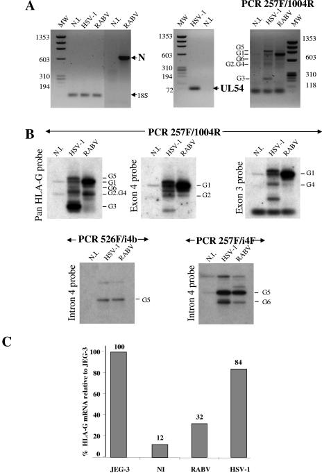
We investigated whether the transcription of HLA-G was upregulated during infection with HSV-1 or RABV by infecting NT2-N and SK-N-SH cells with RABV or HSV-1 and comparing the infected cells with mock-infected cells. RNA was isolated 24 h (RABV) or 18 h (HSV-1) after infection or mock infection. Virus infections were monitored by RT-PCR, using pairs of primers specific for the N gene of RABV, for the UL54 gene of HSV-1, and for the 18S housekeeping gene (Fig. 1A, left and middle; results shown for NT2-N cells only). HLA-G expression in neuronal cells was detected by RT-PCR, using the G.257F/1004R pan-HLA-G primer (Fig. 1A, right). Pan-HLA-G primers detected two bands of PCR products in mock-infected NT2-N cells, five bands in HSV-1-infected cells, and four bands in RABV-infected cells (Fig. 1B, left). Hybridization with exon-specific probes after amplification with appropriate primers (257F/1004R for exon 4-, exon 3-, or pan-HLA-G-specific probes; 526F/i4b or 257F/i4F for an intron 4-specific probe) made it possible to identify HLA-G1, -G2, -G3, -G4, -G5, and -G6 PCR products (Fig. 1B). The HLA-G promoter was repressed in most cells, but the expression of this gene was not totally abolished in human NT2-N neuronal derivatives, because uninfected NT2-N cells produced small amounts of the HLA-G1, -G2, and -G5 transcripts. Moreover, infection with HSV-1 or RABV stimulated the transcription of six isoforms of HLA-G. The two viruses clearly induced different patterns of mRNA production: HSV-1 upregulated mainly HLA-G5 and -G3, whereas RABV mainly upregulated HLA-G1, indicating that NT2-N cells display sophisticated patterns of HLA-G regulation and that HSV-1 and RABV infections modulate the expression of this gene in different ways.
FIG. 1.
HSV-1 and RABV upregulate HLA-G gene transcription. Human postmitotic neurons (NT2-N) and human neuroblastoma (SK-N-SH) cells were mock infected (N.I.) or infected with HSV-1 for 18 h or with RABV for 24 h. (A) HSV-1 and RABV infection by RT-PCR was monitored, using pairs of primers specific for RABV N and for the UL54 gene of HSV-1. 18S, a housekeeping gene, was used for normalization. Results are shown for NT2-N cells. HLA-G transcript levels were analyzed in mock-infected (N.I.) and in HSV-1- or RABV-infected NT2-N cells by RT-PCR, using a pan-HLA-G primer, 257F/1004R. Bands corresponding to HLA-G5, -G1, -G6, -G2/G4, and -G3 were identified. The data shown are representative of at least three independent experiments. (B) The same extracts were used for hybridization. RT-PCR was carried out with the following primer pairs: 257F/1004R, 525F/i4b, and 257F/i4F. The PCR amplification products were hybridized with pan-HLA-G, exon 3-, and exon 4-specific probes for 257F/1004R and with the intron 4-specific probe for the 526/i4b and 57F/i4F primer pairs. Hybridization confirmed that the six HLA-G isoforms were present. (C) Real-time PCR analysis was performed with JEG-3 cells, an HLA-G-positive choriocarcinoma cell line, uninfected SK-N-SH cells, and RABV- or HSV-1-infected SK-N-SH cells. Results are expressed as percentages of the 100-fold increase obtained with JEG-3 cells.
We analyzed the upregulation of HLA-G expression by RABV and HSV-1 in human SK-N-SH neuroblastoma cells, by real-time PCR with a pan-HLA-G primer (Fig. 1C). The levels of real-time PCR amplification in HSV-1- and RABV-infected SK-N-SH cells and in uninfected SK-N-SH cells were expressed as percentages of the amplification observed with JEG-3 cells, a choriocarcinoma cell line expressing HLA-G. Small amounts of HLA-G transcripts were detected in uninfected SK-N-SH cells; the levels of HLA-G transcripts were found to have increased after 24 h of RABV infection and 18 h of HSV-1 infection. HLA-G transcript levels were 32% higher in RABV-infected cells and 84% higher in HSV-1-infected cells than in uninfected SK-N-SH cells (12%).
These data indicate that, in human neuronal cells, the neuronotropic viruses HSV-1 and RABV upregulate the transcription of several HLA-G isoforms, including those encoding the main biologically active isoforms: the membrane bound HLA-G1 and the soluble HLA-G5.
RABV infection upregulates the expression of HLA-G1 and HLA-G5/G6 in neurons.
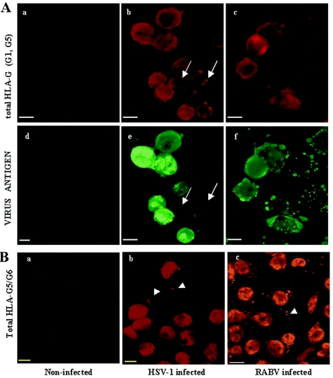
We analyzed the regulation of HLA-G protein levels in human neuronal cells following infection with RABV or HSV-1 by immunocytochemistry with the following MAbs: MEM-G/09 recognizes both the membrane-bound HLA-G1 and the soluble HLA-G5 isoforms, whereas MAb 5A6G7 recognizes the soluble HLA-G5 and HLA-G6 isoforms. HSV-1-, RABV-, and mock-infected SK-N-SH/RA cells were fixed by incubation with PFA, permeabilized, and incubated with an HLA-G-specific MAb. PFA treatment and permeabilization made it possible to detect HLA-G isoforms present in the cytoplasm, as well as those expressed on the plasma membrane. This treatment therefore made it possible to detect all HLA-G molecules (total HLA-G) present in the cells. In our case, the specificity of the two MAbs limited HLA-G detection to isoforms HLA-G1, -G5, and -G6. Under these conditions, HLA-G proteins were clearly detected in the infected cultures (Fig. 2). Both neurotropic viruses increased HLA-G1/5 levels in human neuroblastoma cells, as shown by incubation with MAb MEM-G/09 (Fig. 2Aa to Ac; compare HLA-G levels in uninfected cells [a] and in HSV-1- and RABV-infected cells [b and c]). HSV-1 and RABV upregulated HLA-G5/G6 levels in human neuroblastoma cells, as shown by incubation with MAb 5A6G7 (Fig. 2Ba to Bc; compare HLA-G levels in uninfected cells [a]and in HSV-1- and RABV-infected cells [b and c]). Reactivity to 5A6G7 differed from that to MEM-G/09, as 5A6G7 reacted with nuclei and discrete inclusions within the cytoplasm of infected cells (Fig. 2B), whereas MEM-G/09 strongly stained the cytoplasm and discrete inclusions (Fig. 2Ab and Ac) but did not stain nuclei (Fig. 2Aa). These patterns of reactivity suggest that the HLA-G1 and HLA-G5 have different distributions: HLA-G1 is probably present throughout the cytoplasm, whereas HLA-G5 is located in cytoplasmic inclusions and, possibly, in the nucleus.
FIG. 2.
HSV-1 and RABV increase HLA-G expression in human neuroblastoma and astrocytoma cells. Neuroblastoma SK-N-SH/RA cells were mock infected or infected with HSV-1 for 1 day or with RABV for 2 days. HLA-G expression was monitored by UV microscopy or flow cytometry of fixed and permeabilized cells, using MEM-G/09, a MAb specific for the HLA-G1 and HLA-G5 isoforms (A), and 5A6G7, a MAb specific for the soluble HLA-G5 and G6 isoforms (B). (A) HLA-G (a, b, and c) were detected with MEM-G/09 in uninfected (a), HSV-1-infected (b), and RABV-infected (c) SK-N-SH/RA cells. Viral antigens were detected by using virus-specific Ab in uninfected (d), HSV-1-infected (e), and RABV-infected (f) cells. Both viruses increase HLA-G levels (b and c) over those in uninfected cultures (a). In infected cultures, HLA-G is expressed in both infected (green) and uninfected (arrows) SK-N-SH/RA cells. (B) HLA-G5/G6 was detected in mock-infected (a), HSV-1-infected (b), and RABV-infected (c) SK-N-SH/RA cells. HLA-G was detected in the nuclei and in the cytoplasm. Cytoplasmic accumulation was observed only in the cells of HSV-1- and RABV-infected cultures (arrowheads). In contrast, the nuclei were stained in both uninfected (a) and infected (b and c) neurons. Bars, 10 μm.
Surface HLA-G expression is detected during RABV infection but not during HSV-1 infection.
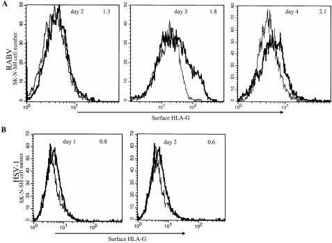
For HLA-G1 and HLA-G5 to exert immunosubversive properties, HLA-G1 must be present at the cell surface and HLA-G5 must be secreted. We therefore determined whether HLA-G was present on the surface of the infected cells or in the supernatant. Cell surface HLA-G expression was analyzed by immunocytochemistry without cell permeabilization. This restricted MAb access to the molecules exposed at the cell surface. HLA-G surface expression was compared in RABV- and HSV-1-infected and uninfected SK-N-SH and U373MG cells by flow cytometry 1, 2, 3, and 4 days after infection, using MEM-G/09 MAb (Fig. 3). In RABV-infected SK-N-SH (Fig. 3A), SFI increased from 1.3 on day 2 to 1.8 on day 3 and 2.1 on day 4. Concomitantly, the percentage of RABV-infected cells detected by flow cytometry on permeabilized cells with an RABV internal antigen-specific Ab increased from 10% on day 2 to 40% on day 3 and 45% on day 4, indicating that HLA-G surface expression increased with the progression of infection. A similar pattern of HLA-G surface expression over time was observed in RABV-infected U373MG cells (data not shown).
FIG. 3.
RABV triggers HLA-G surface expression in neuronal cells, whereas HSV-1 does not. (A) Surface HLA-G expression was detected by flow cytometry with MEM-G/09 MAb on uninfected (line) and RABV-infected (boldface line) SK-N-SH cells 2, 3, and 4 days after infection. Numbers at the top right of the panels indicate the SFI, calculated as described in Materials and Methods. On day 2, the SFI was 1.3. The percentage of cells with an SFI of >5 was 35% in infected cells and 27% in uninfected cells. On day 3, the SFI was 1.8. The percentage of cells with an SFI exceeding 5 was 26% in infected cultures and 14% in uninfected cultures. On day 4, the SFI was 2.1. The percentage of cells with an SFI of >5 was 43% in infected cultures and 20% in uninfected cultures. Percentages of infected cells were 10, 40, and 45 on days 2, 3, and 4, respectively. These kinetic analyses are representative of two independent experiments. (B) In HSV-1-infected cultures, HLA-G expression was detected by flow cytometry as described in the legend to panel A, on the surface of uninfected (line) and HSV-1-infected (boldface line) cells 1 and 2 days postinfection. On day 1, the SFI was 0.8. The percentage of cells with an SFI of >5 was 10% in infected cultures and 12% in uninfected cultures. On day 2, the SFI was 0.6. The percentage of cells with an SFI of >5 was 7% in infected cultures and 12% in uninfected cultures. These data are representative of two independent experiments.
We used the same staining protocol to analyze surface HLA-G expression in HSV-1-infected SK-N-SH cells. In striking contrast to what was observed with RABV, HSV-1 did not trigger the surface expression of HLA-G in SK-N-SH cells (Fig. 3B). SK-N-SH cells were infected (25% of cells infected on day 2 and 48% on day 3); total HLA-G was detected (Fig. 2A) as early as day 1. However, the SFI for HLA-G surface expression was 0.8 and 0.7 on days 1 and 2 postinfection, respectively (Fig. 3B). We were unable to study the kinetics of HLA-G surface expression in HSV-1-infected neuroblastoma cells for a longer period (day 3 or 4) because HSV-1-infected SK-N-SH cells ceased to be viable after 2 days due to the cytopathic effects of the virus.
Levels of HLA-G5 (a soluble isoform) in the supernatants of RABV- and HSV-1-infected cells were determined by means of an immunocapture immunoassay (ELISA/HLA-G5) designed to detect soluble HLA-G5 only. HLA-G5 was not detected in the supernatants of infected NT2-N cells, even in after a 10-fold concentration of the supernatants, suggesting that soluble HLA-G5 was not produced or was produced in amounts too small for detection in our assay conditions (detection threshold, 30 ng/ml). This test provided no reliable information on the possible presence of soluble HLA-G6 or of the secreted form HLA-G1. Although both HLA-G5 and HLA-G6 were recognized by 5A6G7, W6-32, an Ab specific for the monomorphic α chain of HLA molecules, did not detect HLA-G6 when used as a secondary antibody, because this isoform is not associated with the β2 microglobulin chain (45).
Thus, RABV triggers surface expression of the membrane-bound HLA-G1 isoform, whereas HSV-1 does not, and soluble HLA-G5 was not detected in the supernatants of infected neurons. These data suggest that RABV infection triggers the production of membrane-bound HLA-G1 and that HLA-G molecules may be sequestered in HSV-1-infected neuronal cells.
RABV infection upregulated HLA-G expression in both infected and uninfected cells in the neural culture.
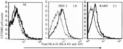
We analyzed the triggering of HLA-G synthesis by HSV-1 using MEM-G/09 MAb in astrocytoma cells, as these cells are highly susceptible to this virus. We monitored cell fluorescence by flow cytometry. As shown in Fig. 4, HSV-1 increased total HLA-G expression in U373MG cells, as in SK-N-SH cells. The SFI was 1.8 on day 1 in the HSV-1-infected culture. In this case, 20% of the cells were infected, as indicated by flow cytometry with an HSV-1 antigen-specific Ab (data not shown). These findings confirm that HSV-1 upregulates HLA-G expression in both neurons and astrocytes.
FIG. 4.
RABV infection upregulates HLA-G in astrocytoma cells despite low levels of infection. Astrocytoma U373MG cells were infected with HSV-1 or RABV or were left uninfected. HLA-G levels were determined by flow cytometry in uninfected (NI panel) and HSV-1- and RABV-infected U373MG (HSV-1 and RABV panels) cells. Panel NI corresponds to the ratio of reactivity of MEM-G/09 MAb with uninfected U373MG (boldface line) versus that of an irrelevant isotype-matched MAb (line). Panels HSV-1 and RABV correspond to the ratio of HLA-G reactivity in infected U373MG (boldface line) to that in day-matched uninfected U373MG cultures (line). The numbers given are SFIs, calculated as described in Materials and Methods. SFI values of >1.25 were considered positive. For the day 1 HSV-1 culture, the SFI = 1.8. The percentage of cells with an SFI of >5 was 79% in infected cultures and 45% in uninfected cultures. For the day 2 RABV culture, the SFI = 2.1. The percentage of cells with an SFI of >5 was 27% in infected cultures and 13% in uninfected cultures. Only 8% of the total U373MG cell population was infected with RABV. These plots are representative of three independent experiments.
Surprisingly, whereas the U373MG cell line displayed only low levels of infection with RABV (<8%), HLA-G was strongly upregulated in RABV-infected cultures, with an SFI of 2.1 for HLA-G expression in cultures infected with RABV for 2 days (Fig. 4, right).
As RABV upregulated HLA-G expression in astrocytoma cells despite the low level of infection, we investigated whether uninfected cells expressed HLA-G when in the neighborhood of RABV-infected cells. Double staining of RABV-infected U373MG cells showed HLA-G expression in 44% (72/165) of U373MG, with only 4% of these cells expressing viral antigen. This phenomenon was not restricted to astrocytoma cell cultures; it was also observed with RABV- and HSV-1 infected SK-N-SH/RA cultures (Fig. 2A). Uninfected cells may emit a strong fluorescent signal for HLA-G (Fig. 2Ab and Ac). Thus, RABV and HSV-1 infections activated HLA-G expression in both infected and neighboring uninfected cells.
We therefore conclude that, upon virus infection, both HLA-G gene transcription and total HLA-G protein synthesis are upregulated in neuronal (SK-N-SH/RA) and neural (U373MG) cells. The membrane-bound isoform HLA-G1 and the HLA-G5/G6 isoforms are included in this upregulation. The pattern of HLA-G expression observed was specific to each virus; HSV-1 upregulated mainly G5 and G3, whereas RABV upregulated mainly G1. These increases in HLA-G synthesis were all mediated by virus infection but HLA-G expression occurs was observed in both infected and neighboring uninfected cells.
HLA-G is expressed in mixed human neuron-astrocyte cultures.
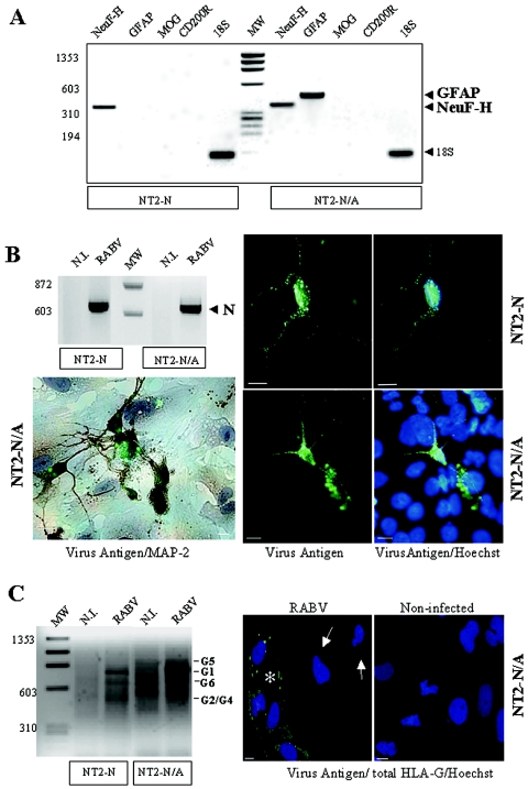
As uninfected cells expressed HLA-G in the presence of infected cells, we investigated whether HLA-G levels also increased in mixed cultures of human postmitotic neurons and astrocytes (NT2-N/A). These two types of cell differ in susceptibility to RABV: neurons are susceptible to RABV infection and astrocytes are not. This mixed culture mimics the physiological conditions of nervous system (NS) infection by this virus in vivo, as RABV primarily infects neurons, with glial cells, including astrocytes, infected poorly if at all (47). The mixed composition of the NT2-N/A cultures was checked by RT-PCR, using primers for GFAP, a marker of astrocytes, and primers for the H protein of neurofilament (NeuF-H), a marker of differentiated neurons (Fig. 5A). Whereas NT2-N cultures produced only NeuF-H transcripts, NT2-N/A cultures were found to produce both GFAP and NeuF-H transcripts, indicating that they were indeed composed of a mixture of astrocytes and/or neurons, whereas NT2-N cultures were composed of neurons only. NT2-N/A cells were as susceptible to RABV as NT2-N (Fig. 5B, top left). Nevertheless, only the cells with a neuronal morphology expressing the neuronal marker MAP-2 were infected with RABV; the astrocytes were not infected, as shown by the results for immunocytochemistry of RABV-infected NT2-N/A (Fig. 5B). Astrocytes, MAP-2-negative cells, display blue staining of the nucleus only, whereas neuron-like cells MAP-2-positive cells (brown staining) displayed green staining, indicating accumulation of the viral antigen in the cytoplasm. RABV infection upregulated HLA-G1, -G6, and -G2/G4 in NT2-N/A and NT2-N cultures (Fig. 5C, left). Nevertheless, NT2-N/A cultures produced larger amounts of HLA-G1 transcripts than NT2-N cultures. We quantified HLA-G amplicon levels in the two cultures by densitometry, with normalization to 18S transcript levels (Fig. 5C). NT2-N/A cultures contained 1.5 times as many HLA-G transcripts as pure NT2-N cultures. A transcriptome analysis with Affymetrix microarrays in RABV-infected NT-2N and RABV-infected NT2-N/A cells indicated that infection resulted in the production of three times more HLA-G transcripts in NT2-N/A cultures than in NT2-N cultures (data not shown). Thus, mixed cultures produced HLA-G more efficiently than pure cultures of neurons. We used double immunocytochemistry to check that the uninfected astrocytes in the mixed culture expressed HLA-G. We used the HLA-G-specific MAb MEM-G/09, together with an RABV Ag-specific Ab. Both uninfected astrocytes and neuron-like RABV-infected cells expressed HLA-G (Fig. 5C). This observation is therefore consistent with RABV infection triggering HLA-G expression in uninfected glial cells surrounding infected neurons. Thus, RABV may induce a local environment adverse to the control of infection by the immune response.
FIG. 5.
RABV infection increases HLA-G expression in infected neurons and in mixed cultures of human neurons and/or astrocytes. (A) Human NT2-N cultures contained only neurons, as demonstrated by detection of the high-molecular-weight neurofilament protein NeuF-H. The absence of glial cell contamination was demonstrated by the absence of GFAP transcripts, a marker of astrocytes; MOG, a marker of oligodendroyctes; and CD200R, a marker of microglia. In contrast (see the right-hand portion of the gel), NT2-N/A culture expressed both NeuF-H and GFAP transcripts, indicating that it contains both neurons and astrocytes. (B) NT2-N and NT2-N/A cultures are susceptible to RABV infection, as shown by PCR amplification of RABV N (647 bp). (Bottom, left) An RABV-infected NT2-N/A culture was doubled stained with an MAP-2-specific Ab (dark brown staining), an antiviral antigen Ab (viral Ag accumulation in green), and Hoechst stain (staining the nuclei blue). Neurons (MAP-2 positive; dark brown) were infected (green), whereas astrocytes (MAP-2 negative) were not. (Right) RABV-infected NT2-N and NT2-N/A cultures were doubled stained with Hoechst stain (blue nuclei) and antiviral antigen Ab (green). In both NT2-N (top) and NT2-N/A cultures (bottom), only neurons (typical neuron morphology) were infected (green), whereas astrocytes (large blue nuclei) were not. (C) HLA-G transcription in infected cells was compared with that in the corresponding mock-infected cells (N.I.) by RT-PCR, using G.257F/G.1004R pan HLA-G primers. RABV increased HLA-G transcript levels in infected NT2-N and NT2-N/A cells. The nature of cells expressing HLA-G in infected NT2-N/A cultures was analyzed by double immunocytochemistry, using an Ab specific for RABV antigen (green) and MEM-G/09, which is specific for the HLA-G1 and 5 isoforms. Nuclei were stained with Hoechst stain (blue). In addition to infected neurons (*), astrocytes (arrows) located close to infected neurons expressed HLA-G (red). Bars, 10 μm.
IFN-β and IFN-γ activate the expression of HLA-G in human neuron cultures and in mixed astrocyte/neuron cultures.
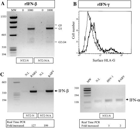
The pattern of HLA-G expression is subject to tight cell-specific transcriptional control. HLA-G expression can be stimulated by IFNs (α, β, and γ). We investigated whether IFN-β regulated HLA-G transcription in NT2-N and NT2-N/A cultures. Treatment for 24 h with recombinant IFN-β (rIFN-β; 1,000 U) increased HLA-G1 transcript levels strongly and G2/G4 and G5 transcript levels to a lesser extent in both NT2-N and NT2-N/A cultures (Fig. 6A). The presence of HLA-G transcripts was confirmed by real-time RT-PCR (data not shown). Thus, RABV infection triggers the transcription of IFN-β in NT2-N and NT2-N/A cultures, leading to an increase in HLA-G1 transcription. It is interesting to note that IFN-β treatment upregulated primarily the HLA-G1 isoform, which is the also the main HLA-G isoform triggered by RABV infection.
FIG. 6.
IFN-β, which is produced by RABV-infected cultures, increases HLA-G expression in NT2-N and NT2-N/A cultures. (A) The treatment of uninfected NT2-N or NT2-N/A cultures with 1,000 IU of rIFN-β stimulated HLA-G transcription in both cell types, as shown by PCR using the pan-HLA-G 257F/1004R primers. (B) The treatment of uninfected SK-N-SH with 500 IU of rIFN-β stimulated HLA-G surface expression as detected by cytofluorometry. The boldface line indicates HLA-G surface expression in IFN-γ-treated cells. The lighter line is used for untreated cells. (C, left) RABV infection increased IFN-β transcription in NT2-N and NT2-N/A cultures, as shown by PCR and real-time PCR (IFN-β expression levels were 121 and 199 times higher than that in uninfected NT2-N and NT2-N/A, respectively). (Right) RABV and HSV-1 infection triggered a modest increase of IFN-α5 transcription in NT2-N culture, as shown by PCR and real-time PCR. Real-time PCR results are given as relative fold increase compared to noninfected cultures (value, 1).
Treatment for 48 h with recombinant IFN-γ (500 IU) increased HLA-G expression at the surface of human neuroblastoma cells SK-N-SH as shown by cytofluorometry (Fig. 6B).
Ability of RABV-infected NT2-N and NT2-N/A cultures to express IFN-β transcripts was analyzed by RT-PCR and real-time PCR (Fig. 6C, left). RABV infection strongly increased IFN-β mRNA production in both NT2-N and NT2-N/A cultures. Real-time PCR analysis indicated that IFN-β transcript levels increased more strongly in RABV-infected NT2-N/A cultures (199 times) than in RABV-infected NT2-N cultures (127 times).
Production of IFN-α and IFN-γ by RABV- or HSV-1-infected NT2-N was also analyzed by RT-PCR and real-time PCR. In contrast to IFN-β, only limited amounts of IFN-α transcripts (Fig. 6C, right) and none for IFN-γ (data not shown) could be detected in the cultures.
These data indicate that expression of HLA-G in human neurons can be upregulated by IFN-β and IFN-γ. IFN-β, the expression of which was strongly increased by RABV infection, possibly plays a role in HLA-G up regulation.
DISCUSSION
HLA-G, which has a restricted tissue distribution (7, 9), was first detected on extravillous cytotrophoblast cells (27). HLA-G has also been observed in healthy brain and eye (29, 35). This study identifies neurons and astrocytes as cells that can express HLA-G on viral infection or in the presence of IFN-β. It also demonstrates that two neuronotropic viruses, RABV and HSV-1, upregulate HLA-G expression in human postmitotic neurons (NT2-N), which are derivatives of human adult neurons. These cells were also found to produce HLA-G transcripts, following exposure to IFN-β. We found that HLA-G1 was preferentially expressed in response to RABV infection or IFN-β treatment, whereas HLA-G5 and -G3 were upregulated in response to HSV-1 infection.
Both RABV and HSV-1 activate HLA-G gene expression in human neurons. IFNs, which modulate HLA-G gene activation, may play a role in this control because HLA-G transcription was upregulated in human NT2-N and NT2-N/A cultures in the presence of IFN-β, and IFN-β was produced by these cells following RABV infection (this study and reference 58). In this hypothesis, the control of HLA-G production by IFN-β would account for the expression of HLA-G by astrocytes in cocultures in which only the neuronal cells were infected. Thus, following the infection of neurons and secretion of IFN-β in the nervous system, cells of the nervous system parenchyma, including astrocytes, could produce HLA-G and establish a local immunosubversive environment. IFN-β is certainly not the only soluble factor involved in HLA-G regulation, as the virus blocks IFN-β production in HSV-1-infected cells, including neurons (20, 58). A role for IFN-α or IFN-γ in HLA-G regulation is unlikely, since only limited or no expression of the two cytokines was detected under our culture conditions. Demonstration of IFN-β in HLA-G regulation by human neuronal cells would require further investigation, including loss-of-function analyses.
The two viruses studied also generate different HLA-G isoform profiles, with RABV inducing mostly HLA-G1 and with HSV-1 inducing mostly HLA-G3 and HLA-G5. Both IFN-β and RABV infections, which induce IFN-β production, triggered the preferential upregulation of the HLA-G1 isoform. In contrast, HSV-1, which blocks IFN-β production, mostly upregulated the HLA-G5 isoform. This suggests that IFN-β and RABV favor the splicing of the HLA-G1 isoform, whereas in the absence of IFN-β and in the presence of unidentified conditions associated with HSV-1 infection, splicing of the HLA-G5 isoform is favored. Another herpesvirus, human cytomegalovirus, does not block IFN production and triggers the production of both HLA-G1 and HLA-G5 by infected alveolar macrophages (49). This finding supports the hypothesis of the involvement of IFN-β in HLA-G1 expression and of an unidentified condition common to the two herpesviruses in the control of HLA-G5 expression. Our observations indicate that RABV and HSV-1 infections can reverse repression of HLA-G and, moreover, differentially regulate the alternative splicing of HLA-G isoforms. Thus, HSV-1 and RABV constitute interesting tools to further analyze the regulation of the HLA-G gene in neurons.
The percentage of cells expressing HLA-G in culture was limited: immunofluorescence analysis indicated that no more than 20% of cells expressed HLA-G. However, these percentages are consistent with virus-mediated HLA-G expression having a physiological role, as demonstrated in another study in which HLA-G1 expression in only about 10% of glioma cells was sufficient to abolish an effective antitumoral response (70). The reasons for this high efficiency are unclear. HLA-G may have an unusually long lifetime at the cell surface, possibly due to the lack of an endocytosis motif in the cytoplasmic domain (16, 52).
Despite the efficient transcription and translation of HLA-G in neuronal cells, HLA-G molecules were not detected at the surface of HSV-1-infected neuronal cells. This may be due to the different nature of HLA-G isoforms triggered by the two viruses, HLA-G1 (RABV) and HLA-G5/G3 (HSV-1). We would therefore have expected to detect HLA-G5 in the cell supernatants. However, HLA-G5-specific ELISA did not detect soluble HLA-G5 in the supernatants of HSV-1-infected cultures, suggesting that HLA-G5 was not secreted or was secreted in amounts too small to be detected. Alternatively, the absence of HLA-G at the cell membrane may result from the blockage of HLA-G trafficking by HSV-1 itself. HSV-1 produces an immediate early protein, ICP47, known to retain both the classical HLA and the nonclassical HLA-G1 heavy chain in the cell reticulum. ICP47 blocks the TAP transporter and therefore the translocation of the peptide into the reticulum (17, 64). HLA-G1 and HLA-G5, like classical HLA molecules, are loaded with a peptide by both TAP-dependent and TAP-independent pathways (30). Impairment of the peptide loading of HLA molecules leads to the rapid degradation of HLA molecules in the reticulum (26, 52). Similar mechanisms may occur in HSV-1-infected human neuronal cells, accounting for our results.
The NS, unlike other organs, has intrinsic mechanisms for controlling its immune response, described as immune privilege (41). Only the eyes, muscles, and testes share this property. Immune privilege is thought to protect the NS against irreversible damage induced by inflammatory or cytotoxic responses. The constitutive production of immunosubversive molecules, such as FasL, by the eye and testis is thought to be a key component of the maintenance of immune privilege in these tissues, as it protects neurons against cytotoxic T cells (21, 22, 38, 48). Functional studies have identified HLA-G as an important mediator of immune tolerance (7, 9, 15, 53, 62, 70, 71). It has also been suggested that HLA-G is involved in immune privilege in the muscles and eyes (29, 73). In this study, RABV was found to upregulate membrane-bound HLA-G1 molecules, thereby potentially impeding host antiviral responses based on T and NK cells. We suggest that HLA-G1 expression on the surface of neural cells during RABV infection protects infected cells against T and NK surveillance, contributing to the establishment of a local subversive environment adverse to the host immune reaction. Thus, HLA-G and FasL may help to maintain NS immune privilege. In contrast, HSV-1 does not seem to use such a mechanism to escape the immune response completely. Defects in surface HLA-G expression or expression at too low a density may maintain immune pressure, leading to latency. Nevertheless, humans experience HSV-1 reactivation when they are stressed or immunocompromised, for example, due to corticosteroid treatment. Stress inhibits the T-cell response to HSV-1 (2), and corticosteroids activate HLA-G transcription (24, 43). This raises questions about the role of HLA-G in HSV-1 reactivation. Future work should focus on whether stress and corticosteroids increase HLA-G transcription enough in cells latently infected with HSV-1 to inhibit the T-cell response.
Acknowledgments
We thank Celine Marcou for excellent technical assistance. We also thank Cécile Pangault (Université de Rennes) for the RT-PCR protocol, Anne Plonquet (Hôpital Henri Mondor, Créteil) for Ntera-2clD/1 cells, Susan Michelson (Institut Pasteur) for HSV-1, and Alain Créange (Hôpital Henri Mondor, Créteil) for Betaferon.
REFERENCES
- 1.Alcami, A., and U. H. Koszinowski. 2000. Viral mechanisms of immune evasion. Trends Microbiol. 8:410-418. [DOI] [PMC free article] [PubMed] [Google Scholar]
- 2.Anglen, C. S., M. E. Truckenmiller, T. D. Schell, and R. H. Bonneau. 2003. The dual role of CD8+ T lymphocytes in the development of stress-induced herpes simplex encephalitis. J. Neuroimmunol. 140:13-27. [DOI] [PubMed] [Google Scholar]
- 3.Baloul, L., S. Camelo, and M. Lafon. 2004. Up-regulation of Fas ligand (FasL) in the central nervous system: a mechanism of immune evasion by rabies virus. J. Neurovirol. 10:372-382. [DOI] [PubMed] [Google Scholar]
- 4.Becher, B., A. Prat, and J. P. Antel. 2000. Brain-immune connection: immuno-regulatory properties of CNS-resident cells. Glia 29:293-304. [PubMed] [Google Scholar]
- 5.Cabello, A., A. Rivero, M. J. Garcia, J. M. Lozano, J. Torre-Cisneros, R. Gonzalez, G. Duenas, M. D. Galiani, A. Camacho, M. Santamaria, R. Solana, C. Montero, J. M. Kindelan, and J. Pena. 2003. HAART induces the expression of HLA-G on peripheral monocytes in HIV-1 infected individuals. Hum. Immunol. 64:1045-1049. [DOI] [PubMed] [Google Scholar]
- 6.Camelo, S., M. Lafage, and M. Lafon. 2000. Absence of the p55 Kd TNF-alpha receptor promotes survival in rabies virus acute encephalitis. J. Neurovirol. 6:507-518. [DOI] [PubMed] [Google Scholar]
- 7.Carosella, E. D., P. Moreau, S. Aractingi, and N. Rouas-Freiss. 2001. HLA-G: a shield against inflammatory aggression. Trends Immunol. 22:553-555. [DOI] [PubMed] [Google Scholar]
- 8.Carosella, E. D., P. Moreau, J. Le Maoult, M. Le Discorde, J. Dausset, and N. Rouas-Freiss. 2003. HLA-G molecules: from maternal-fetal tolerance to tissue acceptance. Adv. Immunol. 81:199-252. [DOI] [PubMed] [Google Scholar]
- 9.Carosella, E. D., P. Paul, P. Moreau, and N. Rouas-Freiss. 2000. HLA-G and HLA-E: fundamental and pathophysiological aspects. Immunol. Today 21:532-534. [PubMed] [Google Scholar]
- 10.Cheung, W. M., W. Y. Fu, W. S. Hui, and N. Y. Ip. 1999. Production of human CNS neurons from embryonal carcinoma cells using a cell aggregation method. BioTechniques 26:946-948, 950-952, 954. [DOI] [PubMed] [Google Scholar]
- 11.Chu, W., Y. Yang, D. E. Geraghty, and J. S. Hunt. 1999. Interferons enhance HLA-G mRNA and protein in transfected mouse fibroblasts. J. Reprod. Immunol. 42:1-15. [DOI] [PubMed] [Google Scholar]
- 12.Colonna, M., F. Navarro, T. Bellon, M. Llano, P. Garcia, J. Samaridis, L. Angman, M. Cella, and M. Lopez-Botet. 1997. A common inhibitory receptor for major histocompatibility complex class I molecules on human lymphoid and myelomonocytic cells. J. Exp. Med. 186:1809-1818. [DOI] [PMC free article] [PubMed] [Google Scholar]
- 13.Contini, P., M. Ghio, A. Poggi, G. Filaci, F. Indiveri, S. Ferrone, and F. Puppo. 2003. Soluble HLA-A,-B,-C and -G molecules induce apoptosis in T and NK CD8+ cells and inhibit cytotoxic T cell activity through CD8 ligation. Eur. J. Immunol. 33:125-134. [DOI] [PubMed] [Google Scholar]
- 14.Cosman, D., N. Fanger, L. Borges, M. Kubin, W. Chin, L. Peterson, and M. L. Hsu. 1997. A novel immunoglobulin superfamily receptor for cellular and viral MHC class I molecules. Immunity 7:273-282. [DOI] [PubMed] [Google Scholar]
- 15.Creput, C., A. Durrbach, C. Menier, C. Guettier, D. Samuel, J. Dausset, B. Charpentier, E. D. Carosella, and N. Rouas-Freiss. 2003. Human leukocyte antigen-G (HLA-G) expression in biliary epithelial cells is associated with allograft acceptance in liver-kidney transplantation. J. Hepatol. 39:587-594. [DOI] [PubMed] [Google Scholar]
- 16.Davis, D. M., H. T. Reyburn, L. Pazmany, I. Chiu, O. Mandelboim, and J. L. Strominger. 1997. Impaired spontaneous endocytosis of HLA-G. Eur. J. Immunol. 27:2714-2719. [DOI] [PubMed] [Google Scholar]
- 17.Easterfield, A. J., B. M. Austen, and O. M. Westwood. 2001. Inhibition of antigen transport by expression of infected cell peptide 47 (ICP47) prevents cell surface expression of HLA in choriocarcinoma cell lines. J. Reprod. Immunol. 50:19-40. [DOI] [PubMed] [Google Scholar]
- 18.Fournel, S., M. Aguerre-Girr, X. Huc, F. Lenfant, A. Alam, A. Toubert, A. Bensussan, and P. Le Bouteiller. 2000. Cutting edge: soluble HLA-G1 triggers CD95/CD95 ligand-mediated apoptosis in activated CD8+ cells by interacting with CD8. J. Immunol. 164:6100-6104. [DOI] [PubMed] [Google Scholar]
- 19.Fournel, S., X. Huc, M. Aguerre-Girr, C. Solier, M. Legros, C. Praud-Brethenou, M. Moussa, G. Chaouat, A. Berrebi, A. Bensussan, F. Lenfant, and P. Le Bouteiller. 2000. Comparative reactivity of different HLA-G monoclonal antibodies to soluble HLA-G molecules. Tissue Antigens 55:510-518. [DOI] [PubMed] [Google Scholar]
- 20.Goodbourn, S., L. Didcock, and R. E. Randall. 2000. Interferons: cell signalling, immune modulation, antiviral response and virus countermeasures. J. Gen. Virol. 81:2341-2364. [DOI] [PubMed] [Google Scholar]
- 21.Green, D. R., and T. A. Ferguson. 2001. The role of Fas ligand in immune privilege. Nat. Rev. Mol. Cell Biol. 2:917-924. [DOI] [PubMed] [Google Scholar]
- 22.Griffith, T. S., T. Brunner, S. M. Fletcher, D. R. Green, and T. A. Ferguson. 1995. Fas ligand-induced apoptosis as a mechanism of immune privilege. Science 270:1189-1192. [DOI] [PubMed] [Google Scholar]
- 23.Hill, A., P. Jugovic, I. York, G. Russ, J. Bennink, J. Yewdell, H. Ploegh, and D. Johnson. 1995. Herpes simplex virus turns off the TAP to evade host immunity. Nature 375:411-415. [DOI] [PubMed] [Google Scholar]
- 24.Ibrahim, E. C., M. Morange, J. Dausset, E. D. Carosella, and P. Paul. 2000. Heat shock and arsenite induce expression of the nonclassical class I histocompatibility HLA-G gene in tumor cell lines. Cell Stress Chaperones 5:207-218. [DOI] [PMC free article] [PubMed] [Google Scholar]
- 25.Jackson, A. C. 2003. Rabies virus infection: an update. J. Neurovirol. 9:253-258. [DOI] [PubMed] [Google Scholar]
- 26.Jun, Y., E. Kim, M. Jin, H. C. Sung, H. Han, D. E. Geraghty, and K. Ahn. 2000. Human cytomegalovirus gene products US3 and US6 down-regulate trophoblast class I MHC molecules. J. Immunol. 164:805-811. [DOI] [PubMed] [Google Scholar]
- 27.Kovats, S., E. K. Main, C. Librach, M. Stubblebine, S. J. Fisher, and R. DeMars. 1990. A class I antigen, HLA-G, expressed in human trophoblasts. Science 248:220-223. [DOI] [PubMed] [Google Scholar]
- 28.Lafon, M. 2004. Subversive neuroinvasive strategy of rabies virus. Arch. Virol. Suppl. 2004:149-159. [DOI] [PubMed] [Google Scholar]
- 29.Le Discorde, M., P. Moreau, P. Sabatier, J. M. Legeais, and E. D. Carosella. 2003. Expression of HLA-G in human cornea, an immune-privileged tissue. Hum. Immunol. 64:1039-1044. [DOI] [PubMed] [Google Scholar]
- 30.Lee, N., A. R. Malacko, A. Ishitani, M. C. Chen, J. Bajorath, H. Marquardt, and D. E. Geraghty. 1995. The membrane-bound and soluble forms of HLA-G bind identical sets of endogenous peptides but differ with respect to TAP association. Immunity 3:591-600. [DOI] [PubMed] [Google Scholar]
- 31.Lefebvre, S., S. Berrih-Aknin, F. Adrian, P. Moreau, S. Poea, L. Gourand, J. Dausset, E. D. Carosella, and P. Paul. 2001. A specific interferon (IFN)-stimulated response element of the distal HLA-G promoter binds IFN-regulatory factor 1 and mediates enhancement of this nonclassical class I gene by IFN-beta. J. Biol. Chem. 276:6133-6139. [DOI] [PubMed] [Google Scholar]
- 32.Lefebvre, S., P. Moreau, V. Guiard, E. C. Ibrahim, F. Adrian-Cabestre, C. Menier, J. Dausset, E. D. Carosella, and P. Paul. 1999. Molecular mechanisms controlling constitutive and IFN-gamma-inducible HLA-G expression in various cell types. J. Reprod. Immunol. 43:213-224. [DOI] [PubMed] [Google Scholar]
- 33.Le Rond, S., J. Le Maoult, C. Creput, C. Menier, M. Deschamps, G. Le Friec, L. Amiot, A. Durrbach, J. Dausset, E. D. Carosella, and N. Rouas-Freiss. 2004. Alloreactive CD4+ and CD8+ T cells express the immunotolerant HLA-G molecule in mixed lymphocyte reactions: in vivo implications in transplanted patients. Eur. J. Immunol. 34:649-660. [DOI] [PubMed] [Google Scholar]
- 34.Lozano, J. M., R. Gonzalez, J. M. Kindelan, N. Rouas-Freiss, R. Caballos, J. Dausset, E. D. Carosella, and J. Pena. 2002. Monocytes and T lymphocytes in HIV-1-positive patients express HLA-G molecule. AIDS 16:347-351. [DOI] [PubMed] [Google Scholar]
- 35.Maier, S., D. E. Geraghty, and E. H. Weiss. 1999. Expression and regulation of HLA-G in human glioma cell lines. Transplant Proc. 31:1849-1853. [DOI] [PubMed] [Google Scholar]
- 36.Malmberg, K. J., V. Levitsky, H. Norell, C. T. de Matos, M. Carlsten, K. Schedvins, H. Rabbani, A. Moretta, K. Soderstrom, J. Levitskaya, and R. Kiessling. 2002. IFN-gamma protects short-term ovarian carcinoma cell lines from CTL lysis via a CD94/NKG2A-dependent mechanism. J. Clin. Investig. 110:1515-1523. [DOI] [PMC free article] [PubMed] [Google Scholar]
- 37.McIntire, R. H., P. J. Morales, M. G. Petroff, M. Colonna, and J. S. Hunt. 2004. Recombinant HLA-G5 and -G6 drive U937 myelomonocytic cell production of TGF-β1. J. Leukoc. Biol. 76:1220-1228. [DOI] [PubMed] [Google Scholar]
- 38.Medana, I., Z. Li, A. Flugel, J. Tschopp, H. Wekerle, and H. Neumann. 2001. Fas ligand (CD95L) protects neurons against perforin-mediated T lymphocyte cytotoxicity. J. Immunol. 167:674-681. [DOI] [PubMed] [Google Scholar]
- 39.Menier, C., B. Saez, V. Horejsi, S. Martinozzi, I. Krawice-Radanne, S. Bruel, C. Le Danff, M. Reboul, I. Hilgert, M. Rabreau, M. L. Larrad, M. Pla, E. D. Carosella, and N. Rouas-Freiss. 2003. Characterization of monoclonal antibodies recognizing HLA-G or HLA-E: new tools to analyze the expression of nonclassical HLA class I molecules. Hum. Immunol. 64:315-326. [DOI] [PubMed] [Google Scholar]
- 40.Michelson, S., P. Dal Monte, D. Zipeto, B. Bodaghi, L. Laurent, E. Oberlin, F. Arenzana-Seisdedos, J. L. Virelizier, and M. P. Landini. 1997. Modulation of RANTES production by human cytomegalovirus infection of fibroblasts. J. Virol. 71:6495-6500. [DOI] [PMC free article] [PubMed] [Google Scholar]
- 41.Miller, D. W. 1999. Immunobiology of the blood-brain barrier. J. Neurovirol. 5:570-578. [DOI] [PubMed] [Google Scholar]
- 42.Moreau, P., F. Adrian-Cabestre, C. Menier, V. Guiard, L. Gourand, J. Dausset, E. D. Carosella, and P. Paul. 1999. IL-10 selectively induces HLA-G expression in human trophoblasts and monocytes. Int. Immunol. 11:803-811. [DOI] [PubMed] [Google Scholar]
- 43.Moreau, P., O. Faure, S. Lefebvre, E. C. Ibrahim, M. O'Brien, L. Gourand, J. Dausset, E. D. Carosella, and P. Paul. 2001. Glucocorticoid hormones upregulate levels of HLA-G transcripts in trophoblasts. Transplant Proc. 33:2277-2280. [DOI] [PubMed] [Google Scholar]
- 44.Moreau, P., G. Mouillot, P. Rousseau, C. Marcou, J. Dausset, and E. D. Carosella. 2003. HLA-G gene repression is reversed by demethylation. Proc. Natl. Acad. Sci. USA 100:1191-1196. [DOI] [PMC free article] [PubMed] [Google Scholar]
- 45.Moreau, P., P. Rousseau, N. Rouas-Freiss, M. Le Discorde, J. Dausset, and E. D. Carosella. 2002. HLA-G protein processing and transport to the cell surface. Cell Mol. Life Sci. 59:1460-1466. [DOI] [PMC free article] [PubMed] [Google Scholar]
- 46.Mouillot, G., C. Marcou, P. Rousseau, N. Rouas-Freiss, E. D. Carosella, and P. Moreau. 2005. HLA-G gene activation in tumor cells involves cis-acting epigenetic changes. Int. J. Cancer 113:928-936. [DOI] [PubMed] [Google Scholar]
- 47.Murphy, F. A. 1977. Rabies pathogenesis. Arch. Virol. 54:279-297. [DOI] [PubMed] [Google Scholar]
- 48.O'Connell, J., A. Houston, M. W. Bennett, G. C. O'Sullivan, and F. Shanahan. 2001. Immune privilege or inflammation? Insights into the Fas ligand enigma. Nat. Med. 7:271-274. [DOI] [PubMed] [Google Scholar]
- 49.Onno, M., C. Pangault, G. Le Friec, V. Guilloux, P. Andre, and R. Fauchet. 2000. Modulation of HLA-G antigens expression by human cytomegalovirus: specific induction in activated macrophages harboring human cytomegalovirus infection. J. Immunol. 164:6426-6434. [DOI] [PubMed] [Google Scholar]
- 50.Pangault, C., G. Le Friec, S. Caulet-Maugendre, H. Lena, L. Amiot, V. Guilloux, M. Onno, and R. Fauchet. 2002. Lung macrophages and dendritic cells express HLA-G molecules in pulmonary diseases. Hum. Immunol. 63:83-90. [DOI] [PubMed] [Google Scholar]
- 51.Paquet-Durand, F., S. Tan, and G. Bicker. 2003. Turning teratocarcinoma cells into neurons: rapid differentiation of NT-2 cells in floating spheres. Brain Res. Dev. Brain Res. 142:161-167. [DOI] [PubMed] [Google Scholar]
- 52.Park, B., S. Lee, E. Kim, S. Chang, M. Jin, and K. Ahn. 2001. The truncated cytoplasmic tail of HLA-G serves a quality-control function in post-ER compartments. Immunity 15:213-224. [DOI] [PubMed] [Google Scholar]
- 53.Paul, P., N. Rouas-Freiss, I. Khalil-Daher, P. Moreau, B. Riteau, F. A. Le Gal, M. F. Avril, J. Dausset, J. G. Guillet, and E. D. Carosella. 1998. HLA-G expression in melanoma: a way for tumor cells to escape from immunosurveillance. Proc. Natl. Acad. Sci. USA 95:4510-4515. [DOI] [PMC free article] [PubMed] [Google Scholar]
- 54.Paul, P., N. Rouas-Freiss, P. Moreau, F. A. Cabestre, C. Menier, I. Khalil-Daher, C. Pangault, M. Onno, R. Fauchet, J. Martinez-Laso, P. Morales, A. A. Villena, P. Giacomini, P. G. Natali, G. Frumento, G. B. Ferrara, M. McMaster, S. Fisher, D. Schust, S. Ferrone, J. Dausset, D. Geraghty, and E. D. Carosella. 2000. HLA-G, -E, -F preworkshop: tools and protocols for analysis of non-classical class I genes transcription and protein expression. Hum. Immunol. 61:1177-1195. [DOI] [PubMed] [Google Scholar]
- 55.Pleasure, S. J., C. Page, and V. M. Lee. 1992. Pure, postmitotic, polarized human neurons derived from NTera 2 cells provide a system for expressing exogenous proteins in terminally differentiated neurons. J. Neurosci. 12:1802-1815. [DOI] [PMC free article] [PubMed] [Google Scholar]
- 56.Ponte, M., C. Cantoni, R. Biassoni, A. Tradori-Cappai, G. Bentivoglio, C. Vitale, S. Bertone, A. Moretta, L. Moretta, and M. C. Mingari. 1999. Inhibitory receptors sensing HLA-G1 molecules in pregnancy: decidua-associated natural killer cells express LIR-1 and CD94/NKG2A and acquire p49, an HLA-G1-specific receptor. Proc. Natl. Acad. Sci. USA 96:5674-5679. [DOI] [PMC free article] [PubMed] [Google Scholar]
- 57.Prehaud, C., S. Lay, B. Dietzschold, and M. Lafon. 2003. Glycoprotein of nonpathogenic rabies viruses is a key determinant of human cell apoptosis. J. Virol. 77:10537-10547. [DOI] [PMC free article] [PubMed] [Google Scholar]
- 58.Préhaud, C., F. Mégret, M. Lafage, and M. Lafon. 2005. Virus infection switches TLR-3-positive human neurons to become strong producers of beta interferon. J. Virol. 79:12893-12904. [DOI] [PMC free article] [PubMed] [Google Scholar]
- 59.Rajagopalan, S., and E. O. Long. 1999. A human histocompatibility leukocyte antigen (HLA)-G-specific receptor expressed on all natural killer cells. J. Exp. Med. 189:1093-1100. [DOI] [PMC free article] [PubMed] [Google Scholar]
- 60.Rebmann, V., A. Busemann, M. Lindemann, and H. Grosse-Wilde. 2003. Detection of HLA-G5 secreting cells. Hum. Immunol. 64:1017-1024. [DOI] [PubMed] [Google Scholar]
- 61.Ristich, V., S. Liang, W. Zhang, J. Wu, and A. Horuzsko. 2005. Tolerization of dendritic cells by HLA-G. Eur. J. Immunol. 35:1133-1142. [DOI] [PubMed] [Google Scholar]
- 62.Rouas-Freiss, N., P. Moreau, C. Menier, and E. D. Carosella. 2003. HLA-G in cancer: a way to turn off the immune system. Semin. Cancer Biol. 13:325-336. [DOI] [PubMed] [Google Scholar]
- 63.Sandhu, J. K., M. Sikorska, and P. R. Walker. 2002. Characterization of astrocytes derived from human NTera-2/D1 embryonal carcinoma cells. J. Neurosci. Res. 68:604-614. [DOI] [PubMed] [Google Scholar]
- 64.Schust, D. J., A. B. Hill, and H. L. Ploegh. 1996. Herpes simplex virus blocks intracellular transport of HLA-G in placentally derived human cells. J. Immunol. 157:3375-3380. [PubMed] [Google Scholar]
- 65.Shiroishi, M., K. Tsumoto, K. Amano, Y. Shirakihara, M. Colonna, V. M. Braud, D. S. Allan, A. Makadzange, S. Rowland-Jones, B. Willcox, E. Y. Jones, P. A. van der Merwe, I. Kumagai, and K. Maenaka. 2003. Human inhibitory receptors Ig-like transcript 2 (ILT2) and ILT4 compete with CD8 for MHC class I binding and bind preferentially to HLA-G. Proc. Natl. Acad. Sci. USA 100:8856-8861. [DOI] [PMC free article] [PubMed] [Google Scholar]
- 66.Solier, C., V. Mallet, F. Lenfant, A. Bertrand, A. Huchenq, and P. Le Bouteiller. 2001. HLA-G unique promoter region: functional implications. Immunogenetics 53:617-625. [DOI] [PubMed] [Google Scholar]
- 67.Thoulouze, M. I., M. Lafage, J. A. Montano-Hirose, and M. Lafon. 1997. Rabies virus infects mouse and human lymphocytes and induces apoptosis. J. Virol. 71:7372-7380. [DOI] [PMC free article] [PubMed] [Google Scholar]
- 68.Wainwright, L. J., A. Lasorella, and A. Iavarone. 2001. Distinct mechanisms of cell cycle arrest control the decision between differentiation and senescence in human neuroblastoma cells. Proc. Natl. Acad. Sci. USA 98:9396-9400. [DOI] [PMC free article] [PubMed] [Google Scholar]
- 69.Wiendl, H., L. Behrens, S. Maier, M. A. Johnson, E. H. Weiss, and R. Hohlfeld. 2000. Muscle fibers in inflammatory myopathies and cultured myoblasts express the nonclassical major histocompatibility antigen HLA-G. Ann. Neurol. 48:679-684. [PubMed] [Google Scholar]
- 70.Wiendl, H., M. Mitsdoerffer, V. Hofmeister, J. Wischhusen, A. Bornemann, R. Meyermann, E. H. Weiss, A. Melms, and M. Weller. 2002. A functional role of HLA-G expression in human gliomas: an alternative strategy of immune escape. J. Immunol. 168:4772-4780. [DOI] [PubMed] [Google Scholar]
- 71.Wiendl, H., M. Mitsdoerffer, V. Hofmeister, J. Wischhusen, E. H. Weiss, J. Dichgans, H. Lochmuller, R. Hohlfeld, A. Melms, and M. Weller. 2003. The non-classical MHC molecule HLA-G protects human muscle cells from immune-mediated lysis: implications for myoblast transplantation and gene therapy. Brain 126:176-185. [DOI] [PubMed] [Google Scholar]
- 72.Wiendl, H., M. Mitsdoerffer, and M. Weller. 2003. Express and protect yourself: the potential role of HLA-G on muscle cells and in inflammatory myopathies. Hum. Immunol. 64:1050-1056. [DOI] [PubMed] [Google Scholar]
- 73.Wiendl, H., M. Mitsdoerffer, and M. Weller. 2003. Hide-and-seek in the brain: a role for HLA-G mediating immune privilege for glioma cells. Semin. Cancer Biol. 13:343-351. [DOI] [PubMed] [Google Scholar]
- 74.Yang, Y., W. Chu, D. E. Geraghty, and J. S. Hunt. 1996. Expression of HLA-G in human mononuclear phagocytes and selective induction by IFN-gamma. J. Immunol. 156:4224-4231. [PubMed] [Google Scholar]


