Abstract
Maedi-visna virus (MVV) is a lentivirus of sheep sharing several key features with the primate lentiviruses. The virus causes slowly progressive diseases, mainly in the lungs and the central nervous system of sheep. Here, we investigate the molecular basis for the differential growth phenotypes of two MVV isolates. One of the isolates, KV1772, replicates well in a number of cell lines and is highly pathogenic in sheep. The second isolate, KS1, no longer grows on macrophages or causes disease. The two virus isolates differ by 129 nucleotide substitutions and two deletions of 3 and 15 nucleotides in the env gene. To determine the molecular nature of the lesions responsible for the restrictive growth phenotype, chimeric viruses were constructed and used to map the phenotype. An L120R mutation in the CA domain, together with a P205S mutation in Vif (but neither alone), could fully convert KV1772 to the restrictive growth phenotype. These results suggest a functional interaction between CA and Vif in MVV replication, a property that may relate to the innate antiretroviral defense mechanisms in sheep.
Maedi-visna virus (MVV) is a member of the lentivirus subfamily of retroviruses, causing encephalitis (visna), pneumonia (maedi), mastitis, and arthritis in sheep (26, 27). The primary target cells of MVV infection are cells of the monocyte/macrophage lineage, and virus expression is activated upon macrophage maturation (6, 11, 22). However, various cell types are permissive for infection by certain MVV strains in vitro. Lentiviruses in general exhibit a highly restricted cell and species tropism. This restriction is largely determined by host factors that either are required for virus replication or have antiviral activity.
Recently, two classes of lentiviral inhibitors were identified. One class is exemplified by APOBEC3G, a cytosine deaminase that induces C-to-U lesions in the viral cDNA minus strand during reverse transcription. This leads either to plus-strand G→A hypermutation in integrated virus DNA (C→T on the minus strand) or possibly also to the degradation of the retroviral cDNA prior to integration (14, 19, 21, 38). The vif gene, which is present in all lentiviruses except equine infectious anemia virus, counteracts APOBEC3G by causing its degradation. Vif is essential for virus infectivity in vivo and for replication in primary cells in vitro (5, 12, 13, 18, 20, 31).
We have shown that MVV lacking Vif is noninfectious in sheep and that replication is cell type dependent. Thus, Vif-deficient MVV replicates poorly in macrophages and sheep choroid plexus (SCP) cells, whereas fetal ovine synovial (FOS) cells appear semipermissive. We have also found an elevated frequency of G→A mutations in Vif-deficient MVV, indicating a function for MVV Vif that is similar to that of human immunodeficency virus type 1 (HIV-1) (18).
The capsid of retroviruses has been shown to be a target for cellular restriction factors in various virus and cell systems. The first of these to be characterized was Fv1, a gene product of an endogenous retrovirus in the mouse genome that blocks certain strains of Friend leukemia virus. The amino acid sequence of the capsid determines whether the virus is sensitive to Fv1 or not (4). A host factor, Lv1, restricting HIV-1 in many monkey cells has recently been identified as tripartite motif protein 5α (TRIM5α), a protein of unknown function (32). A similar restriction has been found in human cells that are resistant to certain murine leukemia virus strains, and the capsid is targeted here as well (15, 35). The restriction factor, Ref1, has also been shown to be a TRIM5α variant (17).
In this study, we examined the different cell tropism and pathogenicity of two molecular clones of MVV (2, 33). One of the clones (KS1) replicates well in SCP cells, but replicates only to low titers in macrophages, and is nonpathogenic in sheep. The other clone (KV1772) replicates to high titers in SCP cells and macrophages and is highly pathogenic in sheep. Both clones have their origin in a transmission experiment where virus was passaged serially through tissue culture and sheep (29), and they differ by 129 nucleotides. By constructing recombinant viruses and a series of amino acid substitution mutations, we mapped the attenuated phenotype of KS1 to two point mutations, one in the CA domain of gag and the other in vif.
MATERIALS AND METHODS
Virus and cells.
The molecularly cloned viruses KV1772 (formerly KV1772kv72/67) and KS1 (formerly LV1-1KS1) have been described previously (2, 29, 30). SCP cells established as described previously (7, 28) were grown at 37°C in a humidified atmosphere of 5% CO2 in Dulbecco's modified Eagle medium (Gibco) supplemented with 2 mM glutamine, 100 IU of penicillin per ml, 100 IU of streptomycin per ml, and either 10% lamb serum (growth medium) or 1% lamb serum (maintenance medium). Macrophage cultures were established as described previously (29).
Transfections were performed by using FOS cells. DNA was transfected with Lipofectamine as specified by the manufacturer (Life Sciences, Inc.). Transfected FOS cells were passaged and incubated in maintenance medium until syncytia appeared (5 to 8 days). Supernatants from transfected cells were also tested for the presence of reverse transcriptase (RT) activity before passage into SCP cells.
RT assay.
Viral particles from 0.5 ml of cell-free supernatants from infected cells were pelleted at 14,000 rpm for 1 h in a microfuge. RT activity was determined as described previously (1).
Real-time PCR assay.
Viral particles from 200 μl of cell-free supernatants from infected cells were pelleted at 14,000 rpm for 1 h in a microfuge. The pellet was dissolved in 10 μl TNE (10 mM Tris, pH 7.5; 100 mM NaCl; 1 mM EDTA) with 0.1% Triton X-100. This lysate was used for generating cDNA with RevertAid M-murine leukemia virus reverse transcriptase (Fermentas) and a primer from the gag gene (V-1818 [5′-CGGGGTACCTTACAACATAGGGGGCGCGG-3′]). Real-time PCR was carried out with a DNA Engine Opticon System (MJ Research) in a final volume of 20 μl. The primers and TaqMan probe were as follows: forward primer, V1636 (5′-TAAATCAAAAGTGTTATAATTGTGGGA-3′), reverse primer, V-1719 (5′-TCCCACAATGATGGCATATTATTC-3′); TaqMan probe, V1665TaqMan (5′-FAM-CCAGGACATCTCGCAAGACAGTGTAGACA-Black Hole Quencher-1-3′). (FAM is 6-carboxyfluorescein.) Calibration curves were derived by running 10-fold dilutions of specific cDNA over a range of 60 to 6 × 10⁷ copies. Each assay included duplicate wells for each dilution of calibration DNA and for each cDNA sample.
Construction of recombinant clones.
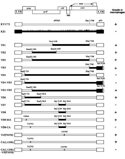
The molecular clone KV1772 is contained in two plasmids as described previously (29). The restriction enzymes used for cloning are indicated in Fig. 1. Mutants were generated by PCR-mediated site-directed mutagenesis, and the mutations were subsequently confirmed by sequencing.
FIG. 1.
Diagrammatic representation of recombinant MVV derived from the molecular clones KV1772 and KS1. The vertical bars indicate the amino acid differences between the two clones. The restriction sites used to construct the viruses are shown. The ability of each virus to grow in macrophages is shown on the right.
Experimental infection of sheep.
Icelandic sheep, 6 months old, were infected intratracheally with 1.0 ml of 104 50% tissue culture infective doses. Virus was injected into the trachea with a needle (23 gauge) under sedation with xylazinum (Xylapan; 0.2 ml intravenously) (34).
Virus isolation from blood and organs.
Virus was isolated from the buffy coat as described previously (33). The following organs were tested for presence of infectious virus by coculture with SCP cells (24): choroid plexus, cerebrum, cerebellum, spleen, cervical, mediastinal and mesenteric lymph nodes, bone marrow, and lungs (one sample from each lobe). If no cytopathic effects were observed, the growth medium was passaged twice, with the last observation after 6 weeks of culture.
RESULTS
The growth attenuation of the KS1 molecular clone is jointly determined by Vif and CA.
To begin to map the determinants of the attenuated replication phenotype of the KS1 clone in macrophages, a series of recombinants were constructed, using restriction fragments from various parts of KS1 and with KV1772 as a backbone. All recombinant viruses were tested for replication in sheep blood-derived macrophages to see if the impaired replication of KS1 was transferred with the respective fragments. The macrophages were infected with equal amounts of virus (as determined by measuring RT activity), and replication was monitored by taking samples daily and quantifying virus by measuring RT activity or using TaqMan-based real-time PCR.
We first tested a fragment comprising env, tat, vif, and a part of pol (VB1) (Fig. 1). There was no effect on replication of the virus in macrophages. Adding further regions of pol, env, and the long terminal repeat did not have any effect either (clones VB2, VB3, VB5, and VB3 to VB5) (Fig. 1). Only when the two mutations in gag were added was the attenuated replication in macrophages transferred (VB4) (Fig. 1). However, replacing a shorter fragment containing the two gag mutations did not result in significantly attenuated replication in macrophages (VB6) (Fig. 1). Only when three mutations in vif were cloned together with the mutations in gag did the virus acquire the attenuated phenotype (VB7) (Fig. 1). The mutations in vif did not have an attenuated effect on their own (VB8) (Fig. 1). It thus appears that there is an interaction between the mutations in gag and vif.
To ask which mutation(s) in gag associated with those in vif, a series of point mutations were introduced by site-directed mutation of KV1772. One of the two mutations in gag was in MA and the other was in CA. To distinguish between these two mutations, they were each constructed separately together with the mutations in vif. The virus with the mutation in MA replicated well in macrophages, whereas the virus with the mutation in CA replicated with kinetics similar to those of the KS1 strain, indicating that only the CA mutation was relevant. We then constructed the P-S mutation in Vif separately [strain Vif(P205S)], the mutation in CA separately [strain CA(L120R)], and the two mutations together [strain CA(L120R)-Vif(P205S)]. The replication kinetics of these strains were compared with the two parent strains in choroid plexus cells and macrophages. All strains replicated similarly in choroid plexus cells, whereas in macrophages CA(L120R) was somewhat affected and KS1 and CA(L120R)-Vif(P205S) replicated more slowly and to a lower titer (Fig. 2). Adaptation of KS1 and CA(L120R)-Vif(P205S) to growth in macrophages was observed upon extended passage, but direct reversion mutants were not detected. We are currently searching for possible second-site suppressor mutations.
FIG. 2.
Replication kinetics of virus production in blood-derived macrophages (A) and sheep choroid plexus cells (B), as measured by TaqMan-based real-time PCR.
Interestingly, the P-S mutation in Vif is in a proline-rich region at the C-terminal end of the protein that has been shown to be important for association with cellular membranes and interaction with the Gag precursor Pr55gag (3, 9, 10), whereas the mutation in CA is an L-R mutation in a position corresponding to amino acid 131 in helix 7 in HIV-1 (8, 36).
In vivo inoculation with the two parental strains and the recombinant virus CA(L120R)-Vif(P205S).
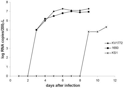
The KS1 strain has repeatedly been shown to be nonpathogenic in sheep, whereas the KV1772 strain is highly pathogenic (33). Of 219 attempts to isolate virus from blood of sheep infected with KS1 in various experiments, virus was isolated only on one occasion. Sequencing of the CA and vif genes of this isolate revealed a reversion of the L-R mutation in CA back to L. This isolate grew well in macrophages (Fig. 3).
FIG. 3.
Replication kinetics of CA revertant (1650), KS1, and 1772 clones in blood-derived macrophages as measured by TaqMan-based real-time PCR.
Three sheep, each 6 months old, were inoculated intratracheally with each of the three virus strains KV1772, KS1, and CA(L120R)-Vif(P205S). Blood was collected for virus isolations once a week for the first 8 weeks and biweekly thereafter until sacrifice after 18 weeks. At sacrifice, virus isolations were attempted from several organs, including brain, lungs, spleen, lymph nodes, and bone marrow. As shown in Table 1, virus was readily recovered from blood and organs of the three sheep that had been inoculated with strain KV1772, whereas no virus was isolated from sheep inoculated with KS1 virus. However, one of the three sheep inoculated with the recombinant CA(L120R)-Vif(P205S) virus was clearly infected. This result was not expected, since the CA(L120R)-Vif(P205S) strain has replication kinetics in macrophages similar to those of the KS1 strain. Sequencing of virus isolates from the infected sheep did not show a reversion of the mutations in CA and vif. One of the blood isolates was tested for replication in macrophages, and it showed attenuated replication similar to that of the CA(L120R)-Vif(P205S) strain (data not shown). We then speculated that this particular sheep might not express a putative virus inhibitor. We therefore infected four additional sheep with the virus CA(L120R)-Vif(P205S) and isolated macrophages from each sheep before infection to test replication of the CA(L120R)-Vif(P205S) strain compared to KV1772. Replication of the CA(L120R)-Vif(P205S) strain was attenuated in macrophages from all of the four sheep (data not shown). The sheep were kept for 18 weeks as before, and attempts were made to isolate virus from the same organs. No virus was isolated from any of the organs, but one isolate was recovered from blood (Table 1).
TABLE 1.
Frequency of virus isolation from KV1772-, KS1-, and CA(L120R)-Vif(P205S)-infected sheep
| Virus strain and sheep no. | No. positive/no. tested in:
|
|||
|---|---|---|---|---|
| Blood | Lympha | Lungs | Central nervous system | |
| KV1772 | ||||
| 2080 | 10/11 | 5/5 | 2/2 | 1/3 |
| 2081 | 8/11 | 5/5 | 2/2 | 0/3 |
| 2082 | 5/11 | 2/5 | 0/2 | 0/3 |
| KS1 | ||||
| 2074 | 0/11 | 0/5 | 0/2 | 0/3 |
| 2075 | 0/11 | 0/5 | 0/2 | 0/3 |
| 2076 | 0/11 | 0/5 | 0/2 | 0/3 |
| CA(L120R)-Vif(P205S) | ||||
| 2077 | 0/11 | 0/5 | 0/2 | 0/3 |
| 2078 | 2/11 | 4/5 | 2/2 | 0/3 |
| 2079 | 0/11 | 0/5 | 0/2 | 0/3 |
| 2089 | 0/11 | 0/5 | 0/2 | 0/3 |
| 2090 | 0/11 | 0/5 | 0/2 | 0/3 |
| 2091 | 1/11 | 0/5 | 0/2 | 0/3 |
| 2092 | 0/11 | 0/5 | 0/2 | 0/3 |
Spleen, bone marrow, cervical, mediastinal, and mesenteric lymph nodes.
DISCUSSION
In this study, we traced the attenuated replication and infectivity of the molecular clone KS1 to two simultaneous mutations, one in the CA domain of gag and the other in vif. The CA mutation affected replication in macrophages somewhat, but with the addition of the vif mutation, replication in macrophages was markedly reduced. The mutation in vif on its own did not affect replication in macrophages. The finding that a reversion of the mutation in CA in the KS1 clone caused a reversion in phenotype further supports the importance of the mutation in the capsid. The CA mutation is an L-R mutation in amino acid 120 of MVV CA. This is a position corresponding to K131 in helix 7 in the N-terminal domain of HIV-1 CA protein. There is a charged amino acid in this position in all lentiviruses except caprine arthritis encephalitis virus and MVV (36). Charge change mutants of adjacent amino acids in helix 7 in HIV-1 have resulted in alteration in cell tropism that seems to be independent of Ref1/TRIM5α (16). These mutations may therefore define an interaction of the capsid with uncharacterized host proteins.
The mutation in the vif gene is a P-to-S mutation in the C terminus, where membrane association, Gag interaction, and Vif multimerization have been mapped for HIV-1; interaction with Gag has also been mapped for caprine arthritis encephalitis virus (3, 9, 10, 25, 37). The interaction with Gag has been mapped to the nucleocapsid part of Gag in vitro, but no interaction has been detected with CA (3, 25). However, Vif has been shown to play a role in the stability of the core of HIV-1 (23), and the interaction between Vif and CA may either be indirect or too transient to be detected by standard methods that are used for detecting protein-protein interactions. It is possible that the two mutations are unrelated and that the effect is additive. However, since the mutation in vif did not have a detectible effect on replication on its own, it is tempting to speculate that there is interaction of CA and Vif in the replication of the virus.
Of seven sheep that were inoculated intratracheally with the virus harboring the two mutations [CA(L120R)-Vif(P205S)], one showed multiorgan infection typical of the wild type virus. However, virus isolates from this sheep retained the two mutations and replication characteristics of the mutant virus [CA(L120R)-Vif(P205S)]. It therefore appears that this sheep lacked host restriction. Unfortunately, the sheep is not available for further examination.
It is becoming increasingly clear that a variety of antiretroviral activities have evolved in mammals. The mutations we have described in this report may define a novel host restriction factor that targets the capsid and is counteracted by Vif. Another possibility would be that the CA mutation slows the growth of the virus by destabilizing the core, thus making it more susceptible to host proteins in general. This might exacerbate the effect of APOBEC3 when Vif is attenuated. We are currently addressing the possibility that APOBEC3 knockdown in sheep macrophages rescues growth of the KS1 MVV.
Acknowledgments
This study was supported by the Icelandic Research Council, the University of Iceland Research Fund, and the Icelandic Research Fund for Graduate Students.
We are indebted to Katherine Staskus and Ashley Haase for the KS1 clone and to Svava Högnadóttir, Steinunn Árnadóttir, and Sigurdur Helgason for expert technical help. We thank Reuben Harris for critically reading the manuscript.
REFERENCES
- 1.Andresdottir, V., X. Tang, G. Agnarsdottir, O. S. Andresson, G. Georgsson, R. Skraban, S. Torsteinsdottir, B. Rafnar, E. Benediktsdottir, S. Matthiasdottir, S. Arnadottir, S. Hognadottir, P. A. Palsson, and G. Petursson. 1998. Biological and genetic differences between lung- and brain-derived isolates of maedi-visna virus. Virus Genes 16:281-293. [DOI] [PubMed] [Google Scholar]
- 2.Andresson, O. S., J. E. Elser, G. J. Tobin, J. D. Greenwood, M. A. Gonda, G. Georgsson, V. Andresdottir, E. Benediktsdottir, H. M. Carlsdottir, and E. O. Mantyla. 1993. Nucleotide sequence and biological properties of a pathogenic proviral molecular clone of neurovirulent visna virus. Virology 193:89-105. [DOI] [PubMed] [Google Scholar]
- 3.Bouyac, M., M. Courcoul, G. Bertoia, Y. Baudat, D. Gabuzda, D. Blanc, N. Chazal, P. Boulanger, J. Sire, R. Vigne, and B. Spire. 1997. Human immunodeficiency virus type 1 Vif protein binds to the Pr55Gag precursor. J. Virol. 71:9358-9365. [DOI] [PMC free article] [PubMed] [Google Scholar]
- 4.DesGroseillers, L., and P. Jolicoeur. 1983. Physical mapping of the Fv-1 tropism host range determinant of BALB/c murine leukemia viruses. J. Virol. 48:685-696. [DOI] [PMC free article] [PubMed] [Google Scholar]
- 5.Desrosiers, R. C., J. D. Lifson, J. S. Gibbs, S. C. Czajak, A. Y. Howe, L. O. Arthur, and R. P. Johnson. 1998. Identification of highly attenuated mutants of simian immunodeficiency virus. J. Virol. 72:1431-1437. [DOI] [PMC free article] [PubMed] [Google Scholar]
- 6.Gendelman, H. E., O. Narayan, S. Kennedy-Stoskopf, P. G. Kennedy, Z. Ghotbi, J. E. Clements, J. Stanley, and G. Pezeshkpour. 1986. Tropism of sheep lentiviruses for monocytes: susceptibility to infection and virus gene expression increase during maturation of monocytes to macrophages. J. Virol. 58:67-74. [DOI] [PMC free article] [PubMed] [Google Scholar]
- 7.Georgsson, G., N. Nathanson, P. A. Palsson, and G. Petursson. 1976. The pathology of visna and maedi in sheep. Front. Biol. 44:61-96. [PubMed] [Google Scholar]
- 8.Gitti, R. K., B. M. Lee, J. Walker, M. F. Summers, S. Yoo, and W. I. Sundquist. 1996. Structure of the amino-terminal core domain of the HIV-1 capsid protein. Science 273:231-235. [DOI] [PubMed] [Google Scholar]
- 9.Goncalves, J., P. Jallepalli, and D. H. Gabuzda. 1994. Subcellular localization of the Vif protein of human immunodeficiency virus type 1. J. Virol. 68:704-712. [DOI] [PMC free article] [PubMed] [Google Scholar]
- 10.Goncalves, J., B. Shi, X. Yang, and D. Gabuzda. 1995. Biological activity of human immunodeficiency virus type 1 Vif requires membrane targeting by C-terminal basic domains. J. Virol. 69:7196-7204. [DOI] [PMC free article] [PubMed] [Google Scholar]
- 11.Gorrell, M. D., M. R. Brandon, D. Sheffer, R. J. Adams, and O. Narayan. 1992. Ovine lentivirus is macrophagetropic and does not replicate productively in T lymphocytes. J. Virol. 66:2679-2688. [DOI] [PMC free article] [PubMed] [Google Scholar]
- 12.Harmache, A., M. Bouyac, G. Audoly, C. Hieblot, P. Peveri, R. Vigne, and M. Suzan. 1995. The vif gene is essential for efficient replication of caprine arthritis encephalitis virus in goat synovial membrane cells and affects the late steps of the virus replication cycle. J. Virol. 69:3247-3257. [DOI] [PMC free article] [PubMed] [Google Scholar]
- 13.Harmache, A., P. Russo, F. Guiguen, C. Vitu, M. Vignoni, M. Bouyac, C. Hieblot, M. Pepin, R. Vigne, and M. Suzan. 1996. Requirement of caprine arthritis encephalitis virus vif gene for in vivo replication. Virology 224:246-255. [DOI] [PubMed] [Google Scholar]
- 14.Harris, R. S., K. N. Bishop, A. M. Sheehy, H. M. Craig, S. K. Petersen-Mahrt, I. N. Watt, M. S. Neuberger, and M. H. Malim. 2003. DNA deamination mediates innate immunity to retroviral infection. Cell 113:803-809. [DOI] [PubMed] [Google Scholar]
- 15.Hatziioannou, T., S. Cowan, S. P. Goff, P. D. Bieniasz, and G. J. Towers. 2003. Restriction of multiple divergent retroviruses by Lv1 and Ref1. EMBO J. 22:385-394. [DOI] [PMC free article] [PubMed] [Google Scholar]
- 16.Hatziioannou, T., S. Cowan, U. K. Von Schwedler, W. I. Sundquist, and P. D. Bieniasz. 2004. Species-specific tropism determinants in the human immunodeficiency virus type 1 capsid. J. Virol. 78:6005-6012. [DOI] [PMC free article] [PubMed] [Google Scholar]
- 17.Hatziioannou, T., D. Perez-Caballero, A. Yang, S. Cowan, and P. D. Bieniasz. 2004. Retrovirus resistance factors Ref1 and Lv1 are species-specific variants of TRIM5α. Proc. Natl. Acad. Sci. USA 101:10774-10779. [DOI] [PMC free article] [PubMed] [Google Scholar]
- 18.Kristbjornsdottir, H. B., V. Andresdottir, V. Svansson, S. Torsteinsdottir, S. Matthiasdottir, and O. S. Andresson. 2004. The vif gene of maedi-visna virus is essential for infectivity in vivo and in vitro. Virology 318:350-359. [DOI] [PubMed] [Google Scholar]
- 19.Lecossier, D., F. Bouchonnet, F. Clavel, and A. J. Hance. 2003. Hypermutation of HIV-1 DNA in the absence of the Vif protein. Science 300:1112. [DOI] [PubMed] [Google Scholar]
- 20.Lockridge, K. M., S. Himathongkham, E. T. Sawai, M. Chienand, and E. E. Sparger. 1999. The feline immunodeficiency virus vif gene is required for productive infection of feline peripheral blood mononuclear cells and monocyte-derived macrophages. Virology 261:25-30. [DOI] [PubMed] [Google Scholar]
- 21.Mangeat, B., P. Turelli, G. Caron, M. Friedli, L. Perrin, and D. Trono. 2003. Broad antiretroviral defence by human APOBEC3G through lethal editing of nascent reverse transcripts. Nature 424:99-103. [DOI] [PubMed] [Google Scholar]
- 22.Narayan, O. 1983. Role of macrophages in the immunopathogenesis of visna-maedi of sheep. Prog. Brain Res. 59:233-235. [DOI] [PubMed] [Google Scholar]
- 23.Ohagen, A., and D. Gabuzda. 2000. Role of Vif in stability of the human immunodeficiency virus type 1 core. J. Virol. 74:11055-11066. [DOI] [PMC free article] [PubMed] [Google Scholar]
- 24.Petursson, G., N. Nathanson, G. Georgsson, H. Panitch, and P. A. Palsson. 1976. Pathogenesis of visna. I. Sequential virologic, serologic, and pathologic studies. Lab. Investig. 35:402-412. [PubMed] [Google Scholar]
- 25.Seroude, V., G. Audoly, P. Gluschankof, and M. Suzan. 2001. Tryptophan 95, an amino acid residue of the Caprine arthritis encephalitis virus Vif protein which is essential for virus replication. Virology 280:232-242. [DOI] [PubMed] [Google Scholar]
- 26.Sigurdsson, B., H. Grimsson, and P. A. Palsson. 1952. Maedi, a chronic progressive infection of sheep's lungs. J. Infect. Dis. 90:233-241. [DOI] [PubMed] [Google Scholar]
- 27.Sigurdsson, B., and P. A. Palsson. 1958. Visna of sheep. A slow demyelinating infection. Br. J. Exp. Pathol. 39:519-528. [PMC free article] [PubMed] [Google Scholar]
- 28.Sigurdsson, B., H. Thormar, and P. A. Palsson. 1960. Cultivation of visna virus in tissue culture. Arch. Gesamte Virusforsch. 10:368-380. [DOI] [PubMed] [Google Scholar]
- 29.Skraban, R., S. Matthiasdottir, S. Torsteinsdottir, G. Agnarsdottir, B. Gudmundsson, G. Georgsson, R. H. Meloen, O. S. Andresson, K. A. Staskus, H. Thormar, and V. Andresdottir. 1999. Naturally occurring mutations within 39 amino acids in the envelope glycoprotein of maedi-visna virus alter the neutralization phenotype. J. Virol. 73:8064-8072. [DOI] [PMC free article] [PubMed] [Google Scholar]
- 30.Staskus, K. A., E. F. Retzel, E. D. Lewis, J. L. Silsby, S. St. Cyr, J. M. Rank, S. W. Wietgrefe, A. T. Haase, R. Cook, and D. Fast. 1991. Isolation of replication-competent molecular clones of visna virus. Virology 181:228-240. [DOI] [PubMed] [Google Scholar]
- 31.Strebel, K., D. Daugherty, K. Clouse, D. Cohen, T. Folks, and M. A. Martin. 1987. The HIV ‘A’ (sor) gene product is essential for virus infectivity. Nature 328:728-730. [DOI] [PubMed] [Google Scholar]
- 32.Stremlau, M., C. M. Owens, M. J. Perron, M. Kiessling, P. Autissier, and J. Sodroski. 2004. The cytoplasmic body component TRIM5α restricts HIV-1 infection in Old World monkeys. Nature 427:848-853. [DOI] [PubMed] [Google Scholar]
- 33.Torsteinsdottir, S., G. Agnarsdottir, S. Matthiasdottir, B. Rafnar, V. Andresdottir, O. S. Andresson, K. Staskus, G. Petursson, P. A. Palsson, and G. Georgsson. 1997. In vivo and in vitro infection with two different molecular clones of visna virus. Virology 229:370-380. [DOI] [PubMed] [Google Scholar]
- 34.Torsteinsdottir, S., S. Matthiasdottir, N. Vidarsdottir, V. Svansson, and G. Petursson. 2003. Intratracheal inoculation as an efficient route of experimental infection with maedi-visna virus. Res. Vet. Sci. 75:245-247. [DOI] [PubMed] [Google Scholar]
- 35.Towers, G., M. Bock, S. Martin, Y. Takeuchi, J. P. Stoye, and O. Danos. 2000. A conserved mechanism of retrovirus restriction in mammals. Proc. Natl. Acad. Sci. USA 97:12295-12299. [DOI] [PMC free article] [PubMed] [Google Scholar]
- 36.von Schwedler, U. K., T. L. Stemmler, V. Y. Klishko, S. Li, K. H. Albertine, D. R. Davis, and W. I. Sundquist. 1998. Proteolytic refolding of the HIV-1 capsid protein amino-terminus facilitates viral core assembly. EMBO J. 17:1555-1568. [DOI] [PMC free article] [PubMed] [Google Scholar]
- 37.Yang, S., Y. Sun, and H. Zhang. 2001. The multimerization of human immunodeficiency virus type-I (HIV-1) Vif protein: a requirement for Vif function in the viral life-cycle. J. Biol. Chem. 276:4889-4893. [DOI] [PMC free article] [PubMed] [Google Scholar]
- 38.Zhang, H., B. Yang, R. J. Pomerantz, C. Zhang, S. C. Arunachalam, and L. Gao. 2003. The cytidine deaminase CEM15 induces hypermutation in newly synthesized HIV-1 DNA. Nature 424:94-98. [DOI] [PMC free article] [PubMed] [Google Scholar]


