Abstract
A sensitive and specific immunoassay for the simultaneous detection of Clostridium botulinum type C (BoNT/C) and type D neurotoxin was developed. Goat anti-mouse immunoglobulin G was bound to polyethylene disks in a small disposable column used for this assay. The sample was preincubated together with monoclonal antibodies specific for the heavy chain of BoNT/C and D and affinity-purified, biotinylated polyclonal antibodies against these neurotoxins. This complex was captured on the assay disk. Streptavidin-poly-horseradish peroxidase was used as a conjugate, and a precipitating substrate allowed the direct semiquantitative readout of the assay, if necessary. For a more accurate quantitative detection, the substrate can be eluted and measured in a photometer. Depending on the preincubation time, a sensitivity of 1 mouse lethal dose ml−1 was achieved in culture supernatants.
Clostridium botulinum is a gram-positive, anaerobic, and spore-forming soil bacterium that can be found in the guts of domestic animals (9, 10). The fact that it produces the most toxic metabolite known to humans brought it to the attention of medical microbiologists in the early days of this field (27). Nowadays, seven distinct botulinum neurotoxins (BoNTs) are known. In the order of their discovery, they have been named types A to G. However, the capability to produce BoNTs is not limited to C. botulinum only. Some strains of Clostridium baratii (8) and Clostridium butyricum (3, 21) are toxigenic as well.
The prevalence of C. botulinum in the soil might explain where the bacterium enters the food chain (20), leading to the best-known form of the disease in the worst case, food-borne botulism (25). The bacterium multiplies in food or feed under favorable conditions and produces the toxin, which is orally taken up by the host. The typical signs of flaccid paralysis develop, which are caused by the inhibition of acetylcholine release at the synapses (19).
In animal husbandry and for wildlife, types C and D botulism are predominant. Throughout the world, millions of waterfowl have reportedly died from botulism caused by BoNT type C (BoNT/C) (24). BoNT/C and D are pathogenic for our domestic animals, with sometimes dramatic losses in the affected farms (6, 15). The losses of cattle reported from Brazil amount to five million animals over the past 10 years (18).
However, the disease may present as a toxico infection as well. With the shaker foal syndrome in horses, it was shown that the bacteria colonize the gut and produce the toxin in the host animal (23). Visceral botulism in cattle (5) and equine grass sickness (7) might have a toxico infectious botulinum etiology as well.
The recent concerns for the use of botulinum neurotoxins by bioterrorists (2) again highlighted the fact that only a limited number of tools are available to detect BoNTs. The mouse bioassay, still the most common method, needs to be replaced for obvious reasons. Recently, alternative in vitro tests (13, 17) have become commercially available. However, these assays are limited to the BoNT types that are pathogenic for humans, namely, types A, B, and E. Rocke et al. (22) developed an assay for the diagnosis of type C botulism in birds, and Thomas (26) developed an enzyme-linked immunosorbent assay (ELISA) for the detection of BoNT/C and D.
The major aim of the work presented here was to develop a highly sensitive and specific diagnostic test for the detection of BoNT/C and D in one assay with a direct semiquantitative readout.
MATERIALS AND METHODS
All reagents and chemicals were purchased from Merck, Darmstadt, Germany, unless otherwise stated.
Purified toxin.
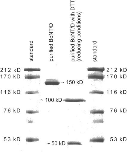
The 150-kDa neurotoxins of BoNT/C and D were produced and purified as previously described (16). Briefly, type C strain 003-9 and type D strain CB-16 (kindly provided by S. Kozaki, Osaka Prefecture University, Japan) (Table 1) were used. The cultures were grown anaerobically in 10-liter batch cultures in a protein-rich medium (1% peptone from casein [pancreatically digested], 1% meat extract, 0.3% yeast extract, 0.1% soluble starch, 0.5% d-glucose, 0.5% sodium chloride, 0.3% sodium acetate, and 0.05% l-cysteine-HCl) at 37°C. When maximum toxin titers had been reached (usually after 4 days), microfiltration followed by ultrafiltration was used to separate the bacteria from the supernatant in the first step and to concentrate and further purify the toxin in the second step. In four consecutive chromatographic purification runs, highly purified BoNT/C and D were obtained (Fig. 1). These steps included hydrophobic interaction at pH 8.0, anion exchange at pH 8.0, anion exchange at pH 6.0, and finally a size exclusion run. The biological activity was quantified in the mouse bioassay according to relevant guidelines (1, 11).
TABLE 1.
Identification and source of the C. botulinum and C. tetani strains used for specificity testing
| Species | Type (C. botulinum) | Group (C. botulinum) | Sourcea | Strain identification |
|---|---|---|---|---|
| C. botulinum | A | I | NCTC | 7272 |
| A | I | CECT | 551 | |
| A | I | OPU | 62A | |
| B | I | NCTC | 7273 | |
| B | I | CECT | 4610 | |
| B | I | OPU | Okra | |
| C | III | NCTC | 8264 | |
| C | III | NRZ | REB1455 | |
| C | III | OPU | 003-9 | |
| D | III | NCTC | 8265 | |
| D | III | IP | 1873D | |
| D | III | OPU | CB-16 | |
| E | II | NCTC | 8266 | |
| E | II | CECT | 4611 | |
| E | II | UH | CB-S-21E | |
| F | I | NCTC | 10281 | |
| F | II | OPU | ||
| F | II | NZRCC | 83-4304 | |
| C. tetani | NCTC | 279 | ||
| DSMZ | 1285 |
NCTC, National Collection of Type Cultures, London, United Kingdom; CECT, Colección Espan̈ola de Cultivos Tipo, Valencia, Spain; OPU, S. Kozaki, Osaka Prefecture University, Department of Veterinary Science, Osaka, Japan; NRZ, Nationales Referenzzentrum für Clostridien, Erfurt, Germany; IP, Institut Pasteur, Paris, France; UH, H. Korkeala, University of Helsinki, Department of Food and Environmental Hygiene, Helsinki, Finland; NZRCC, New Zealand Reference Culture Collection, Porirua, New Zealand; DSMZ, Deutsche Sammlung von Mikroorganismen und Zellkulturen, Braunschweig, Germany.
FIG. 1.
Sodium dodecyl sulfate-polyacrylamide gel electrophoresis of purified BoNT/D under nonreducing (lane 2) and reducing (with dithiothreitol [DTT] treatment) (lane 3) conditions, which separate the heavy and light chains. kD, kilodaltons.
Toxoid.
The toxoid was prepared by adding formalin to the purified neurotoxin to a final concentration of 0.4%. This solution was kept for 10 days at 35°C.
Culture supernatants.
For specificity testing, well-defined and toxin-typed C. botulinum cultures, types A to F (three strains per type), and two Clostridium tetani cultures were grown in RCM (Oxoid, Wesel, Germany) for 4 days at 37°C. The anaerobic incubation atmosphere was adjusted to 90% N2, 5% H2, and 5% CO2 with a gas exchange system (Mart Microbiology, Lichtenvoorde, The Netherlands). The cultures were centrifuged, and the supernatants were sterile filtered with disposable Minisart 0.2-μm filters (Sartorius, Göttingen, Germany). The toxicity was again determined in the mouse bioassay and diluted to 10,000 mouse lethal doses (MLD) ml−1, if possible. In type F cultures, only 100 MLD ml−1 were achieved. C. tetani cultures were used undiluted. More detailed information on the cultures and the origin of the strains is given in Table 1.
Mouse bioassay.
The biological activity of the purified neurotoxins and the culture supernatants was quantified in NMRI mice (Harlan Winkelmann, Borchen, Germany) weighing 18 to 25 g according to AOAC (1) and German Deutsches Institut für Normung (11) guidelines. To keep the number of animals used at a minimum, the biological activity was determined in critical steps only. Medium and buffer controls were included with each step. The sample was diluted 1:10 in gelatin-phosphate buffer (50 mM phosphate-buffered saline [PBS], 0.2% gelatin, pH 6.2). Serial 10-fold dilutions were then made in gelatin-phosphate buffer. A total of 0.5 ml of each dilution was injected intraperitoneally into two mice. In between the highest positive and the first negative dilutions, additional twofold dilutions were made. The animals were observed for clinical signs or death over a period of 4 days. The toxicity is given in MLD per milliliter, which corresponds to the reciprocal value of the highest dilution at which both mice showed typical signs and died. For toxin typing, monovalent antisera supplied by the CDC, Atlanta, Ga., or id-dlo, Lelystad, The Netherlands, were used. In accordance with the suppliers' instructions, an aliquot of the antiserum was mixed with the purified neurotoxin or the culture supernatant and incubated for 30 min at 37°C before being injected into the mice. The neutralization was considered successful if the animals with the homologous antiserum survived and if those with the heterologous antiserum died.
Monoclonal antibodies.
Monoclonal antibodies were raised against BoNT/D toxoid and purified by ABION (Jülich, Germany). In the final screening, clones were checked against the purified toxins (types C and D) and the culture supernatants including BoNT/A to BoNT/F in a direct ELISA and by Western blot analysis. For the ELISA procedure, 96-well PolySorb plates (NUNC, Wiesbaden, Germany) were coated with 100 μl purified BoNT/C and D at a concentration of 1 μg ml−1 or with culture supernatant diluted 1:40 in 0.1 M bicarbonate buffer, pH 9.6, at 4°C for 12 h. Plates were blocked with 200 μl skim milk buffer per well (blocking buffer, 15 mM PBS, pH 7.2, 0.05% Tween 20, 5.0% skim milk powder) at 37°C for 2 h. Plates were washed (washing buffer [15 mM PBS, pH 7.2, 0.05% Tween 20]) three times. The same washing conditions were applied throughout the protocol. The plates were stored at −20°C until further use. For testing of the hybridomas, 100 μl of the hybridoma culture supernatants was pipetted into the wells. The plates were incubated at 37°C for 1 h and washed. Anti-mouse immunoglobulin G (IgG), peroxidase labeled (Sigma, Taufkirchen, Germany), was diluted 1:5,000 in 15 mM PBS, pH 7.2, and an aliquot of 100 μl was pipetted into each well and again incubated at 37°C for 30 min. Plates were washed, and the TMB substrate (Sigma) was prepared and added according to the manufacturer's instructions. After 10 min, the reaction was stopped with 50 μl of 1 M H2SO4. The plates were read in the photometer at 450 nm.
The Western blot analysis was performed according to a standard protocol. Briefly, separate 7.5% sodium dodecyl sulfate-polyacrylamide gels were run with purified BoNT/C and BoNT/D under reducing (with dithiothreitol) and nonreducing conditions in a Mini-Protean II system (Bio-Rad, Munich, Germany). The proteins in the gels were transferred to nitrocellulose membranes with a Mini Trans-Blot cell (Bio-Rad) according to the manufacturer's instructions. The following steps used the same buffers as those in the ELISA. The membranes were incubated in blocking buffer at 37°C for 1 h. Three washing steps followed at room temperature for 5 min each. The membranes were cut into strips, transferred to small tubes, and incubated with the undiluted hybridoma culture supernatants at 37°C for 60 min. The strips were washed five times at room temperature for 5 min. Anti-mouse IgG, alkaline phosphate labeled (Sigma), was diluted 1:30,000 in 15 mM PBS, pH 7.2, pipetted into the tubes, and incubated at 37°C for 1 h. After five washing steps of 5 min each, BCIP (5-bromo-4-chloro-3-indolylphosphate)-Nitro Blue Tetrazolium substrate (Sigma) was added according to the manufacturer's instructions. The strips were incubated for 5 to 10 min depending on the color intensity of the bands, washed twice, and dried on a glass plate.
The results of the ELISA and Western blot analyses are summarized in Table 2. Clones 21 and 23 were selected for further work because they were specific for the heavy chain in BoNT/C and D and showed no cross-reactivity with the culture supernatants of the other types.
TABLE 2.
Screening of positive clones in ELISA and Western blot analysesa
| Assay | No. of supernatants/total no.b for Clone
|
||||
|---|---|---|---|---|---|
| 11 | 12 | 21 | 22 | 23 | |
| ELISA | |||||
| Purified neurotoxin types | |||||
| C | + | + | + | + | + |
| D | + | + | + | + | + |
| Culture supernatant types | |||||
| A | 3/3 | 3/3 | 0/3 | 0/3 | 0/3 |
| B | 3/3 | 3/3 | 0/3 | 0/3 | 0/3 |
| C | 3/3 | 3/3 | 3/3 | 3/3 | 3/3 |
| D | 3/3 | 3/3 | 3/3 | 3/3 | 3/3 |
| E | 0/3 | 0/3 | 0/3 | 0/3 | 0/3 |
| F | 0/3 | 0/3 | 0/3 | 0/3 | 0/3 |
| Western blot | |||||
| Purified neurotoxin types | |||||
| C, 150-kDa molecule | ND | ND | + | − | + |
| C, heavy chain | ND | ND | + | − | + |
| C, light chain | ND | ND | − | − | − |
| D, 150-kDa molecule | ND | ND | + | − | + |
| D, heavy chain | ND | ND | + | − | + |
| D, light chain | ND | ND | − | − | − |
+, Absorbance above assay cutoff (ELISA) or distinct band visible (Western blot); −, absorbance below assay cutoff (ELISA) or no distinct band visible (Western blot); ND, not determined.
Number of positively tested culture supernatants/number of total culture supernatants tested.
Polyclonal antibodies.
Goats were immunized with the BoNT/D toxoid to obtain polyclonal serum. The raw serum was produced by ABION. It was further purified by affinity chromatography. Briefly, purified toxin type D was bound to Sepharose CL 6B (Amersham Pharmacia, Freiburg, Germany) by coupling with sodium-m-periodate (Sigma). Sodium-borohydride (Sigma) was used as a reducing agent. Proteins that bound unspecifically were washed off with 0.5 M NaCl. The antibodies were eluted with 0.3 M citric acid, pH 2.1. Na2CO3 was added to the eluate to give a pH of 7.2.
To avoid antibody-antigen complexes which coeluted from the affinity column, size exclusion chromatography was performed as a second step.
The polyclonal antibodies showed cross-reactivity with BoNT/C (data not shown). Therefore, separate BoNT/C polyclonal antibodies were not produced.
Biotinylation of antibodies.
For the biotin-streptavidin-horseradish peroxidase (HRP) detection approach, the affinity-purified polyclonal antibodies were biotinylated. To prepare a biotin stock solution, 2 mg biotinamidocaproate N-hydroxysuccinimide ester (Sigma) was dissolved in 1 ml dimethyl sulfoxide. The storage buffer of the antibodies was exchanged to carbonate buffer (0.1 M Na2CO3, 0.1 M NaHCO3, pH 8.5). The ratio was calculated (mbiotin = mantibodies × biotinylation degree × Mbiotin/Mantibodies [where m is the amount and M is the molecular mass of biotin or antibodies]), and both solutions were mixed. The reaction mixture was incubated for 3 h at room temperature on a rotary shaker. Excess biotin was washed out on a PD-10 Sephadex G-25 M column (Amersham Pharmacia). The antibodies were eluted with PBS (20 mM Na2HPO4 · 2H2O, 6 mM NaH2PO4 · H2O, 120 mM NaCl, pH 7.3). Bovine serum albumin (BSA) was added (15 mg ml−1) to stabilize the antibody solution. The biotinylation degree was determined and calculated using a commercial kit according to the manufacturer's instructions (Immuno Pure HABA; Perbio Science, Bonn, Germany).
Immunoaffinity assay columns.
The immunoaffinity assay consisted of small disposable plastic columns (65 mm by 7 mm) that were packed with three sintered polyethylene disks at the bottom (Fig. 2). All assay components were purchased from ABION. The disk in the middle (assay disk) was noncovalently coated with the various capture antibodies tested: The antibody stock solutions were diluted in carbonate buffer (50 mM Na2CO3, 50 mM NaHCO3, pH 9.5), and the disks were added. After the reaction mixture was incubated on a rotary shaker for 16 h at room temperature, the disks were allowed to settle and the supernatant was siphoned off. The reaction was stopped by the addition of BSA to a final concentration of 1%. The same incubation conditions were applied for 1 h, and then the buffer was exchanged for carbonate buffer plus 0.1% BSA. The stop disks were blocked with the 0.1% BSA solution only. The disks were transferred to the columns as shown in Fig. 2. The columns were washed with PBS and stored at 4°C until further use.
FIG. 2.
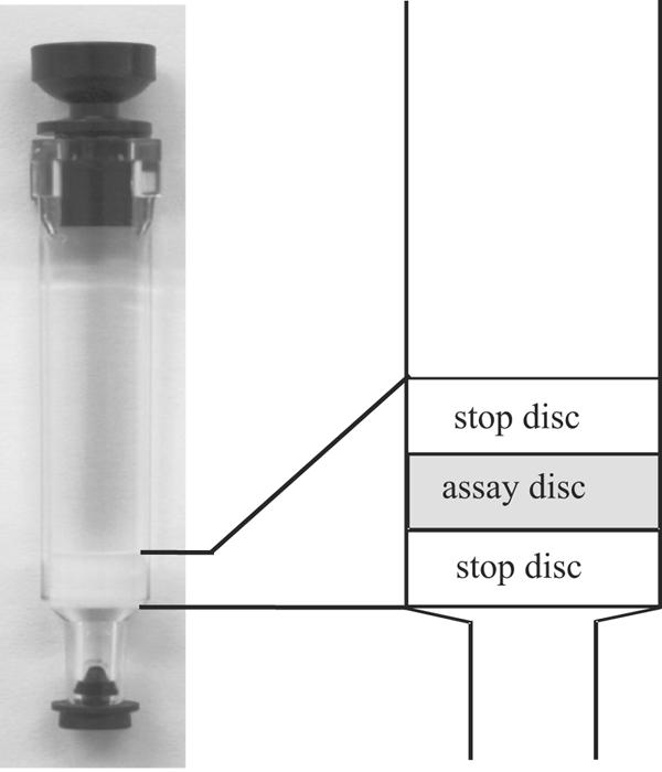
Disposable immunoaffinity assay column (65 mm by 7 mm). Three sintered polyethylene disks are at the bottom of the column. The assay disk in the middle is coated with the capture reagent (capture antibodies).
Calculation of the cutoff.
The cutoff was calculated by the method described previously by Frey et al. (14). The formula is expressed as the standard deviation multiplied by a factor that is based on the number of negative controls and the confidence level. In this study, a confidence level of 99% was assumed.
Basic assay setup protocol.
Various assay parameters were modified and tested to define a basic assay protocol and layout of the test columns. The initial testing parameters to be checked were capture antibodies (monoclonal or polyclonal anti-BoNT antibodies and anti-mouse antibodies), detection antibodies (monoclonal or polyclonal anti-BoNT antibodies and secondary antibodies), concentrations of these antibodies (capture antibody, anti-BoNT antibodies, and secondary antibodies), blocking buffers (casein, bovine serum albumin, and skim milk), buffer pH, antibody labels (HRP, alkaline phosphate, and biotin), substrates, sample volumes, and incubation times and temperatures.
In these preliminary investigations (data not shown), the following assay setup proved to be the best procedure for further optimization (Fig. 3).
FIG. 3.
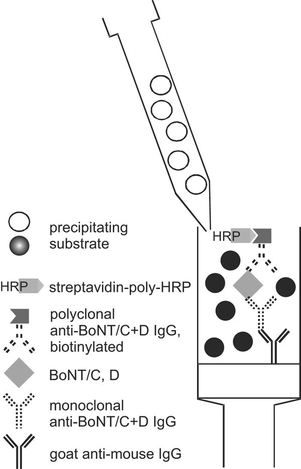
Assay setup. The sample is preincubated with monoclonal anti-BoNT/C and D and biotinylated polyclonal anti-BoNT/C and D (anti-BoNT/C+D) antibodies. During preincubation, a complex with BoNT is formed, which is captured by the anti-mouse IgG coated on the assay disk of the column. Streptavidin-poly-HRP binds to the biotinylated antibody and precipitates the substrate on the assay disk.
The assay disks were coated with goat anti-mouse IgG (Sigma) with a concentration of 10 μg per disk. The biotinylated polyclonal antibodies (biotinylation degree of between 2 and 6) and the monoclonal antibodies were mixed with the sample and incubated at room temperature on a rotary shaker. A total of 500 μl of this solution was pipetted onto a test column that had been washed previously with 750 μl of 2.5% casein buffer (SDT, Jülich, Germany). After the sample-antibody solution had run through the column, it was washed three times with 750 μl 2.5% casein buffer. A total of 500 μl of streptavidin-poly-HRP (SDT) was pipetted onto the column, followed by two wash steps with 2.5% casein buffer and one step with deionized H2O using 750 μl each. A 100-μl volume of precipitating HRP substrate (BM-Blue; Roche, Mannheim, Germany) was added, and residuals were washed out with 750 μl deionized H2O.
At this stage, the test could be read out semiquantitatively based on the color intensity of the assay disk compared with a standard dilution row. In addition, the reaction was stopped by adding 300 μl of 8% H2SO4. This eluate was collected in a microtiter plate and measured in a plate reader at 450 nm.
Optimization of the assay setup and protocol.
The basic assay setup that was developed was further optimized. Antibody concentrations, streptavidin conjugates (SDT), and preincubation times of the antibodies with the toxin/sample were modified to increase the sensitivity of the assay. Table 3 gives an overview of the various modifications tested.
TABLE 3.
Optimization of the assay setup and protocol
| Test parameter | Modification | ||
|---|---|---|---|
| Antibody concn (μg/ml) | |||
| monoclonal antibody | 0.1 | 0.1 | 0.3 |
| biotinylated polyclonal antibody | 0.05 | 0.1 | 0.1 |
| Streptavidin-HRP conjugates | Poly-HRP 25a | Poly-HRP 40a | Poly-HRP 80a |
| Preincubation time (h) of antibodies with toxin/ sample at room temp | 2 | 4 | 16 |
Poly-HRP 25, 40, and 80 were supplied by SDT. These conjugates varied in the degree of condensation, with poly-HRP 80 having the highest degree.
Specificity.
For specificity testing, the filtered and adjusted culture supernatants were used (BoNT/A to E, 10,000 MLD ml−1; BoNT/F, 100 MLD ml−1 [C.tetani undiluted culture supernatant]).
Precision.
Intra-assay and interassay precision were assessed with dilution rows (1 to 1,000 MLD ml−1) of types C and D culture supernatants of the strains which were used to generate the antibodies. In both precision tests, 5 dilutions were measured, either in one run (intra-assay) or in five runs (interassay).
RESULTS
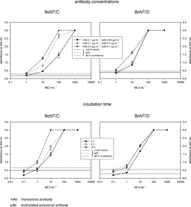
The antibody concentrations and the preincubation time of the antibodies with the toxin/sample had an impact on the assay performance, as shown in Fig. 4. The best assay sensitivity was achieved with 0.3 μg ml−1 monoclonal antibodies and 0.1 μg ml−1 biotinylated polyclonal antibodies for both toxin types. For type D, 1 MLD ml−1 was detected with a 2-h preincubation time. The sensitivity was slightly increased when the solution was allowed to incubate for 4 h: 0.5 MLD ml−1 could be detected. A further increase with a 16-h incubation time was not observed. For type C, however, a shift in sensitivity was achieved after 16 h, when 0.5 MLD ml−1 were found to be above the calculated assay cutoff.
FIG. 4.
Variations of antibody concentrations and incubation times and their effect on the sensitivity and assay performance for the detection of BoNT/C and D. The best assay sensitivity is achieved with 0.3 μg ml−1 monoclonal antibodies and 0.1 μg ml−1 biotinylated polyclonal antibodies for BoNT/C and D. With BoNT/D, 4-h incubations showed the highest sensitivity; with BoNT/C, 16-h incubations showed the highest sensitivity.
Streptavidin-poly-HRP conjugates 25, 40, and 80 (SDT) were tested, differing in the degree of condensation of HRPmolecules. Streptavidin-poly-HRP 80 proved to have thebest signal-to-noise ratio for the detection of BoNT/C andD.
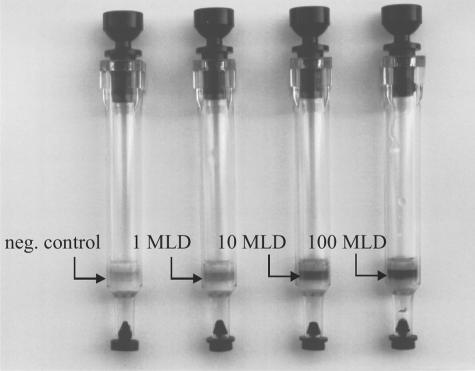
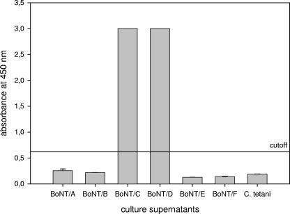
Figure 5 shows a dilution row of a BoNT/D culture supernatant. In specificity testing, strong positive signals were observed with all strains of types C and D, whereas all other strains were below the assay cutoff (Fig. 6).
FIG. 5.
Dilution of standardized BoNT/D culture supernatant in the optimized assay (toxicity is given as MLD per milliliter). Note the slight background in the negative (neg.) control.
FIG. 6.
Assay specificity tested with C. botulinum and C. tetani culture supernatants. BoNT/C and D cultures gave positive results. The other culture supernatants were below the assay cutoff.
Intra-assay relative coefficients of variation (CVs) ranged from 2.1% to 6.1% in type C and from 2.1 to 5.8 in type D, with the lower concentrations having the higher CV. For interassay variations, relative CVs from 3.8% to 8.2% (type C) and from 2.5% to 7.6% (type D) were calculated. Again, the lower concentrations had the higher CVs.
DISCUSSION
The mouse bioassay is still considered to be the “gold standard” for the detection of botulinum neurotoxins. It can detect the toxin in the lower picograms-per-milliliter range. Although this approach does not totally reflect the natural route of intoxication, it is the method of choice to measure the biological activity of the 150-kDa toxin with its heavy and light chains. For obvious reasons, the mouse bioassay needs to be replaced. It is time-consuming and expensive, and most important of all, it does not suit our modern understanding of animal welfare and protection.
Despite the fact that antigen-antibody-based in vitro assays reflect the antigenicity of the sample material and not necessarily the biological activity, they can be an important and helpful diagnostic tool. In all cases, when the biological activity does not need to be quantified or proved, the in vitro tests can deliver adequate data.
Thomas previously described an ELISA for the detection of BoNT/C and D (26). Rocke et al. developed an immunostick ELISA for the diagnosis of type C botulism in wild birds (22). Both diagnostic tests use polyclonal antibodies for capture and detection. These assay setups proved to be highly sensitive for type C and/or type D toxins, and the results were comparable with the mouse bioassay. Thomas (26), however, observed a disagreement between the ELISA results and the mouse bioassay in some samples. Since the polyclonal antibodies used in this assay were directed against the toxic and nontoxic components of the whole toxin complex, the different results may reflect the proportion of these components present in the sample (4, 26), with the mouse bioassay detecting the toxin only and the in vitro assay finding epitopes on the toxic and nontoxic proteins of the toxin complex.
The immunoaffinity chromatographic column assay developed here focused on the 150-kDa toxin only. Cross-reactivity with epitopes on the nontoxic proteins was avoided by using a monoclonal capture antibody, which was specific for the heavy chains of BoNT/C and D. The antibodies used enable the simultaneous detection of BoNT/C and D in one test, which facilitates the use of the assay. There is no need to keep separate columns, reagents, or antibodies in stock. Toxin typing cannot be performed. However, for a laboratory confirmation of botulism in animals or the detection of toxigenic C.botulinum, it is of secondary interest to know whether type C or D is present in the sample.
In its final assay setup, the toxin/sample solution was preincubated with the monoclonal and biotinylated polyclonal antibodies to achieve maximum sensitivity. The solution was applied to the column, and washing steps, the addition of streptavidin-poly-HRP 80 as a signal amplifier, and the precipitating substrate followed. The toxin can be detected semiquantitatively with the naked eye if a standard toxin dilution is run in parallel with the sample (Fig. 5). For more precise data, either the precipitated substrate was eluted and the absorbance was measured photometrically or a photometer for thedirect readout of the columns was used (Senova, Jena, Germany). With these assay conditions, 0.5 MLD ml−1 were detected after a 4-h preincubation for type D. For type C, the solution had to be incubated for 16 h to reach a comparable sensitivity. It is possible to lower this incubation period down to 30 min, if the loss in sensitivity is acceptable.
Crude culture supernatant was used for the optimization work and the sensitivity and specificity testing of this in vitro approach for two reasons. First, clinical, environmental, or culture samples do not contain the pure toxin, but they do contain the complex including the hemagglutinating and nontoxic nonhemagglutinating proteins as does the culture supernatant (19). Second, whether the nontoxic components affect the assay sensitivity had to be determined. It was shown that the data obtained in the in vitro assay corresponded well to the in vivo toxicity measured in the mouse bioassay. However, the dynamic range of the assay is narrow, which was already observed in an ELISA for the detection of BoNT/A, B, and E (12).
The column design of the assay offers several advantages compared to conventional ELISA techniques for the detection of botulinum neurotoxins. The toxin-antibody complex is captured out of the sample matrix. Larger sample volumes can thus be applied, and the sensitivity is increased. Sample components that might interfere with the immunological reaction are washed out if they are small enough to pass the assay disk. This is always the case when sterile filtered sample material/culture supernatant is used. Since the reagents were added in flowthrough, the column assay itself usually did not exceed 15 min. If the maximum sensitivity is not needed and the preincubation can be cut down to 30 min, the assay delivers a result within 45 min.
Systems for the automation of the assay have become available (Senova). However, it must be pointed out that the test is limited to smaller sample numbers and does not aim to be used in high-throughput laboratories.
In summary, an immunoaffinity chromatographic column test that detects BoNT/C and D as purified toxin or in pure culture supernatants was developed. Antibodies cross-reacting between BoNT/C and D have been used, enabling the simultaneous detection of the toxins. The sensitivity achieved is equal to that of the mouse bioassay. Further investigations will have to prove whether this assay is able todetect toxigenic C. botulinum types C and D by toxin identification in clinical and/or environmental samples or BoNT/C and D directly in complex clinical and environmental sample matrices.
This system should be readily adapted to the detection of the other toxin types. An additional and future development of the immunoaffinity columns might be the small-scale purification of BoNT/C and D, e.g., out-of-sample matrices, for use in other assays.
Acknowledgments
This work was supported by grant 0311128 from the Bundesministerium für Bildung und Forschung (Germany).
We are especially grateful to S. Kozaki at the Osaka Prefecture University, Japan, who provided us with the C. botulinum types C and D used in this work. We thank A. Leunig and M. Schmidt for their excellent laboratory work.
REFERENCES
- 1.AOAC. 1979. Clostridium botulinum and its toxins in foods. Official method 977.26. AOAC, Gaithersburg, Md.
- 2.Arnon, S. S., R. Schechtler, and T. V. Inglesby. 2001. Botulinum toxin as a biological weapon: medical and public health management. JAMA 285:1059-1070. [DOI] [PubMed] [Google Scholar]
- 3.Aureli, P., L. Fenicia, B. Pasolini, M. Gianfranceschi, L. M. McCroskey, and C. L. Hatheway. 1986. Two cases of type E infant botulism caused by neurotoxigenic Clostridium butyricum in Italy. J. Infect. Dis. 154:207-211. [DOI] [PubMed] [Google Scholar]
- 4.Betley, M. J., and H. Sugiyama. 1979. Noncorrelation between mouse toxicity and serologically assayed toxin in Clostridium botulinum type A culture fluids. Appl. Environ. Microbiol. 38:297-300. [DOI] [PMC free article] [PubMed] [Google Scholar]
- 5.Böhnel, H., B. Schwagerick, and F. Gessler. 2001. Visceral botulism—a new form of bovine Clostridium botulinum toxication. J. Vet. Med. A 48:373-383. [DOI] [PubMed] [Google Scholar]
- 6.Cobb, S. P., R. A. Hogg, D. J. Challoner, M. M. Brett, C. T. Livesey, R. T. Sharpe, and T. O. Jones. 2002. Suspected botulism in dairy cows and its implications for the safety of human food. Vet. Rec. 150:5-8. [DOI] [PubMed] [Google Scholar]
- 7.Collier, D. S. J., S. O. Collier, and P. D. Rossdale. 2001. Grass sickness—the same old suspects but still no convictions! Equine Vet. J. 33:540-542. [DOI] [PubMed] [Google Scholar]
- 8.Collins, M. D., and A. K. East. 1998. Phylogeny and taxonomy of the food-borne pathogen Clostridium botulinum and its neurotoxins. J. Appl. Microbiol. 84:5-17. [DOI] [PubMed] [Google Scholar]
- 9.Dahlenborg, M., E. Borch, and P. Radström. 2001. Development of a combined selection and enrichment PCR procedure for Clostridium botulinum types B, E, and F and its use to determine prevalence in fecal samples from slaughtered pigs. Appl. Environ. Microbiol. 67:4781-4788. [DOI] [PMC free article] [PubMed] [Google Scholar]
- 10.Dahlenborg, M., E. Borch, and P. Radström. 2002. Prevalence of Clostridium botulinum types B, E and F in faecal samples from Swedish cattle. Int. J. Food Microbiol. 82:105-110. [DOI] [PubMed] [Google Scholar]
- 11.Deutsches Institut für Normung. 1992. Mikrobiologische Untersuchung von Fleisch und Fleischerzeugnissen; Nachweis von Clostridium botulinum und Botulinum-Toxin. DIN 10102. Beuth, Berlin, Germany.
- 12.Doellgast, G. J., M. X. Triscott, G. A. Beard, J. D. Bottoms, T. Cheng, B. H. Roh, M. G. Roman, P. A. Hall, and J. E. Brown. 1993. Sensitive enzyme-linked immunosorbent assay for detection of Clostridium botulinum neurotoxins A, B, and E using signal amplification via enzyme-linked coagulation assay. J. Clin. Microbiol. 31:2402-2409. [DOI] [PMC free article] [PubMed] [Google Scholar]
- 13.Ferreira, J. L., S. Maslanka, E. Johnson, and M. Goodnough. 2003. Detection of botulinal neurotoxins A, B, E, and F by amplified enzyme-linked immunosorbent assay: collaborative study. J. AOAC Int. 86:314-331. [PubMed] [Google Scholar]
- 14.Frey, A., J. Di Canzio, and D. Zurakowski. 1998. A statistically defined endpoint titer determination method for immunoassays. J. Immunol. Methods 221:35-41. [DOI] [PubMed] [Google Scholar]
- 15.Galey, F. D., R. Terra, R. Adaska, M. A. Etchebarne, B. Puschner, E. Fisher, R. H. Whitlock, T. Rocke, T. Willoughby, and E. Tor. 2000. Type C botulism in dairy cattle from feed contaminated with a dead cat. J. Vet. Diagn. Investig. 12:204-209. [DOI] [PubMed] [Google Scholar]
- 16.Gessler, F., and H. Böhnel. 1999. Production and purification of Clostridium botulinum type C and D neurotoxin. FEMS Immunol. Med. Microbiol. 24:361-367. [DOI] [PubMed] [Google Scholar]
- 17.Gessler, F., and H. Böhnel. 2004. Nachweis von Botulinum-Neurotoxinen—ein methodischer Über- und Ausblick. Tierärztl. Umsch. 59:5-9. [Google Scholar]
- 18.Große-Herrenthey, A. 2004. Personal communication.
- 19.Johnson, E. A., and M. Bradshaw. 2001. Clostridium botulinum and its neurotoxins: a metabolic and cellular perspective. Toxicon 39:1703-1722. [DOI] [PubMed] [Google Scholar]
- 20.Lund, B. M., and M. W. Peck. 2000. Clostridium botulinum, p. 1057-1109. In B. M. Lund, A. C. Baird-Parker and G. W. Gould (ed.), The microbiological safety and quality of food. Aspen, Gaithersburg, Md.
- 21.Meng, X., T. Karasawa, K. Zou, X. Kuang, X. Wang, C. Lu, C. Wang, K. Yamakawa, and S. Nakamura. 1997. Characterization of a neurotoxigenic Clostridium butyricum strain isolated from the food implicated in an outbreak of food-borne type E botulism. J. Clin. Microbiol. 35:2160-2162. [DOI] [PMC free article] [PubMed] [Google Scholar]
- 22.Rocke, T. E., S. R. Smith, and S. W. Nashold. 1998. Preliminary evaluation of a simple in vitro test for the diagnosis of type C botulism in wild birds. J. Wildl. Dis. 34:744-751. [DOI] [PubMed] [Google Scholar]
- 23.Rooney, J. R., and M. E. Prickett. 1976. New puzzler for equine practitioners: shaker foal syndrome. Mod. Vet. Pract. 48:44-45. [Google Scholar]
- 24.Sandler, R. J., T. E. Rocke, M. D. Samuel, and T. M. Yuill. 1993. Seasonal prevalence of Clostridium botulinum type C in sediments of a northern California wetland. J. Wildl. Dis. 29:533-539. [DOI] [PubMed] [Google Scholar]
- 25.Smith, L. D. S., and H. Sugiyama. 1988. Botulism, the organism, its toxin, the disease. C. C. Thomas, Springfield, Ill.
- 26.Thomas, R. J. 1991. Detection of Clostridium botulinum types C and D toxin by ELISA. Aust. Vet. J. 68:111-113. [DOI] [PubMed] [Google Scholar]
- 27.Van Ermengem, E. P. 1897. Über einen neuen Bacillus und seine Beziehung zum Botulismus. Z. Hyg. Infektionskrankh. 26:1-56. [Google Scholar]


