Abstract
Previous studies demonstrated that neonatal mice up to about a week old are less susceptible than adult mice to infection by intraperitoneal inoculation with mouse-passaged scrapie. In peripherally inoculated adult mice, scrapie replicates in lymphoid tissues such as the spleen before invading the central nervous system. Here, we investigated scrapie susceptibility in neonatal mice in more detail, concentrating on spleen involvement. First, we demonstrated that neonatal mice are about 10 times less susceptible than adults to intraperitoneal scrapie inoculation. Then we injected mice intraperitoneally with a scrapie dose that produced disease in all mice inoculated at 10 days or older but in only about a third of neonatally inoculated mice. In this experiment, spleens collected 70 days after scrapie injection of mice 10 days old or older almost all contained pathological prion protein, PrPSc, and those that were bioassayed all contained high infectivity levels. In contrast, at this early stage, only two of six spleens from neonatally inoculated mice had detectable, low infectivity levels; no PrPSc was detected, even in the two spleens. Therefore, neonatal mice have an impaired ability to replicate scrapie in their spleens, suggesting that replication sites are absent or sparse at birth but mature within 10 days. The increase in susceptibility with age correlated with the first immunocytochemical detection of the normal cellular form of prion protein, PrPc, on maturing follicular dendritic cell networks. As lymphoid tissues are more mature at birth in sheep, cattle, and humans than in mice, our results suggest that in utero infection with scrapie-like agents is theoretically possible in these species.
Transmissible spongiform encephalopathies (TSEs, or prion diseases) are often acquired by dietary, environmental, or iatrogenic exposure to infection. For example, consumption of contaminated meat and bone meal by cattle has been implicated as the major driver of the bovine spongiform encephalopathy (BSE) epidemic (55). BSE has spread to a number of other species, including humans (as variant Creutzfeldt-Jakob disease [vCJD]), probably also by a dietary route (44, 57). There is evidence that scrapie in sheep (14, 49) and chronic wasting disease (CWD) in mule deer (40, 41) can be acquired by contact or environmental exposure, although the mechanism of transmission in each case has not been established. Furthermore, there have been several instances of the iatrogenic transmission of sporadic CJD in humans and scrapie in sheep and goats, associated with the injection of contaminated pharmaceuticals (10, 56). Recently, there have also been two probable transmissions of vCJD by blood transfusion (31, 47).
Following the experimental TSE infection of animals by feeding or by other peripheral routes, the infectious agent usually replicates and accumulates in secondary lymphoid tissues at an early stage, long before it becomes detectable in the central nervous system (27). Studies in several rodent models have shown that, following peripheral inoculation, this lymphoid replication phase is critical for the subsequent spread of infection to the nervous system (19, 20). Replication in the lymphoid tissues is accompanied by the accumulation of an abnormally folded form of the host prion protein, PrP, which can be detected by its relative protease resistance (15). The presence of this pathological form, termed PrPSc, is generally regarded as a marker for infection. PrPSc is detected in the lymphoid tissues of patients with vCJD (24, 54), deer with CWD (50), and sheep with natural scrapie (53) both at the clinical phase of the disease and preclinically. In the case of vCJD and sheep scrapie, infectivity in lymphoid tissues has also been detected directly by bioassay in mice (8, 22).
In the lymphoid tissues of mice experimentally infected with scrapie, PrPSc accumulation is intimately associated with the processes of the follicular dendritic cells (FDCs) of the lymphoid follicles (7, 26). In the naturally occurring diseases vCJD, CWD, and scrapie, the site of pathological PrP accumulation within lymphoid tissues is also consistent with an FDC association (1, 24, 50). There are now multiple lines of evidence from mouse TSE models that FDCs are critical for the replication and accumulation of infectivity in the lymphoid tissues (33). Mice that have genetically or transgenically induced deficiencies of the immune system that lead to an absence of mature FDCs also fail to replicate scrapie in their spleens and, as a result, are relatively difficult to infect by a peripheral route (7, 19, 28, 37). Furthermore, treatments that temporarily dedifferentiate FDCs (35, 36, 38, 42) or interfere with their function (29, 34) also interfere with scrapie replication in the lymphoid tissues, reduce susceptibility to infection, and impair the spread of infection to the central nervous system.
Studies in transgenic mice deficient in PrP have shown that expression of this protein is required for an animal to be susceptible to TSE infection (9, 39). In the lymphoid tissues of uninfected mice, high levels of the normal cellular form of PrP, PrPc, are present on FDCs (6). A series of studies on the ME7 mouse scrapie model, using bone marrow grafting between PrP-expressing and PrP-deficient mice, has indicated that replication in lymphoid tissues and overall susceptibility depend on PrP expression by cells such as FDCs that are radiation resistant and not derived from bone marrow (7). In this model, expression of PrP by bone marrow-derived cells such as lymphocytes has little or no influence on the disease.
vCJD has occurred predominantly in young adults, suggesting that age-related factors may influence susceptibility and/or levels of exposure to BSE (21). Furthermore, epidemiological evidence indicates that most cattle developing BSE were infected as calves (3). Similarly, it has been suggested that young lambs are more susceptible than adults to natural infection with scrapie (11). These indications of possible age-related susceptibility factors in humans, cattle, and sheep prompted us to reexamine age-related variation in the susceptibility of mice to peripheral scrapie inoculation. Using doses that were 100% lethal to adult mice, it was found previously that some neonatally inoculated mice survived peripheral scrapie challenge (46). Furthermore, the neonatally inoculated mice that developed scrapie did so with a wider incubation period range than those inoculated when they were older. Susceptibility to scrapie and the incubation period range were found to become adult-like about a week after birth (46). We now have the opportunity to reexamine this phenomenon in light of recent insights into scrapie pathogenesis in peripheral lymphoid tissues, in particular, concerning the involvement of FDCs in pathogenesis.
The aims of the present study were, first, to determine more precisely the relative susceptibilities of mice peripherally inoculated with scrapie as newborns and as adults and, second, to investigate the ability of the neonatal spleen to sustain scrapie agent replication. We also investigated how disease susceptibility relates to the developmental maturation of the spleen, particularly in terms of the expression of PrPc on maturing FDC networks. A major function of FDCs in adult mice is to trap antigens in the form of immune complexes via antibody or complement receptors (52). As antigen trapping via complement receptors has been implicated in the scrapie infection of lymphoid tissues (29, 34), we also examined markers for this function within the developing mouse spleen.
MATERIALS AND METHODS
Mice.
C57BL/Dk (hereafter referred to as C57BL) mice were used in all experiments. Male and female mice were used in approximately equal numbers.
Scrapie inoculations. (i) Experiment 1.
Brain from a clinically affected C57BL mouse, infected with the ME7 strain of mouse-passaged scrapie, was homogenized in physiological saline at a 10−1 dilution, and serial 10-fold dilutions were prepared in saline to a 10−5 dilution. Groups of C57BL mice, aged either 0 to 1 day (neonates) or 19 to 43 days (adults), were injected intraperitoneally (i.p.) with 20 μl of inoculum at each dilution (neonates to 10−4, adults to 10−5). Immediately after inoculation, the abdomens of neonatal mice were sprayed with Novoseal in order to prevent inoculum leakage (inoculum leakage was not a problem in older mice). Mice were scored for signs of clinical scrapie and sacrificed at a standard clinical end point which was used to determine the incubation period (13). Mice that did not develop scrapie were maintained until they showed signs of intercurrent illness or old age. Brains were collected from all mice, and scrapie diagnosis was confirmed histopathologically from the presence or absence of vacuolar degeneration (18). The relative susceptibility of newborn and adult mice was estimated from the dilutions of inoculum resulting in the infection of 50% of mice (ID50s) measured in each age group. For this analysis, all mice with positive histopathological scrapie diagnosis were counted as infected and mice that survived beyond 550 days postinoculation with no clinical or histopathological signs of scrapie were counted as survivors. The ID50 was calculated by the Spearman-Kärber method (17) from the proportion infected at each dilution.
(ii) Experiment 2.
Groups of C57BL mice aged 0 to 1, 10, 14, or 29 to 46 days were inoculated i.p. with 20 μl of a 10−2 dilution of ME7 brain homogenate as described above. Twelve mice from each group were killed at 70 days postinjection (d.p.i.) and their spleens collected for PrPSc detection by Western blotting and for infectivity bioassay (in the oldest group, only from mice inoculated at 29 days old). Remaining mice were assessed for clinical signs as described above and killed at the terminal stage of disease. Mice that did not show clinical scrapie signs were sacrificed with intercurrent disease or at 590 to 600 days after injection, when the experiment was terminated. Brains from clinically affected scrapie mice and from apparent survivors were assessed histopathologically as described above. Spleens from neonatally injected mice that developed clinical disease and from apparent survivors were collected and frozen for PrPSc immunoblot analysis.
(iii) Experiment 3.
Three groups of C57BL mice, aged 0 to 2, 9 to 11, and 20 to 37 days, were inoculated intracerebrally (i.c.) with 20 μl of a 10−2 dilution of ME7 brain homogenate and assessed for clinical and histopathological signs of scrapie as described above.
(iv) Statistical analysis.
Comparisons between incubation periods were made using MINITAB statistical software.
Infectivity bioassays.
Individual half spleens from experiment 2, collected at 70 d.p.i., were homogenized in physiological saline at a 5% concentration, and 20 μl was injected i.c. into groups of 12 C57BL adult indicator mice. Bioassay mice were clinically and neuropathologically assessed as described above, and the titer in each spleen was estimated from the mean incubation period by using a standard dose-incubation period response curve, derived from titration data for ME7-infected spleen tissue injected i.c. into C57BL mice. This curve showed a linear relationship between the log10 dose (ID50 units/mouse) and incubation period (days) over the relevant dose range (R2 = 0.9998, intercept = 14.4057 log10 ID50 units, slope = −0.05347 log10 ID50 units/day). The titer of infectivity (y) in each spleen was calculated using the formula y = −0.05347x + 14.4057 + 3.0 log10 i.c. ID50 units/g, where x is the mean incubation period (days) in the bioassay mice.
PrPSc immunoblotting.
PrPSc was purified from halved spleens from experiment 2 by using a differential extraction method as previously described (15). Briefly, spleen homogenates were treated with 50 μg/ml proteinase K (Sigma). PrPSc was extracted and sedimented using 2% Sarkosyl solution followed by high-speed ultracentrifugation. The detergent-soluble fraction containing PrPc was discarded, and the pellets were dried and resuspended in sodium dodecyl sulfate buffer. Samples were run on a 12% sodium dodecyl sulfate-polyacrylamide gel and then transferred to polyvinylidene difluoride membranes, where they were probed overnight using a 1/5,000 dilution of rabbit anti-mouse polyclonal antibody, 1B3 (16). An alkaline phosphatase-conjugated goat anti-rabbit secondary antibody (Jackson Immunoresearch) was applied at a 1/10,000 dilution to detect antibody binding, and the immunoblots were developed using 5-bromo-4-chloro-3-indolylphosphate (BCIP)-nitroblue tetrazolium (Sigma).
Immunocytochemistry. (i) Tissues.
Spleens were collected from six to nine uninfected C57BL mice at each of the following ages: 1 day, 4 days, 8 days, 10 days, 14 days, 26 days, and 30 days. As negative controls, similar numbers of spleens were collected at the same ages from transgenic mice in which the Prnp gene had been disrupted, preventing PrP expression (129/Ola background) (39). The spleens were frozen immediately in liquid nitrogen and embedded in optimum cutting temperature compound. Cryostat sections (10 μm) were cut, fixed in acetone, and dried.
(ii) PrP immunolabeling.
After blocking nonspecific binding sites with normal goat serum, sections were labeled with a polyclonal rabbit PrP-specific antibody, 1B3 (16). Adjacent control sections were incubated with normal rabbit serum. After washing, slides were incubated with Alexa 488-conjugated goat anti-rabbit serum (Molecular Probes Inc.), washed, and mounted in fluorescent mounting medium (Dako).
(iii) Double PrP and FDC-M1 immunolabeling.
Following PrP labeling as described above, irrelevant binding sites were blocked with normal mouse serum and the sections were incubated with FDC-M1 rat monoclonal antibody (30) (a gift from M. Kosco-Vilbois, Serono, Geneva, Switzerland), which recognizes an uncharacterized epitope on immature and mature FDCs (5). Adjacent control sections were incubated with normal rat serum. After washing, biotinylated mouse anti-rat antibody (Jackson) was applied, followed, after washing, by streptavidin-conjugated Cy3 (Jackson). Slides were mounted as described above.
(iv) Double PrP and CR1 or FDC-M2 immunolabeling.
The same procedure described above was used for PrP/CR1 or PrP/FDC-M2 labeling, substituting complement receptor 1 (CR1)-specific rat monoclonal antibody (Pharmingen) or FDC-M2 rat monoclonal antibody (a gift from M. Kosco-Vilbois) for the FDC-M1 antibody. The CR1-specific antibody (CD35) recognizes complement receptor 1, while the FDC-M2 antibody recognizes complement component 4 (C4) (51).
(v) Confocal microscopy.
The sections were viewed using a Leica TCS NT microscope.
RESULTS
Susceptibility of neonatal and adult mice to peripheral scrapie infection.
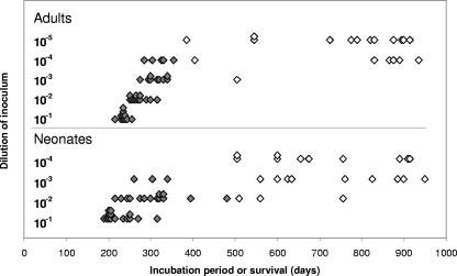
Intraperitoneal inoculation of adult mice with serial 10-fold dilutions of ME7 scrapie inoculum (experiment 1) resulted in a 100% incidence of scrapie in the 10−1 and 10−2 dilution groups (Fig. 1). Only a proportion of mice in the 10−3 and 10−4 dilution groups developed scrapie, and all mice injected with a 10−5 dilution survived without showing clinical or neuropathological signs of scrapie. As expected, the incubation period lengthened with dilution. In contrast, in neonatally i.p. injected mice, a 100% scrapie incidence was seen only in the 10−1 dilution group. Some mice survived in the 10−2 and 10−3 dilution groups, and no mice developed disease in the 10−4 dilution group (Fig. 1). Again, incubation periods tended to increase with dilution. For analysis, survivors were defined as those mice that survived beyond 550 d.p.i., showing no clinical or neuropathological signs of scrapie, i.e., at least 50 days longer than the last mouse in the experiment that developed clinical scrapie. Based on the proportion of infected mice at each dilution and using the Spearman-Kärber method, the ID50 was 10−2.6 ± 100.2 for neonatal mice compared to 10−4.0 ± 100.2 for adult mice. Thus, neonatal mice are about 10-fold less susceptible to i.p. scrapie challenge than adult mice.
FIG. 1.
Incubation period of disease or survival time following i.p. inoculation of adult mice and neonatal mice with 10-fold dilutions of ME7 scrapie-infected brain homogenate. Shaded symbols represent individual terminally affected mice with a histolopathologically confirmed scrapie diagnosis. Unshaded symbols represent mice with a negative clinical and histopathological diagnosis that survived beyond the last clinical case in their challenge age group.
Effect of age on susceptibility to peripheral scrapie infection.
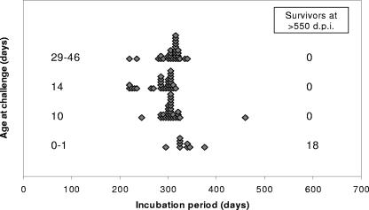
Mice of various ages were inoculated i.p. with a 10−2 dilution of ME7 scrapie brain homogenate (experiment 2). All mice inoculated at 10, 14, and 29 to 46 days old developed scrapie symptoms and vacuolar scrapie pathology, with mean incubation periods of 308 ± 6, 284 ± 6, and 305 ± 5 days, respectively (Fig. 2; Table 1). There were no survivors following inoculation of mice of these ages, although one mouse injected at 10 days old developed scrapie following a prolonged incubation period of 460 days, suggesting that 10-day-old mice are not all fully susceptible. In contrast, injection of 0- to 1-day-old mice gave 18 of 27 survivors after 550 d.p.i., showing no clinical or histopathological signs of scrapie. The nine mice in this group that developed scrapie (Fig. 2; Table 1) showed typical clinical symptoms and spongiform change in the brain following a mean incubation period of 334 ± 7 days, which was significantly longer than each of the mean incubation periods in the older age groups by at least 26 days (e.g., comparing the 0- to 1-day and 10-day-old groups, P < 0.05, using a two-tailed t test). In contrast to the results following i.p. inoculation, when 0- to 2-day-old, 9- to 11-day-old, and 20- to 37-day-old mice were injected i.c. with a 10−2 ME7 scrapie brain homogenate (experiment 3), a 100% incidence of disease was seen in all three groups. There were also, for i.c. injected mice, no significant differences in incubation periods between the three age groups, as follows: 162 ± 2 days (n = 10) in mice injected as newborns, 158 ± 2 days (n = 13) in mice injected at 9 to 11 days old, and 160 ± 2 days (n = 9) in mice injected at 20 to 27 days old.
FIG. 2.
Incubation period of disease following i.p. inoculation of 0- to 1-, 10-, 14-, and 29- to 46-day-old mice with a 10−2 dilution of ME7 scrapie-infected brain homogenate. Shaded symbols represent individual terminally affected mice with a histopathologically confirmed scrapie diagnosis. The numbers of mice surviving to beyond 550 days with a negative histopathological scrapie diagnosis are indicated.
TABLE 1.
Intraperitoneal inoculation of mice of different ages with ME7 scrapie, showing the proportion of mice that developed disease, mean incubation periods, and the proportion in which PrPSc was detected in spleen by immunoblotting
| Age at inoculation (days) | Proportion of mice with clinical scrapie | Incubation period (mean ± SEM) (days) | Proportion of mice with detectable PrPSc in their spleensa
|
||
|---|---|---|---|---|---|
| 70 d.p.i. | Terminal scrapie | Survivors | |||
| 0-1 | 9/27b | 334 ± 7 | 0/12 | 9/9 | 1/18b |
| 10 | 28/28 | 308 ± 6 | 11/12 | NT | NA |
| 14 | 28/28 | 284 ± 6 | 12/12 | NT | NA |
| 29-46 | 29/29 | 305 ± 5 | 12/12 | NT | NA |
NT, not tested; NA, not applicable.
A total of 18 of 27 mice, referred to as survivors, remained free of clinical signs of scrapie beyond 550 d.p.i.; no TSE-related pathology was detected in their brains.
PrPSc accumulation in the spleens of mice inoculated at different ages.
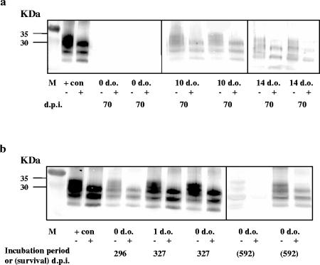
As expected from previous studies in adult mice (15, 34, 36, 38), PrPSc was detected by immunoblotting in all spleens taken at 70 d.p.i. from mice inoculated i.p. at 29 days old (Table 1). PrPSc was also detected in spleens collected at 70 d.p.i. from all mice inoculated at 14 days old and from 11 of 12 mice inoculated at 10 days old. However, PrPSc could not be detected in any of the spleens collected at this time point from neonatally injected mice (Fig. 3a; Table 1). Spleens collected at the clinical end point from mice that had been inoculated as neonates and eventually developed clinical scrapie all showed accumulations of PrPSc (Fig. 3b; Table 1). PrPSc was also detected in the spleen of one clinically negative neonatally injected mouse 592 days postinjection. The spleens of all other surviving mice in this group were negative for PrPSc (Fig. 3b; Table 1).
FIG. 3.
Immunoblot analysis of spleen tissue taken from mice inoculated i.p. with ME7 scrapie at 0 to 1, 10, or 14 days old (d.o.). Samples were treated in the presence (+) or the absence (−) of proteinase K (PK) prior to electrophoresis. (a) At 70 d.p.i, no PK resistant accumulations of PrPSc were detected in the spleens of mice inoculated neonatally, but, at this stage, PrPSc could be detected in the spleens of mice injected at 10 and 14 days old (also at 29 days old [not shown]). (b) PrPSc was detected in the spleens of clinically affected mice inoculated as neonates (left hand panel). PrPSc was undetectable in the spleens of neonatally inoculated, clinically negative mice, except in one which had readily detectable PK-resistant accumulations at 592 d.p.i. (right hand panel). All gels included spleen extracts from a clinically affected mouse inoculated as an adult as a positive control (+con). Lane M contains molecular size markers.
Infectivity levels in spleens collected at 70 days after i.p. inoculation.
Previous studies of ME7 in adult mice have shown that infectivity titers in the spleen reach plateau levels by 70 days after an i.p. injection (12). Four spleens, randomly selected from those collected at 70 d.p.i., were bioassayed individually from each of the groups of mice inoculated at 10, 14, and 29 days old by i.c. injection into groups of indicator mice. The 100% incidence of disease and short incubation periods in the indicator mice showed that all of these spleens contained high levels of infectivity, with individual titers ranging from 5.4 to 7.2 log10 i.c. ID50 units/g of tissue (Table 2). There were no consistent differences between the titers measured in the spleens of mice inoculated at the three different ages. In contrast, at 70 d.p.i., infectivity was detected in only two out of six spleens bioassayed from mice inoculated at 0 to 1 day old (Table 2). Levels of infectivity detected in these two spleens were at least 10-fold lower (4.5 log10 i.c. ID50 units/g) than those in spleens of mice inoculated at 10 days or older. The same two spleens were found to be PrPSc negative by immunoblotting (see above) This inconsistency between the bioassay and immunoblot results may simply indicate that accumulation of PrPSc lags behind the accumulation of infectivity in the early stages of pathogenesis, as has been observed in the spleens of mice infected as adults (36).
TABLE 2.
Bioassays of individual half spleens collected from C57BL mice 70 days after intraperitoneal injection with ME7 scrapie
| Age at inoculation (days) | Spleen no. | Proportion of assay mice with scrapie pathologya | Incubation period (mean ± SEM) (days) | No. of pathology negative assay mice after 400 days | Estimated titer (log10 i.c. ID50 units/g)b |
|---|---|---|---|---|---|
| 0-1 | 1 | 0 | 9 | ||
| 2 | 0 | 11 | |||
| 3 | 0 | 8 | |||
| 4 | 10/10 | 242 ± 5 | 0 | 4.5 | |
| 5 | 0 | 12 | |||
| 6 | 10/10 | 242 ± 8 | 0 | 4.5 | |
| 10 | 7 | 11/11 | 221 ± 6 | 0 | 5.6 |
| 8 | 12/12 | 206 ± 8 | 0 | 6.4 | |
| 9 | 11/11 | 195 ± 3 | 0 | 7.0 | |
| 10 | 12/12 | 197 ± 2 | 0 | 6.9 | |
| 14 | 11 | 11/11 | 209 ± 6 | 0 | 6.2 |
| 12 | 10/10 | 224 ± 6 | 0 | 5.4 | |
| 13 | 12/12 | 207 ± 6 | 0 | 6.3 | |
| 14 | 12/12 | 198 ± 4 | 0 | 6.8 | |
| 29 | 15 | 10/10 | 215 ± 8 | 0 | 5.9 |
| 16 | 12/12 | 199 ± 3 | 0 | 6.8 | |
| 17 | 10/10 | 199 ± 4 | 0 | 6.8 | |
| 18 | 11/11 | 190 ± 4 | 0 | 7.2 |
Mice dying of intercurrent disease before 160 days postinjection and mice for which no pathology report was possible are excluded from the data.
Limit of the assay was approximately 3.0 log10 i.c. ID50 units/g.
PrPc and FDC immunolabeling in the spleens of uninfected young mice.
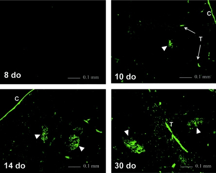
From the results described above, we concluded that the relative resistance of newborn mice to peripheral scrapie challenge is likely to be related to the inefficiency with which scrapie initially replicates in their spleens. As TSE replication depends on the expression of PrP (9, 39), we determined the time at which PrPc becomes detectable in the spleens of uninfected young mice by immunofluorescent labeling. No PrPc was detected in the spleens of 1-, 4-, or 8-day-old mice (six mice per age group). Small foci of PrPc labeling were seen within developing spleen follicles in four out of nine 10-day-old mice tested, and more extensive labeling was seen in the follicles of all mice 14 days old and older (Fig. 4). From 10 days onward, PrPc labeling was also seen in the spleen capsule and trabeculae, as has been reported previously for adult mice (32). No labeling was seen in control spleen sections from age-matched PrP-deficient mice or in control spleen sections from PrP-expressing mice where the PrP-specific antibody had been replaced with normal rabbit serum (not shown).
FIG. 4.
PrPc detection by immunofluorescent labeling in the spleens of uninfected mice at 8, 10, 14, and 30 days old (do). Labeling was first seen in a proportion of mice at 10 days, associated with some developing follicles (arrowheads), the spleen capsule (C), and trabeculae (T).
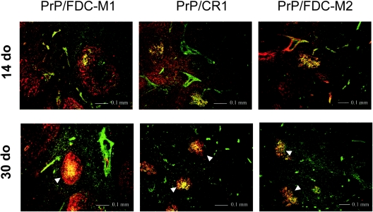
To investigate the cellular association of PrPc within the follicles, sections were double labeled for PrP and other markers that are known to be associated with FDCs in adult mice (the epitopes recognized by the FDC-M1 and FDC-M2 monoclonal antibodies and also CR1:CD35). The first of these FDC markers to appear in the spleen was the FDC-M1 epitope, showing a perivascular punctate pattern in 1-day-old mice. By 4 days, diffuse FDC-M1 labeling was seen around the periphery of the immature white pulp. This labeling pattern was closely similar to that reported in mice of this age by other groups and possibly indicates the presence of FDC precursors (5, 25). At 10 to 14 days old, diffuse or dispersed cellular FDC-M1 labeling was still seen around the periphery of the white pulp (Fig. 5). At this age, most FDC-M1-labeled cells were unlabeled by the PrP-specific antibody, but some discrete, strongly FDC-M1-positive cell clusters were also PrP positive (Fig. 5). All PrP-positive cell clusters seen within the follicles were also FDC-M1 positive. With increasing age, FDC-M1 labeling became more localized within the follicles, showing an adult-like appearance by 26 to 30 days and a much more precise colocalization with PrP labeling (Fig. 5). These results suggest that the appearance of PrPc on FDCs is related to maturation events in the developing follicle. However, further interpretation based on FDC-M1 labeling is limited by the fact that the epitope recognized by the FDC-M1 antibody is uncharacterized.
FIG. 5.
Double immunofluorescent labeling using PrP- (green) and FDC-M1-, CR1-, or FDC-M2 (red)-specific antibodies in the spleens of 14- and 30-day-old mice. Colocalization of PrP and FDC-M1, CR1, or FDC-M2 (yellow) is seen on FDC networks in developing follicles (arrowheads).
The FDC-M2 antibody recognizes a form of complement C4, which, like CR1, is a marker for the complement-dependent antigen-trapping function of FDCs (51). Labeling with both the CR1 and FDC-M2 antibodies was first seen in mice 14 days old, slightly later than PrPc labeling but on precisely the same FDC-M1-positive cell clusters within the follicles (Fig. 5). In 26- to 30-day-old mice, as in older mice, more numerous discrete clusters of FDCs within the follicles labeled strongly with FDC-M1, FDC-M2, and CR1-specific antibodies, in each case showing a precise colocalization with PrP labeling (Fig. 5). In this study, the FDC-M1-, FDC-M2-, and CR1-labeling patterns seen in PrP-deficient mice were similar to those seen in PrP-expressing mice of the same ages (not shown).
DISCUSSION
We demonstrate here that neonatal mice are about 10 times less susceptible to peripheral ME7 scrapie infection than young adult mice, becoming almost fully susceptible by 10 days of age. Our data further suggest that the relative resistance of neonatal mice is related to an impaired ability of their spleens to sustain infection. It is therefore likely that critical cellular sites in the spleen that are necessary for the establishment of infection are not fully developed at birth but mature within 10 days in most mice. In mice inoculated as adults, FDCs of the lymphoid follicles have been shown to be critical for ME7 scrapie replication in peripheral lymphoid tissues (35-38). Furthermore, there is evidence that replication is likely to depend on PrP expression by FDCs in adult mice (7). In this study, we demonstrate that the transition to an adult-like scrapie susceptibility correlates with the first detection of PrPc on maturing splenic FDC networks at about 10 days old. However, the expression of PrPc must be put into the context of other maturational events that are occurring at this time.
FDCs trap and retain intact antigens, complexed to antibody and/or complement, for presentation to B cells (52). This process is thought to be central to the selection of high-affinity B-cell clones, isotype switching of antibodies, and the generation of B-cell memory. FDCs may trap immune complexes via CR1 and CR2 or via the antibody receptor FcγRII. In previous studies, mice deficient in complement components or their receptors were shown to be relatively resistant to peripherally injected scrapie (29, 34), suggesting that trapping via complement receptors facilitates the infection of lymphoid tissues. On the other hand, a deficiency in antibodies or their receptors has no effect on scrapie pathogenesis, implying that lymphoid tissue infection does not depend on antigen trapping via antibody receptors (29).
In a study elsewhere on the ontogeny of FDCs in BALB/c mice, FDC-M1 labeling, presumably of FDC precursors, first appeared at postnatal day 3, followed by the complement receptor labeling of a few FDC-M1-positive clusters by day 7 (using an antibody that recognized both CR1 and CR2, CR1/2, or CD21/35) (5). At day 10, CR1/2 labeling was more prominent and complement C4, recognized by the FDC-M2 antibody, was evident on a few FDC clusters. Trapping of injected immune complexes was demonstrable at day 7. In another study elsewhere in C57BL/6N mice, complement receptors (CR1/2) were first seen at day 12 in the spleen, somewhat later than in the BALB/c study (25). In the present study in a different C57BL mouse line (C57BL/Dk), CR1 and C4 were first detected on FDCs at 14 days of age. There is evidence that CR2 appears slightly earlier than CR1 in mouse lymph nodes (25); though not documented, this may also be the case in the spleen, perhaps accounting for the relatively late detection of complement receptors in our study. Discrepancies in timing between the three studies may also be due to genetic differences between the mouse strains used, environmental factors, or technical differences in the detection methods used. Nevertheless, the consensus of these studies is that antigen trapping by splenic FDCs via complement receptors becomes active in mice during the second postnatal week.
We found that PrP antibody labeled a subset of FDC-M1-positive cells in developing spleen follicles from 10 days onward. The simplest explanation for the increase in susceptibility to scrapie at this time is therefore that it is related primarily to the appearance of PrPc on FDCs. However, markers for complement-dependent antigen trapping (CR1 and C4) appeared only slightly later, colocalizing precisely with PrP. This raises the possibility that maturation of this trapping function also contributes to the increase in scrapie susceptibility in these young mice. The close temporal and spatial association of the appearance of PrPc with markers for complement-dependent antigen trapping might also suggest an involvement of PrPc in this function.
Newborn mice are clearly not absolutely resistant to peripheral scrapie challenge (46). In the study reported here, about a third of the mice injected as neonates accumulated infectivity in their spleens in the first few weeks after inoculation and the same proportion went on to develop clinical scrapie. Immunoblot analysis of the spleens from neonatally inoculated mice at the end point of disease demonstrated high levels of PrPSc, similar to those in mice inoculated as adults. These results suggest that the early pathogenesis of scrapie in the neonatally inoculated mice that go on to develop clinical disease involves replication in the spleen, as is the case in adults. However, there is a slight delay in disease progression in these neonatally inoculated mice, as shown by the relatively low levels of infectivity in the spleen at 70 d.p.i., the failure to detect PrPSc at this stage, and the slightly extended incubation periods.
There are several possible explanations as to how the spleens of some neonatally inoculated mice become infected. It may simply be the case that functionally immature FDCs in the spleen are infected by a relatively inefficient mechanism. Another possibility is that the spleens of these mice are not infected directly but become infected after an initial replication phase in other lymphoid tissues such as lymph nodes. In adult mice, splenectomy before peripheral inoculation with ME7 scrapie dramatically lengthens the incubation period, showing that neuroinvasion occurs most rapidly via the spleen in this model (20). However, splenectomized mice still develop disease, following agent replication in other lymphoid tissues and neuroinvasion from these sites. In mice, there is evidence that follicles with CR1/2 networks appear in some lymph nodes a few days earlier than in the spleen (25). Therefore, it is possible that initial replication in lymph nodes is relatively more significant for disease progression in very young mice than in adults. Indeed, the effect of splenectomy on scrapie incubation period in newborn mice is not as clear-cut as the effect in adults (20).
Whether spleens of neonatally injected mice become infected directly or via other lymphoid tissues, there is still likely to be an interval of at least a week between inoculation and the first appearance of PrPc and functional FDCs in any lymphoid tissue. During this time, it is possible that infectivity is sequestered elsewhere and becomes available to FDCs once they are sufficiently mature to support replication of the agent. In adult mice, there is often a “zero phase” following peripheral scrapie inoculation before increasing levels of infectivity can be detected in the spleen by bioassay (27). This apparent delay may reflect the limits of sensitivity of the bioassay, but it could also represent a period during which infectivity is sequestered. Whether or not a neonatally inoculated mouse becomes infected may depend on the balance between the degradation of infectivity by cells such as macrophages or dendritic cells and the sequestration of infectivity in a compartment from which it can gain access to functionally mature FDCs several days later. Alternatively, initial infection of some neonatally inoculated mice might occur independently of the lymphoid system, as has been demonstrated in immunodeficient adult mice injected peripherally with high doses of scrapie (19, 37). A plausible explanation for this phenomenon in adults is that it involves direct infection of the peripheral or central nervous system, and this could also be the case in some neonatally inoculated mice, with infectivity spreading from the nervous system to the lymphoid tissues at a later stage.
As humans, sheep, and cattle are more immunologically mature at birth than mice, our observations are not directly relevant to postnatal age-related susceptibility factors in the natural TSEs of these species. However, they are relevant when considering the possibility of in utero infection. Immunohistochemical studies of fetal sheep lymph nodes have demonstrated the presence of FDCs in the primary follicles during the last month of gestation (23). Similarly, in humans, FDC networks have been observed in immature B-cell follicles in lymph nodes from the 16th gestational week (4) and in spleen from the 26th gestational week (43). Although we are not aware of any studies of prenatal PrPc expression in the lymphoid tissues of humans and sheep, there is therefore a theoretical possibility of in utero infection if the fetus were exposed to infectivity. In sheep, there is evidence of maternal transmission in natural scrapie (11, 14), but it is not clear whether transmission from ewe to lamb occurs before, during, or after birth. Both infectivity and substantial accumulations of PrPSc have been demonstrated in the placentas of pregnant scrapie-affected ewes (2, 45, 48), but it is not known whether this results in exposure of the developing fetus. There is no evidence at present of maternal transmission in acquired forms of CJD.
The evidence presented here regarding the age-related susceptibility of mice to scrapie clearly demonstrates that developmental factors in the immune system influence the establishment of infection in lymphoid tissues. Moreover, the timing of the developmental maturation of scrapie susceptibility correlates with the functional maturation of FDCs and with the first appearance of PrPc on these cells. This again highlights the importance of functional FDCs in the early stages of TSE infection and leads to the prediction that deficits in FDC function, for example, in old age, will be associated with a reduction in susceptibility to peripherally acquired TSE infection.
Acknowledgments
This work was funded by the Biotechnology and Biological Sciences Research Council.
We thank Irene McConnell, Jenny Beaton, Nicola McAllister, and Anne Suttie for their excellent technical assistance, Jill Sales for help with statistical analysis, and Marie Kosco-Vilbois for her generous gift of the FDC-M1 and FDC-M2 antibodies. We also acknowledge the invaluable help and advice received from Neil Mabbott, Karen Brown, Patricia McBride, and John Fazakerley.
REFERENCES
- 1.Andreoletti, O., P. Berthon, E. Levavasseur, D. Marc, F. Lantier, E. Monks, J.-M. Elsen, and F. Schelcher. 2002. Phenotyping of protein-prion (PrPsc)-accumulating cells in lymphoid and neural tissues of naturally scrapie-affected sheep by double labeling immunohistochemistry. J. Histochem. Cytochem. 50:1357-1370. [DOI] [PubMed] [Google Scholar]
- 2.Andreoletti, O., C. Lacroux, A. Chabert, L. Monnereau, G. Tabouret, F. Lantier, P. Berthon, F. Eychenne, S. Lafond-Benestad, J.-M. Elsen, and F. Schelcher. 2002. PrPSc accumulation in placentas of ewes exposed to natural scrapie: influence of foetal PrP genotype and effect on ewe-to-lamb transmission. J. Gen. Virol. 83:2607-2616. [DOI] [PubMed] [Google Scholar]
- 3.Arnold, M. E., and J. W. Wilesmith. 2004. Estimation of the age-dependent risk of infection to BSE of dairy cattle in Great Britain. Prev. Vet. Med. 66:35-47. [DOI] [PubMed] [Google Scholar]
- 4.Asano, S., Y. Akaike, T. Muramatsu, M. Mochizuki, T. Tsuda, and H. Wakasa. 1993. Immunohistological detection of the primary follicle (PF) in human fetal and newborn lymph node anlages. Pathol. Res. Pract. 189:921-927. [DOI] [PubMed] [Google Scholar]
- 5.Balogh, P., Y. Aydar, J. G. Tew, and A. K. Szakal. 2001. Ontogeny of the follicular dendritic cell phenotype and function in the postnatal murine spleen. Cell. Immunol. 214:45-53. [DOI] [PubMed] [Google Scholar]
- 6.Brown, K. L., D. L. Ritchie, P. A. McBride, and M. E. Bruce. 2000. Detection of PrP in extraneural tissues. Microsc. Res. Tech. 50:40-45. [DOI] [PubMed] [Google Scholar]
- 7.Brown, K. L., K. Stewart, D. L. Ritchie, N. A. Mabbott, A. Williams, H. Fraser, W. I. Morrison, and M. E. Bruce. 1999. Scrapie replication in lymphoid tissues depends on prion protein-expressing follicular dendritic cells. Nature Med. 5:1308-1312. [DOI] [PubMed] [Google Scholar]
- 8.Bruce, M. E., I. McConnell, R. G. Will, and J. W. Ironside. 2001. Detection of variant Creutzfeldt-Jakob disease infectivity in extraneural tissues. Lancet 358:208-209. [DOI] [PubMed] [Google Scholar]
- 9.Bueler, H., A. Aguzzi, A. Sailer, R.-A. Greiner, P. Autenried, M. Aguet, and C. Weissmann. 1993. Mice devoid of PrP are resistant to scrapie. Cell 73:1339-1347. [DOI] [PubMed] [Google Scholar]
- 10.Caramelli, M., G. Ru, C. Casalone, E. Bozzetta, P. L. Acutis, A. Calella, and G. Forloni. 2001. Evidence for the transmission of scrapie to sheep and goats from a vaccine against Mycoplasma agalactiae. Vet. Rec. 148:531-536. [DOI] [PubMed] [Google Scholar]
- 11.Diaz, C., Z. G. Vitezica, R. Rupp, O. Andreoletti, and J. M. Elsen. 2005. Polygenic variation and transmission factors involved in the resistance/susceptibility to scrapie in a Romanov flock. J. Gen. Virol. 86:849-857. [DOI] [PubMed] [Google Scholar]
- 12.Dickinson, A. G., V. M. H. Meikle, and H. Fraser. 1969. Genetical control of the concentration of ME7 scrapie agent in the brain of mice. J. Comp. Pathol. 79:15-22. [DOI] [PubMed] [Google Scholar]
- 13.Dickinson, A. G., V. M. H. Meikle, and H. Fraser. 1968. Identification of a gene which controls the incubation period of some strains of scrapie agent in mice. J. Comp. Pathol. 78:293-299. [DOI] [PubMed] [Google Scholar]
- 14.Dickinson, A. G., J. T. Stamp, and C. C. Renwick. 1974. Maternal and lateral transmission of scrapie in sheep. J. Comp. Pathol. 84:19-25. [DOI] [PubMed] [Google Scholar]
- 15.Farquhar, C. F., J. Dornan, R. A. Somerville, A. M. Tunstall, and J. Hope. 1994. Effect of Sinc genotype, agent isolate and route of infection on the accumulation of protease-resistant PrP in non-central nervous system tissues during the development of murine scrapie. J. Gen. Virol. 75:495-504. [DOI] [PubMed] [Google Scholar]
- 16.Farquhar, C. F., R. A. Somerville, and L. A. Ritchie. 1989. Post-mortem immunodiagnosis of scrapie and bovine spongiform encephalopathy. J. Virol. Methods 24:215-221. [DOI] [PubMed] [Google Scholar]
- 17.Finney, D. J. 1978. Statistical method in biological assay, 3rd ed. Charles Griffin and Co., London, United Kingdom.
- 18.Fraser, H. 1993. Diversity in the neuropathology of scrapie-like diseases in animals. Br. Med. Bull. 49:792-809. [DOI] [PubMed] [Google Scholar]
- 19.Fraser, H., K. L. Brown, K. Stewart, I. McConnell, P. McBride, and A. Williams. 1996. Replication of scrapie in spleens of SCID mice follows reconstitution with wild-type mouse bone marrow. J. Gen. Virol. 77:1935-1940. [DOI] [PubMed] [Google Scholar]
- 20.Fraser, H., and A. G. Dickinson. 1978. Studies of the lymphoreticular system in the pathogenesis of scrapie: the role of spleen and thymus. J. Comp. Pathol. 88:563-573. [DOI] [PubMed] [Google Scholar]
- 21.Ghani, A., N. M. Ferguson, C. A. Donnelly, and R. M. Anderson. 2003. Factors determining the pattern of the variant Creutzfeldt-Jakob disease (vCJD) epidemic in the UK. Proc. R. Soc. Lond. B 270:689-698. [DOI] [PMC free article] [PubMed] [Google Scholar]
- 22.Hadlow, W. J., R. C. Kennedy, and R. E. Race. 1982. Natural infection of Suffolk sheep with scrapie virus. J. Infect. Dis. 146:657-664. [DOI] [PubMed] [Google Scholar]
- 23.Halleraker, M., C. M. Press, and T. Landsverk. 1994. Development and cell phenotypes in primary follicles of foetal sheep lymph nodes. Cell Tissue Res. 275:51-62. [DOI] [PubMed] [Google Scholar]
- 24.Head, M. W., D. L. Ritchie, N. Smith, V. McLoughlin, W. Nailon, S. Samad, S. Masson, M. Bishop, L. McCardle, and J. W. Ironside. 2004. Peripheral tissue involvement in sporadic, iatrogenic, and variant Creutzfeldt-Jakob disease: an immunohistochemical, quantitative, and biochemical study. Am. J. Pathol. 164:143-153. [DOI] [PMC free article] [PubMed] [Google Scholar]
- 25.Hoshi, H., K. Horie, K. Tanaka, H. Nagata, S. Aizawa, M. Hiramoto, T. Ryouke, and H. Aijima. 2001. Patterns of age-dependent changes in the numbers of lymph follicles and germinal centres in somatic and mesenteric lymph nodes in growing C57Bl/6 mice. J. Anat. 198:189-205. [DOI] [PMC free article] [PubMed] [Google Scholar]
- 26.Jeffrey, M., G. McGovern, C. M. Goodsir, K. L. Brown, and M. E. Bruce. 2000. Sites of prion protein accumulation in scrapie-infected mouse spleen revealed by immuno-electron microscopy. J. Pathol. 190:323-332. [DOI] [PubMed] [Google Scholar]
- 27.Kimberlin, R. H., and C. A. Walker. 1979. Pathogenesis of mouse scrapie: dynamics of agent replication in spleen, spinal cord and brain after infection by different routes. J. Comp. Pathol. 89:551-562. [DOI] [PubMed] [Google Scholar]
- 28.Klein, M. A., R. Frigg, E. Flechsig, A. J. Raeber, U. Kalinke, H. Bluethmann, F. Bootz, M. Suter, R. M. Zinkernagel, and A. Aguzzi. 1997. A crucial role for B cells in neuroinvasive scrapie. Nature 390:687-690. [DOI] [PubMed] [Google Scholar]
- 29.Klein, M. A., P. S. Kaeser, P. Schwarz, H. Weyd, I. Xenarios, R. M. Zinkernagel, M. C. Carroll, J. S. Verbeek, M. Botto, M. J. Walport, H. Molina, U. Kalinke, H. Acha-Orbea, and A. Aguzzi. 2001. Complement facilitates early prion pathogenesis. Nature Med. 7:488-492. [DOI] [PubMed] [Google Scholar]
- 30.Kosco, M. H., E. Pflugfelder, and D. Gray. 1992. Follicular dendritic cell-dependent adhesion and proliferation of B cells in vitro. J. Immunol. 148:2331-2339. [PubMed] [Google Scholar]
- 31.Llewelyn, C. A., P. E. Hewitt, R. S. G. Knight, K. Amar, S. Cousens, J. MacKenzie, and R. G. Will. 2004. Possible transmission of variant Creutzfeldt-Jakob disease by blood transfusion. Lancet 363:417-421. [DOI] [PubMed] [Google Scholar]
- 32.Lotscher, M., M. Recher, L. Hunziker, and M. A. Klein. 2003. Immunologically induced, complement-dependent up-regulation of the prion protein in the mouse spleen: follicular dendritic cells versus capsule and trabeculae. J. Immunol. 170:6040-6047. [DOI] [PubMed] [Google Scholar]
- 33.Mabbott, N. A., and M. E. Bruce. 2002. Follicular dendritic cells as targets for intervention in transmissible spongiform encephalopathies. Semin. Immunol. 14:285-293. [DOI] [PubMed] [Google Scholar]
- 34.Mabbott, N. A., M. E. Bruce, M. Botto, M. J. Walport, and M. B. Pepys. 2001. Temporary depletion of complement component C3 or genetic deficiency of C1q significantly delays onset of scrapie. Nature Med. 7:485-487. [DOI] [PubMed] [Google Scholar]
- 35.Mabbott, N. A., F. Mackay, F. Minns, and M. E. Bruce. 2000. Temporary inactivation of follicular dendritic cells delays neuroinvasion of scrapie. Nature Med. 6:719-720. [DOI] [PubMed] [Google Scholar]
- 36.Mabbott, N. A., G. McGovern, M. Jeffrey, and M. E. Bruce. 2002. Temporary blockade of the tumor necrosis factor receptor signaling pathway impedes the spread of scrapie to the brain. J. Virol. 76:5131-5139. [DOI] [PMC free article] [PubMed] [Google Scholar]
- 37.Mabbott, N. A., A. Williams, C. F. Farquhar, M. Pasparakis, G. Kollias, and M. E. Bruce. 2000. Tumor necrosis factor alpha-deficient, but not interleukin-6-deficient, mice resist peripheral infection with scrapie. J. Virol. 74:3338-3344. [DOI] [PMC free article] [PubMed] [Google Scholar]
- 38.Mabbott, N. A., J. Young, I. McConnell, and M. E. Bruce. 2003. Follicular dendritic cell dedifferentiation by treatment with an inhibitor of the lymphotoxin pathway dramatically reduces scrapie susceptibility. J. Virol. 77:6845-6854. [DOI] [PMC free article] [PubMed] [Google Scholar]
- 39.Manson, J. C., A. R. Clarke, P. A. McBride, I. McConnell, and J. Hope. 1994. PrP gene dosage determines the timing but not the final intensity or distribution of lesions in scrapie pathology. Neurodegeneration 3:331-340. [PubMed] [Google Scholar]
- 40.Miller, M. W., and E. S. Williams. 2003. Horizontal prion transmission in mule deer. Nature 425:35-36. [DOI] [PubMed] [Google Scholar]
- 41.Miller, M. W., E. S. Williams, N. T. Hobbs, and L. L. Wolfe. 2004. Environmental sources of prion transmission in mule deer. Emerg. Infect. Dis. 10:1003-1006. [DOI] [PMC free article] [PubMed] [Google Scholar]
- 42.Montrasio, F., R. Frigg, M. Glatzel, M. Klein, F. Mackay, A. Aguzzi, and C. Weissmann. 2000. Impaired prion replication in spleens of mice lacking functional follicular dendritic cells. Science 288:1257-1259. [DOI] [PubMed] [Google Scholar]
- 43.Namikawa, R., T. Mizuno, H. Matsuoka, H. Fukami, R. Ueda, G. Itoh, M. Matsuyama, and T. Takahashi. 1986. Ontogenic development of T and B cells and non-lymphoid cells in the white pulp of human spleen. Immunology 57:61-69. [PMC free article] [PubMed] [Google Scholar]
- 44.Nathanson, N., J. Wilesmith, G. A. Wells, and C. Griot. 1999. Bovine spongiform encephalopathy and related diseases, p. 431-463. In S. B. Prusiner (ed.), Prion biology and diseases. Cold Spring Harbor Laboratory Press, Cold Spring Harbor, N.Y.
- 45.Onodera, T., T. Ikeda, Y. Murumatsu, and M. Shinagawa. 1993. Isolation of scrapie agent from the placenta of sheep with natural scrapie in Japan. Microbiol. Immunol. 37:311-316. [DOI] [PubMed] [Google Scholar]
- 46.Outram, G. W., A. G. Dickinson, and H. Fraser. 1973. Developmental maturation of susceptibility to scrapie in mice. Nature 241:536-537. [DOI] [PubMed] [Google Scholar]
- 47.Peden, A. H., M. W. Head, D. L. Ritchie, J. E. Bell, and J. W. Ironside. 2004. Preclinical vCJD after blood transfusion in a PRNP codon 129 heterozygous patient. Lancet 364:527-529. [DOI] [PubMed] [Google Scholar]
- 48.Race, R., A. Jenny, and D. Sutton. 1998. Scrapie infectivity and proteinase K-resistant prion protein in sheep placenta, brain, spleen, and lymph node: implications for transmission and antemortem diagnosis. J. Infect. Dis. 178:949-953. [DOI] [PubMed] [Google Scholar]
- 49.Ryder, S., G. Dexter, S. Bellworthy, and S. Tongue. 2004. Demonstration of lateral transmission of scrapie between sheep kept under natural conditions using lymphoid tissue biopsy. Res. Vet. Sci. 76:211-217. [DOI] [PubMed] [Google Scholar]
- 50.Sigurdson, C. J., C. Barillas-Mury, M. W. Miller, B. Oesch, L. J. van Keulen, J. P. Langeveld, and E. A. Hoover. 2002. PrPCWD lymphoid cell targets in early and advanced chronic wasting disease of mule deer. J. Gen. Virol. 83:2617-2628. [DOI] [PubMed] [Google Scholar]
- 51.Taylor, P. R., M. C. Pickering, M. C. Kosco-Vilbois, M. J. Walport, M. Botto, S. Gordon, and L. Martinez-Pomares. 2002. The follicular dendritic cellrestricted epitope, FDC-M2, is complement C4; localization of immune complexes in mouse tissues. Eur. J. Immunol. 32:1888-1896. [DOI] [PubMed] [Google Scholar]
- 52.van den Berg, T. K., K. Yoshida, and C. D. Dijkstra. 1995. Mechanism of immune complex trapping by follicular dendritic cells. Curr. Top. Microbiol. Immunol. 201:49-67. [DOI] [PubMed] [Google Scholar]
- 53.van Keulen, L. J. M., B. E. C. Schreuder, R. H. Meloen, G. Mooij-Harkes, M. E. W. Vromans, and J. P. M. Langeveld. 1996. Immunohistochemical detection of prion protein in lymphoid tissues of sheep with natural scrapie. J. Clin. Microbiol. 34:1228-1231. [DOI] [PMC free article] [PubMed] [Google Scholar]
- 54.Wadsworth, J. D. F., S. Joiner, A. Hill, T. A. Campbell, P. J. Luthert, and J. Collinge. 2001. Tissue distribution of protease resistant prion protein in variant Creutzfeldt-Jakob disease using a highly sensitive immunoblotting assay. Lancet 358:171-180. [DOI] [PubMed] [Google Scholar]
- 55.Wilesmith, J. W. 1994. An epidemiologists view of bovine spongiform encephalopathy. Phil. Trans. R. Soc. Lond. B 343:357-361. [DOI] [PubMed] [Google Scholar]
- 56.Will, R. G., M. P. Alpers, D. Dormont, L. B. Schonberger, and J. Tateishi. 1999. Infectious and sporadic prion diseases, p. 465-507. In S. B. Prusiner (ed.), Prion biology and diseases. Cold Spring Harbor Laboratory Press, Cold Spring Harbor, N.Y.
- 57.Will, R. G., J. W. Ironside, M. Zeidler, S. N. Cousens, K. Estibeiro, A. Alperovitch, S. Poser, M. Pocchiari, A. Hofman, and P. G. Smith. 1996. A new variant of Creutzfeldt-Jakob disease in the UK. Lancet 347:921-925. [DOI] [PubMed] [Google Scholar]


