Abstract
Undirected mismatch repair initiated by the incorporation of the base analog 2-aminopurine kills DNA-methylation-deficient Escherichia coli dam cells by DNA double-strand breakage. Subsequently, the chromosomal DNA is totally degraded, resulting in DNA-free cells.
The DNA mismatch repair (MMR) system has evolved as an error correction mechanism following DNA replication, but it acts also as an editor of homologous recombination (14). Intriguingly, MMR can kill bacterial and mammalian cells by inactivating cellular DNA bearing mismatches recognized by this DNA repair system (14). Several widely used chemotherapeutic agents kill tumor and normal cells by this mechanism, providing a ground for direct selection of mismatch repair-deficient mutator cells resistant to such treatments and additionally prone to evolution of new cancers (1, 3).
In enterobacteria, such cell killing requires the absence of DNA Dam methylation (adenine methylation at GATC sequences), allowing the MMR machinery to produce lethal double-strand breaks (DSB) (9). Such mismatch-stimulated killing (6) is so efficient that mismatch repair-deficient mutants (mutS, mutL, and mutH) were isolated as survivors of Escherichia coli dam mutants treated with the base analogue 2-aminopurine (2-AP) (8). The incorporation of 2-AP in DNA leads to the 2-AP · T and 2-AP · C mispairs that are recognized by the MMR.
The mismatch-stimulated killing requires functional mismatch recognition (MutS), repair-initiating protein matchmaker (MutL), nickase cutting the unmethylated strand of hemimethylated double-stranded GATC sequence (MutH endonuclease), and helicase II (UvrD) proteins (5, 6, 14). Because a single mismatch is sufficient to cause mismatch-stimulated killing (6), a coincident attack by MutH on GATC sequences (flanking the mismatch) on opposite strands, followed by convergent unwinding by helicase II, probably causes the DSB. Even without any exogenous introduction of mismatched bases, the RecABCD-dependent repair of DSBs is required for the viability of dam mutants, suggesting that at least one replication error (mismatch) is generated in each replication cycle (8, 13). The viability of the dam recA mutS (mutL or mutH) triple mutants provides the argument that mismatch repair is the potential killer of dam mutants also, under normal replication conditions (8, 11).
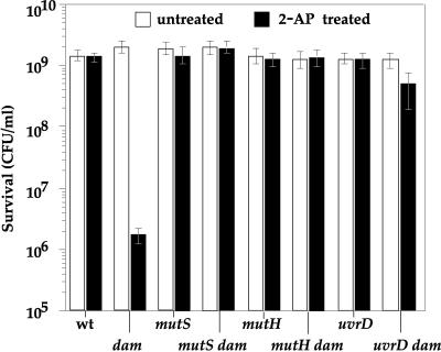
Here, we have studied the fate of E. coli dam cells exposed to the lethal effects of 2-AP. The survival analysis of 2-AP-treated dam cells (Table 1 shows a list of strains) shows that the MutS, MutH, and UvrD proteins are responsible for 103-fold viability loss in dam cells treated with 2-AP (Fig. 1), which corroborates previously published data (8). Flow cytometry and fluorescent microscopy were used to monitor the cellular DNA content. For the flow cytometry analysis, an overnight culture made in LB was diluted 1,000-fold in LB, and bacteria were grown at 37°C until reaching an optical density at 600 nm of 0.05. The culture was split into two; the first (control) culture continued to grow up to an optical density of 0.4 before addition of 250 μg/ml of rifampin. Rifampin prevents initiation of replication but allows the finalization of the replication cycles. After 90 min of incubation at 37°C, 0.4 ml of culture was withdrawn and mixed with 1.6 ml of cold methanol (100%) and left on ice for 1 h. After centrifugation, the resulting pellet was resuspended in 1.5 ml of 0.01 M Tris (pH 7.4), 0.01 M MgCl2, and DAPI (4′,6′-diamidino-2-phenylindole) (0.3 μg/ml) (15). Flow cytometry was performed with an Epics-ELITE (Coulter). The second test culture was treated with 1 mg/ml of 2-AP for 2 h 30 min and then centrifuged (2 min at room temperature) to eliminate the 2-AP before the rifampin treatment, as described above.
TABLE 1.
Strains used in this study
| Strain | Relevant genotype | Genotype or phenotype | Origin/reference |
|---|---|---|---|
| JJC520 | Wild type | deoA21 lac624lacY1 Cmr | 2 |
| MK1 | dam | As JJC520 but Δdam-16::Kanr | P1 transduction of Δdam-16::Kanr from E. coli Genetic Stock Center strain no. 7308 |
| MK2 | mutS | As JJC520 but mutS Ω(Smr/Spr) | P1 transduction of mutS Ω(Smr/Spr)/(4) |
| MK3 | dam mutS | As MK1 but ΔmutS Ω(Smr/Spr) | As above |
| MK4 | recD | As JJC520 but recD1901::Tn10 | P1 transduction of recD1901::Tn10 from E. coli Genetic Stock Center strain no. 7429 |
| MK5 | dam recD | As MK1 but recD1901::Tn10 | As above |
| MK6 | Wild type | As JJC520 but mhpC281::Tn10 Cms | P1 transduction of mhpC281::Tn10 from E. coli Genetic Stock Center strain no. 7333 |
| MK7 | dam | As MK6 but dam-13::Tn9 | P1 transduction of dam13::Tn9 from GM2199 strain kindly provided by M.G. Marinus |
| MK8 | mutH | As MK6 but mutH471::Tn5 | P1 transduction of mutH471::Tn5/(12) |
| MK9 | dam mutH | As MK7 but mutH471::Tn5 | As above |
| MK10 | uvrD | As MK6 but uvrD::phleo | P1 transduction of uvrD::phleo/(16) |
| MK11 | dam uvrD | As MK7 but uvrD::phleo | As above |
FIG. 1.
Survival of 2-AP treated cells. Overnight cultures grown in M9 minimal medium-glucose were diluted and plated on M9 minimal medium-glucose containing (or not containing) 500 μg/ml of 2-AP and incubated at 37°C. Each point represents the mean (± standard error) of six independent experiments. wt, wild type.
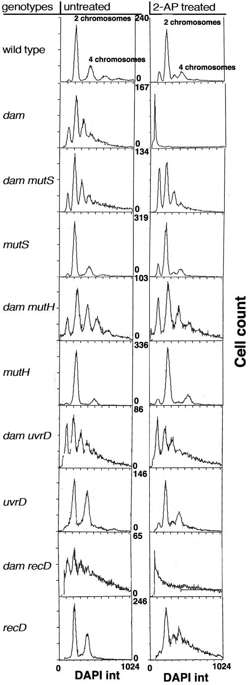
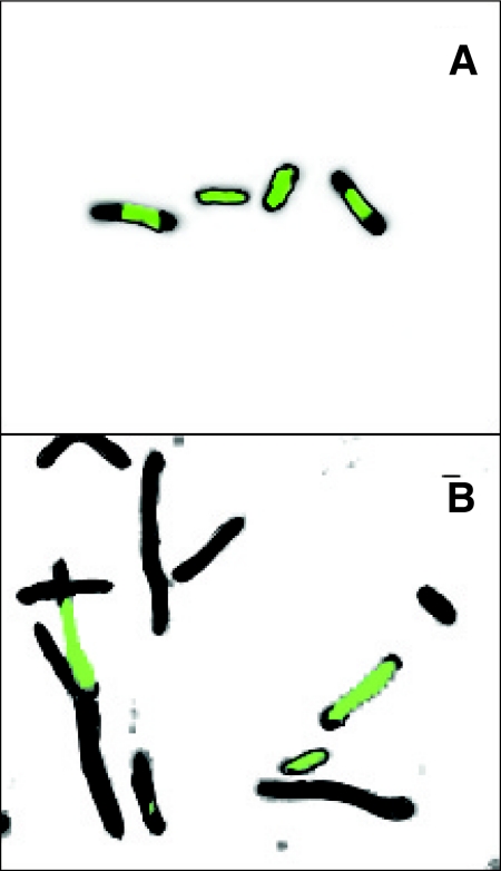
Flow cytometry analysis shows that the 2-AP pretreatment of the wild-type strain produced, besides the two major peaks containing two and four chromosomes, also two small peaks corresponding to one and three chromosomes, indicating induction of significant asynchrony at the DNA replication initiation level or chromosome degradation (Fig. 2). In contrast, in the dam mutant, the same treatment resulted in disappearance of all peaks, indicating the loss of the complete set of chromosomes. The only detectable peak was composed of cells deprived of most of their DNA, which was confirmed by the fluorescence microscopy (Fig. 3). The chromosomal degradation in the 2-AP-treated dam mutant is dependent on active MutS, MutH, and UvrD proteins (Fig. 2).
FIG. 2.
Flow cytometry analysis of DNA content in different strains with or without 2-AP treatment. DAPI int (integral) is the area signal of the pulses produced at the detector as the cell passes through the measurement region.
FIG. 3.
Production of DNA-free bacteria after 2-AP treatment in the dam mutant. The E. coli dam-mutant culture was treated with 2-AP as described in the text, and then cells were visualized using phase-contrast and fluorescence microscopy. DNA was stained with DAPI. (A) Untreated cells; (B) 2-AP treated cells. DAPI-stained DNA is green, and DNA-less cell space and DNA-less cells are black.
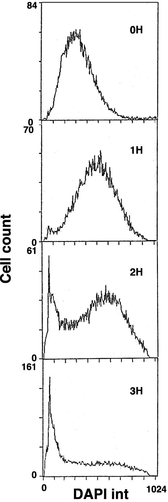
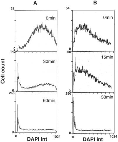
As this result was obtained after successive 2-AP and rifampin treatments, we wondered whether 2-AP treatment alone could induce chromosome degradation in dam mutants. The flow cytometry analysis of the dam cells incubated with 2-AP for various periods showed that the peak of bacteria without DNA started to emerge after 2 h of treatment and it became dominant after 3 h (Fig. 4). Therefore, the rifampin treatment does not participate in DNA degradation. However, rifampin affects the kinetics of chromosome degradation (Fig. 5A). This was demonstrated by treating cells for 1 h with 2-AP, which is insufficient to produce the peak of DNA-less bacteria, and by subsequently incubating cells with rifampin. We observed that a 30-min postincubation was sufficient to produce a substantial fraction of DNA-free bacteria, indicating that the addition of rifampin apparently accelerates DNA degradation (Fig. 5A).
FIG. 4.
Kinetics of chromosome degradation in 2-AP-treated dam cells without addition of rifampin. dam-mutant culture was treated with 2-AP and incubated at 37°C for different times as indicated. Samples were precipitated with methanol and analyzed as described in the text. DAPI int (integral) is the area signal of the pulses produced at the detector as the cell passes through the measurement region.
FIG. 5.
The effect of variable time of rifampin and 2-AP treatment on chromosome degradation in dam-mutant cells. (A) dam culture treated with 2-AP for 1 h at 37°C was centrifuged, resuspended, and incubated in LB containing rifampin. At different times, samples were precipitated with methanol and analyzed as described in the text. (B) 2-AP was added to a dam culture and, after various times, 0.4 ml of culture was centrifuged for 2 min at room temperature and the pellet resuspended in LB containing rifampin, followed by incubation for 90 min at 37°C before methanol treatment. Finally, cells were analyzed as described in the text. DAPI int (integral) is the area signal of the pulses produced at the detector as the cell passes through the measurement region.
In this regimen, we treated bacteria with 2-AP for more than two generations (generation time was 30 min), which might produce DNA with two complementary strands containing 2-AP. Hence, the question arises whether the incorporation of this base analogue into one of the strands is sufficient to trigger degradation of DNA in a dam mutant. Fixing the rifampin treatment to 90 min, we varied the time of 2-AP treatment, withdrawing samples, centrifuging them at room temperature for 2 min in order to eliminate the 2-AP, and then resuspending them in LB containing rifampin. Figure 5B shows that the incorporation of the 2-AP for the time span of one generation is largely enough to trigger the chromosome degradation. Even the short exposure of cells to the 2-AP (2 min required for centrifugation) could alter the formation of mature chromosomes (Fig. 5B). This result shows that the incorporation of 2-AP on only one DNA strand is sufficient to trigger DNA degradation.
Finally, as the RecBCD-dependent recombination machinery is required for the viability of dam mutants (10, 17), we tested whether extensive chromosome degradation results from its exonuclease V activity. The analysis of the DNA content of 2-AP-treated dam recD cells (lacking exonuclease V activity) indicates that degradation depends only partially on exonuclease V activity (Fig. 2). This is in contrast to DNA breakdown initiated by radiation-induced DSBs, where the global degradation depends on RecBCD (7). The MMR-generated DSBs are expected to be different from DSBs induced by radiation or restriction enzymes. Initiated by MutH nicking, on opposite strands, of GATC sequences flanking a 2-AP mismatch, the DSB would result from convergent-DNA unwinding by UvrD helicase. The DNA ends with single-stranded tails are better substrates for single-strand exonucleases (e.g., RecJ, ExoI, ExoVII, and ExoX) than for the double-strand exonuclease RecBCD. All these nucleases may alternate in the global DNA breakdown. The described efficiency of producing DNA-free E. coli cells offers a facile method for experimentation with bacterial cells devoid of their genes.
Acknowledgments
We were indebted to M.-C. Gendron (Institut J. Monod) for flow cytometry measurements and M. Selva for technical help.
REFERENCES
- 1.Bardelli, A., D. P. Cahill, G. Lederer, M. R. Speicher, K. W. Kinzler, B. Vogelstein, and C. Lengauer. 2001. Carcinogen-specific induction of genetic instability. Proc. Natl. Acad. Sci. USA 98:5770-5775. [DOI] [PMC free article] [PubMed] [Google Scholar]
- 2.Bierne, H., D. Vilette, S. D. Ehrlich, and B. Michel. 1997. Isolation of a dnaE mutation which enhances RecA-independent homologous recombination in the Escherichia coli chromosome. Mol. Microbiol. 24:1225-1234. [DOI] [PubMed] [Google Scholar]
- 3.Bignami, M., I. Casorelli, and P. Karran. 2003. Mismatch repair and response to DNA-damaging antitumour therapies. Eur. J. Cancer 39:2142-2149. [DOI] [PubMed] [Google Scholar]
- 4.Brégeon, D., I. Matic, M. Radman, and F. Taddei. 1999. Inefficient mismatch repair: genetic defects and down regulation. J. Genet. 78:21-28. [Google Scholar]
- 5.Burdett, V., C. Baitinger, M. Viswanathan, S. T. Lovett, and P. Modrich. 2001. In vivo requirement for RecJ, ExoVII, ExoI, and ExoX in methyl-directed mismatch repair. Proc. Natl. Acad. Sci. USA 98:6765-6770. [DOI] [PMC free article] [PubMed] [Google Scholar]
- 6.Doutriaux, M.-P., R. Wagner, and M. Radman. 1986. Mismatch-stimulated killing. Proc. Natl. Acad. Sci. USA 83:2576-2578. [DOI] [PMC free article] [PubMed] [Google Scholar]
- 7.Emmerson, P. T. 1968. Recombination deficient mutants of Escherichia coli K12 that map between thyA and argA. Genetics 60:19-30. [DOI] [PMC free article] [PubMed] [Google Scholar]
- 8.Glickman, B. W., and M. Radman. 1980. Escherichia coli mutator mutants deficient in methylation-instructed DNA mismatch correction. Proc. Natl. Acad. Sci. USA 77:1063-1067. [DOI] [PMC free article] [PubMed] [Google Scholar]
- 9.Lobner-Olesen, A., O. Skovgaard, and M. G. Marinus. 2005. Dam methylation: coordinating cellular processes. Curr. Opin. Microbiol. 8:154-160. [DOI] [PubMed] [Google Scholar]
- 10.Marinus, M. G. 2000. Recombination is essential for viability of an Escherichia coli dam (DNA adenine methyltransferase) mutant. J. Bacteriol. 182:463-468. [DOI] [PMC free article] [PubMed] [Google Scholar]
- 11.McGraw, B. R., and M. G. Marinus. 1980. Isolation and characterization of Dam+ revertants and suppressor mutations that modify secondary phenotypes of dam-3 strains of Escherichia coli K-12. Mol. Gen. Genet. 178:309-315. [DOI] [PubMed] [Google Scholar]
- 12.Pang, P. P., A. S. Lundberg, and G. C. Walker. 1985. Identification and characterization of the mutL and mutS gene products of Salmonella typhimurium LT2. J. Bacteriol. 163:1007-1015. [DOI] [PMC free article] [PubMed] [Google Scholar]
- 13.Peterson, K. R., and D. W. Mount. 1993. Analysis of the genetic requirements for viability of Escherichia coli K-12 DNA adenine methylase (dam) mutants. J. Bacteriol. 175:7505-7508. [DOI] [PMC free article] [PubMed] [Google Scholar]
- 14.Schofield, M. J., and P. Hsieh. 2003. DNA mismatch repair: molecular mechanisms and biological function. Annu. Rev. Microbiol. 57:579-608. [DOI] [PubMed] [Google Scholar]
- 15.Skarstad, K., H. B. Steen, and E. Boye. 1985. Escherichia coli DNA distributions measured by flow cytometry and compared with theoretical computer simulations. J. Bacteriol. 163:661-668. [DOI] [PMC free article] [PubMed] [Google Scholar]
- 16.Veaute, X., S. Delmas, M. Selva, J. Jeusset, E. LeCam, I. Matic, F. Fabre, and M. A. Petit. 2005. UvrD helicase, unlike Rep helicase, dismantles RecA nucleoprotein filaments in Escherichia coli. EMBO J. 24:180-189. [DOI] [PMC free article] [PubMed] [Google Scholar]
- 17.Wang, T.-C. V., and K. C. Smith. 1986. Inviability of dam recA and dam recB cells of Escherichia coli is correlated with their inability to repair DNA double-strand breaks produced by mismatch repair. J. Bacteriol. 165:1023-1025. [DOI] [PMC free article] [PubMed] [Google Scholar]


