Abstract
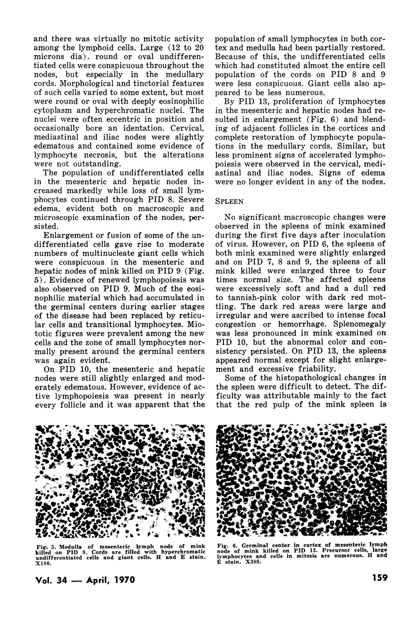
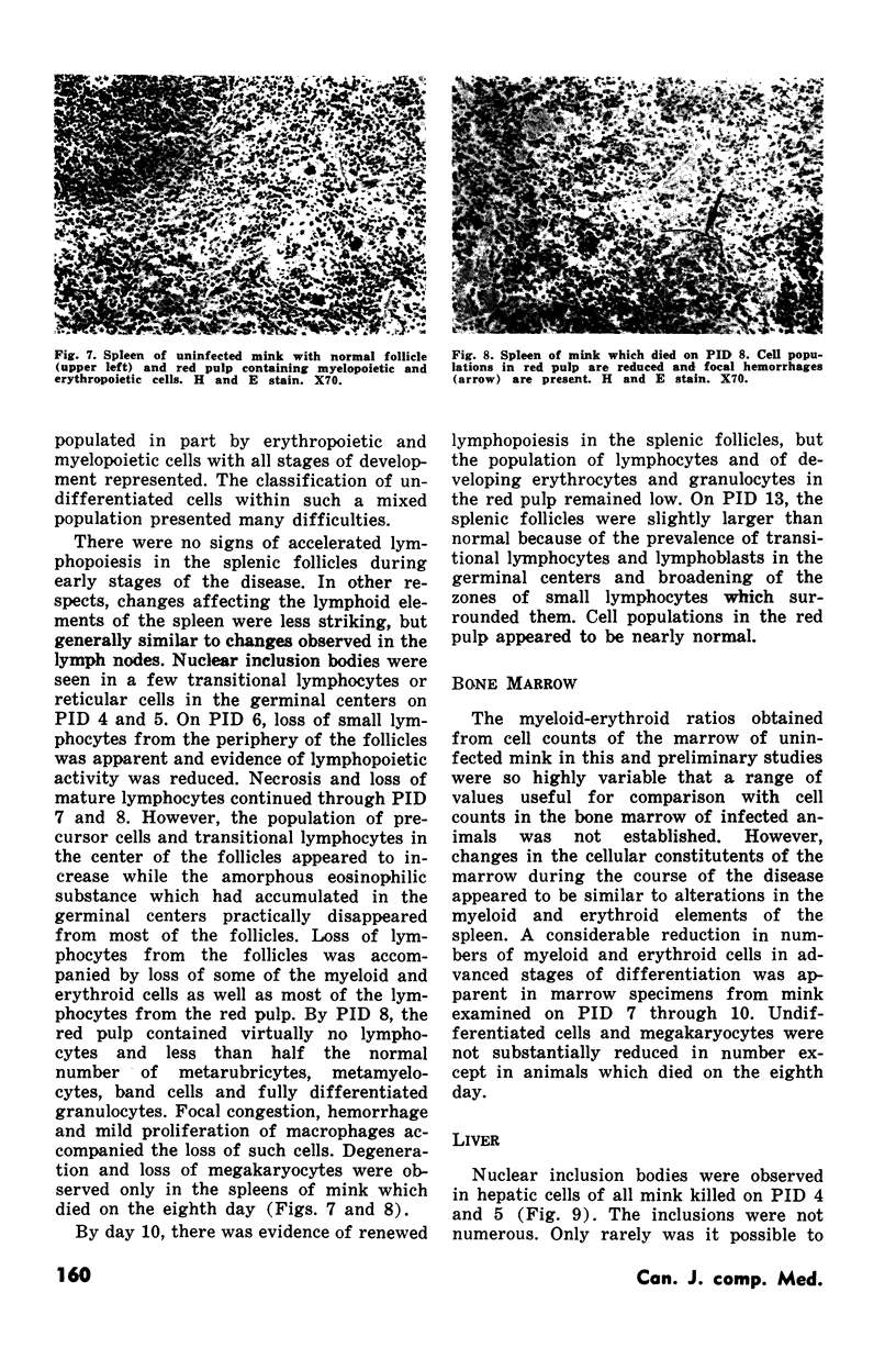
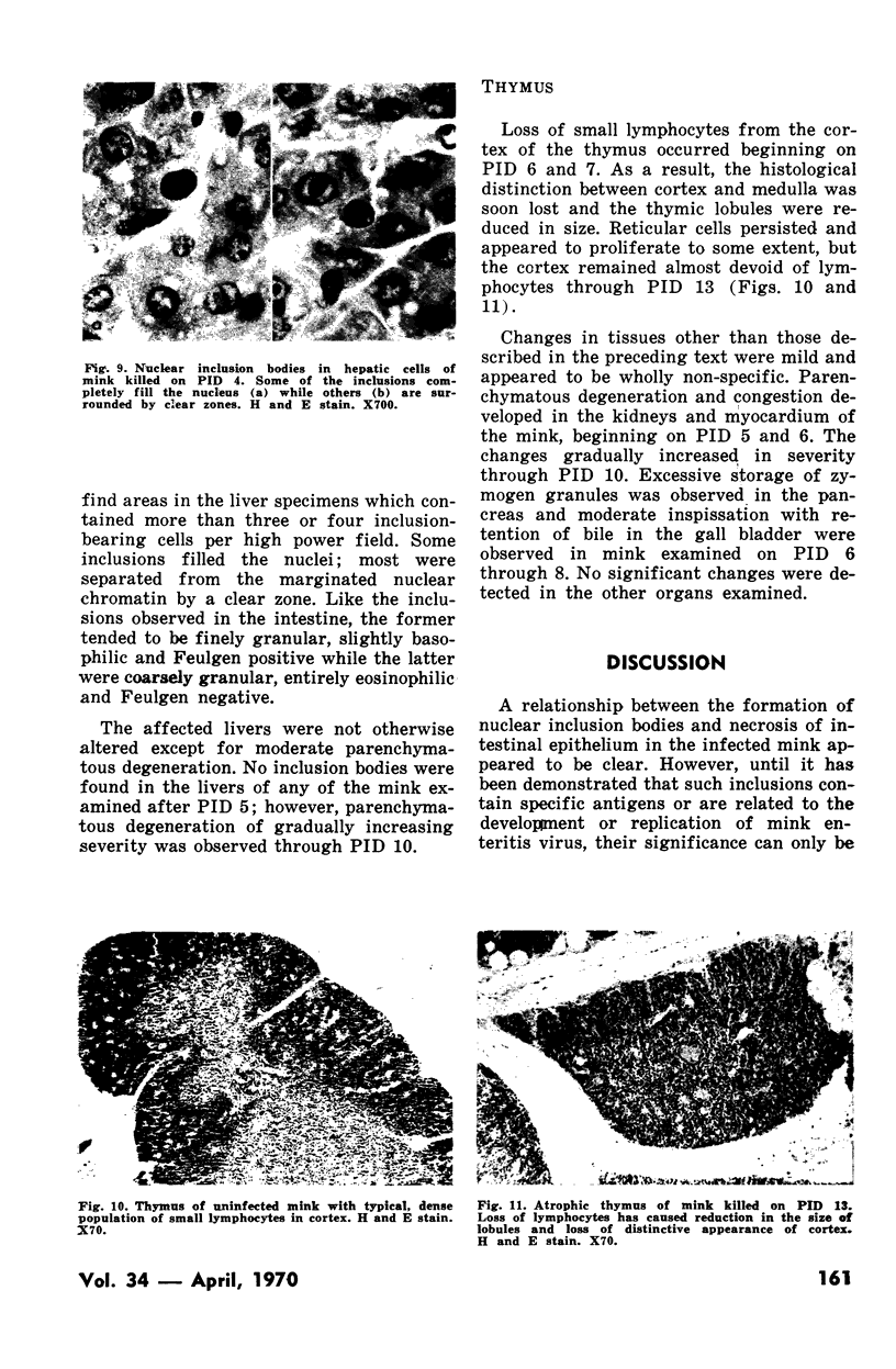
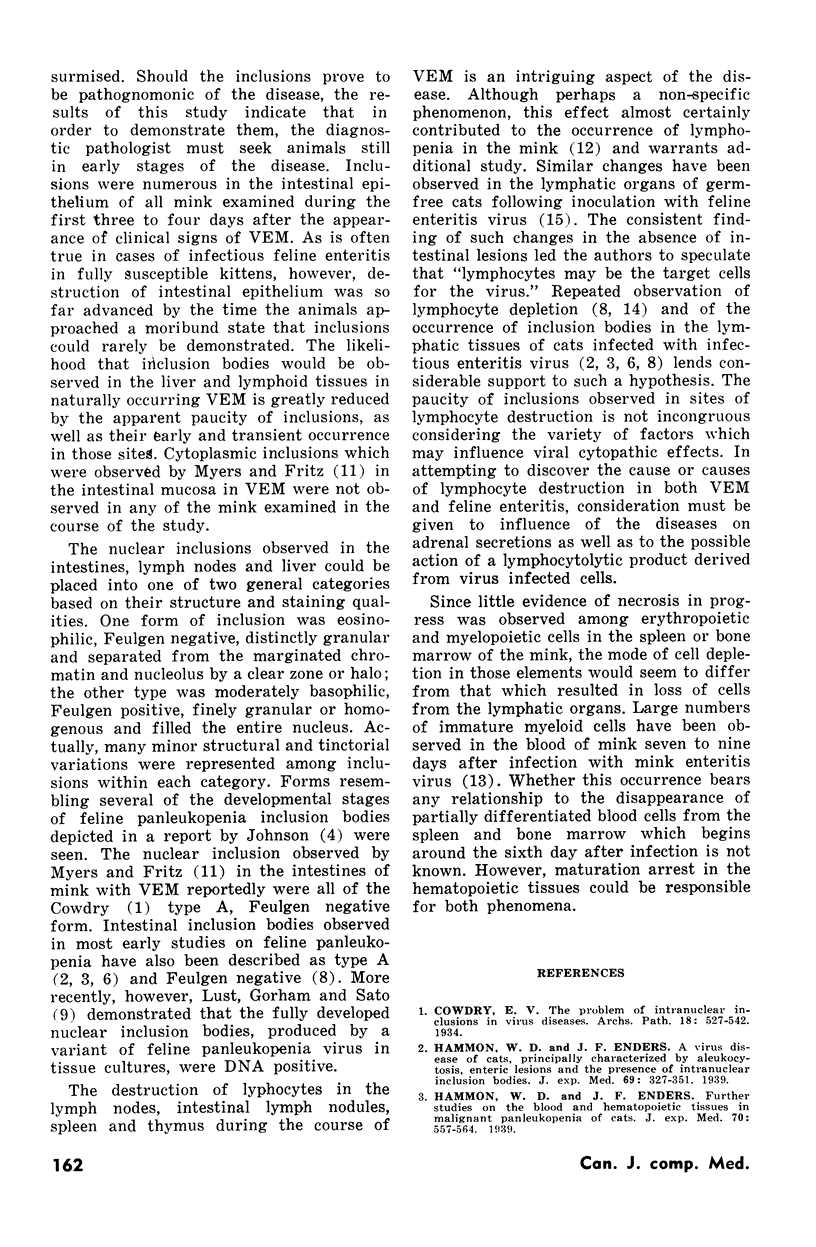
The lesions which characterize viral enteritis of mink (VEM) were studied in twenty-six, ten-week-old mink which had been infected by force feeding a tissue suspension containing a Wisconsin strain of mink enteritis virus. The pathogenesis of the lesions was reconstructed from gross and histopathological changes observed in animals which were selected randomly from the group each day for necropsy during the course of the disease.
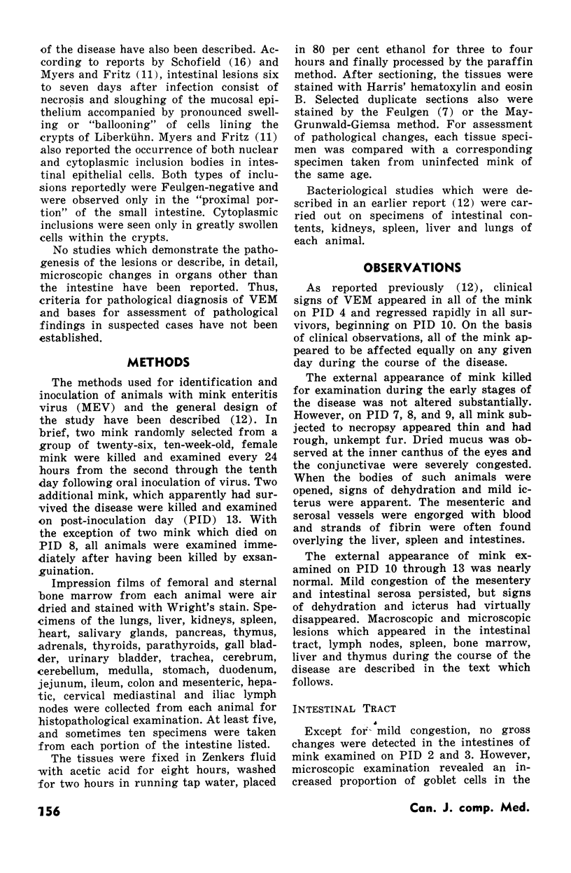
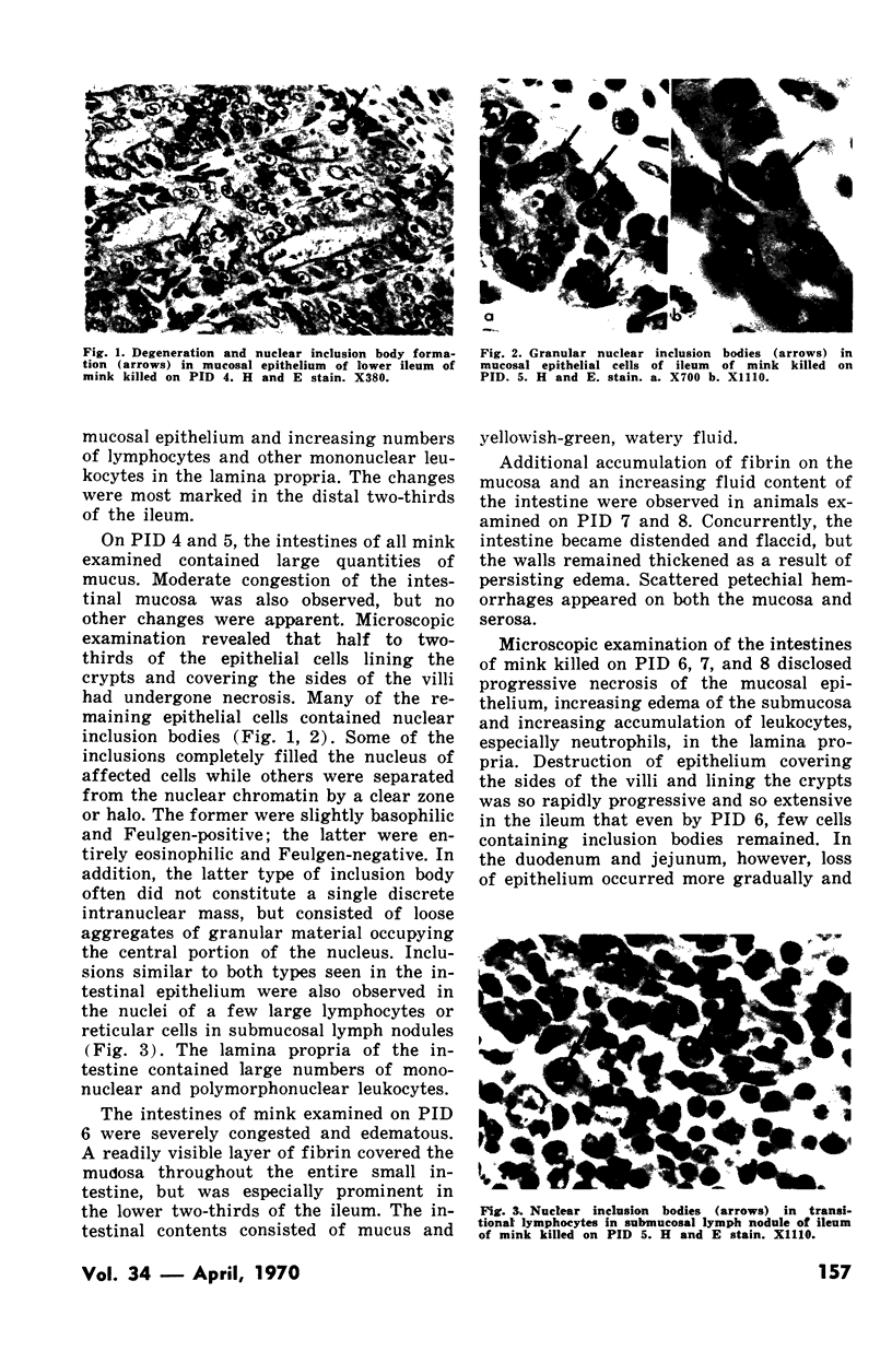
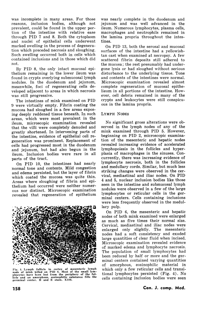
Alterations were observed in the tissues of all mink examined from post-inoculation day (PID) 4 through 13. The principal macroscopic lesions which consisted of fibrinous enteritis, enlargement and hemorrhage of the spleen and edema of mesenteric and hepatic lymph nodes were most conspicuous on PID 7 and 8. Histopathological changes including necrosis and desquamation of intestinal epithelium, depletion of mature lymphocytes in lymph nodes, thymus and spleen and loss of partly differentiated myeloid and erythroid cells from spleen and bone marrow also reached full development on PID 7 and 8. However, nuclear inclusion bodies which were presumed to be a product of the causative agent and, therefore, of diagnostic significance were most prevalent on PID 3, 4 and 5. The inclusions were observed in mucosal epithelial cells of the intestine, parenchymal cells of the liver and in lymphocyte precursor cells of the spleen, intestinal lymph nodules and masenteric and hepatic lymph nodes.
Full text
PDF
Images in this article
Selected References
These references are in PubMed. This may not be the complete list of references from this article.
- Johnson R. H. Feline panleucopaenia virus. II. Some features of the cytopathic effects in feline kidney monolayers. Res Vet Sci. 1965 Oct;6(4):472–481. [PubMed] [Google Scholar]
- Lawrence J. S., Syverton J. T., Shaw J. S., Smith F. P. Infectious feline agranulocytosis. Am J Pathol. 1940 May;16(3):333–354.9. [PMC free article] [PubMed] [Google Scholar]
- Lucas A. M., Riser W. H. Intranuclear Inclusions in Panleukopenia of Cats: A Correlation with the Pathogenesis of the Disease and Comparison with Inclusions of Herpes, B-Virus, Yellow Fever, and Burns. Am J Pathol. 1945 May;21(3):435–465. [PMC free article] [PubMed] [Google Scholar]
- Lust S. J., Gorham J. R., Sato N. Occurrence of intranuclear inclusions in cell cultures infected with infectious feline enteritis virus. Am J Vet Res. 1965 Sep;26(114):1163–1166. [PubMed] [Google Scholar]
- Macpherson L. W. Feline Enteritis Virus - Its Transmission To Mink Under Natural And Experimental Conditions. Can J Comp Med Vet Sci. 1956 Jun;20(6):197–202. [PMC free article] [PubMed] [Google Scholar]
- Myers W. L., Fritz T. E. Histopathologic Changes in Virus Enteritis of Mink. Can J Comp Med Vet Sci. 1959 Aug;23(8):246–249. [PMC free article] [PubMed] [Google Scholar]
- Reynolds H. A. Some clinical and hematological features of virus enteritis of mink. Can J Comp Med. 1969 Apr;33(2):155–159. [PMC free article] [PubMed] [Google Scholar]


