

Abstract
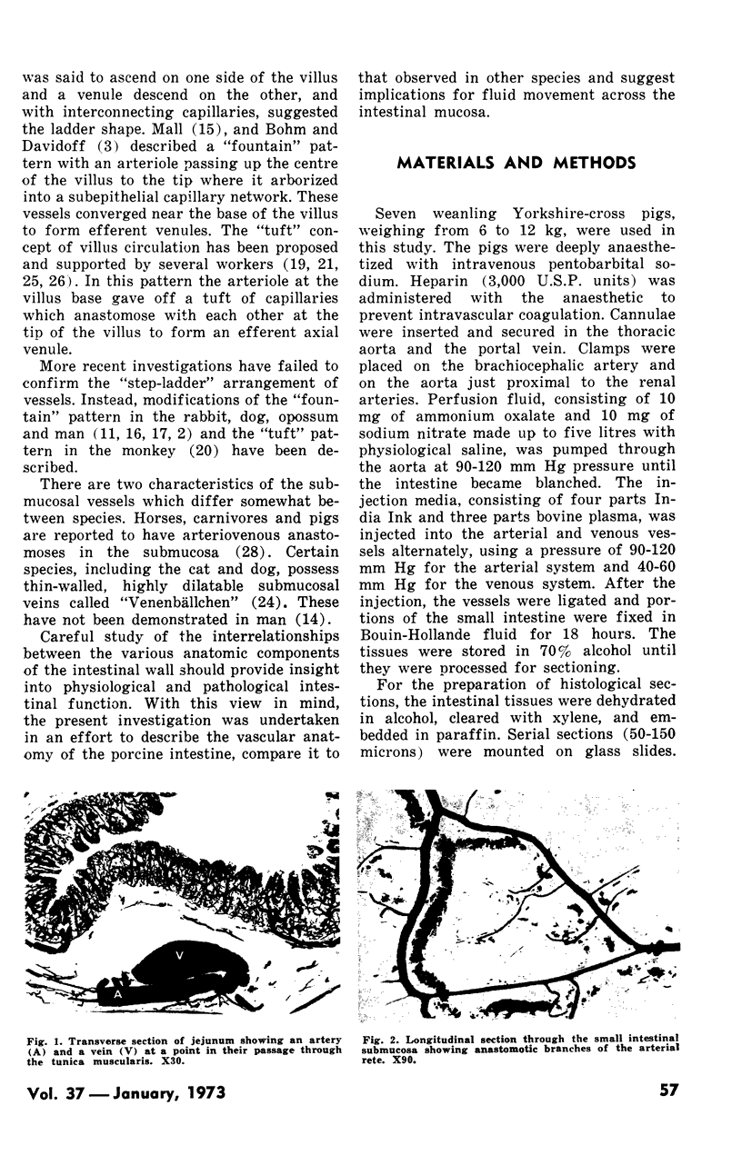
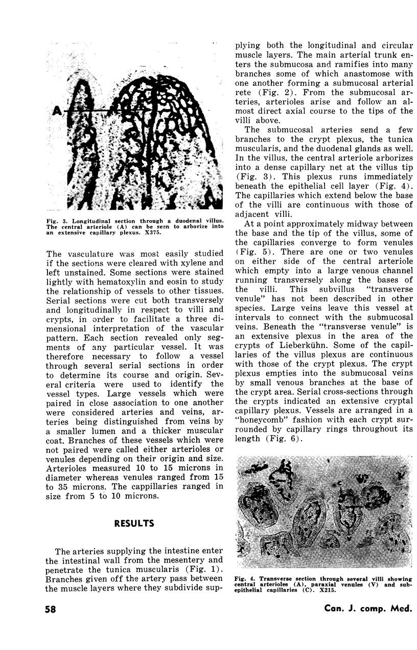
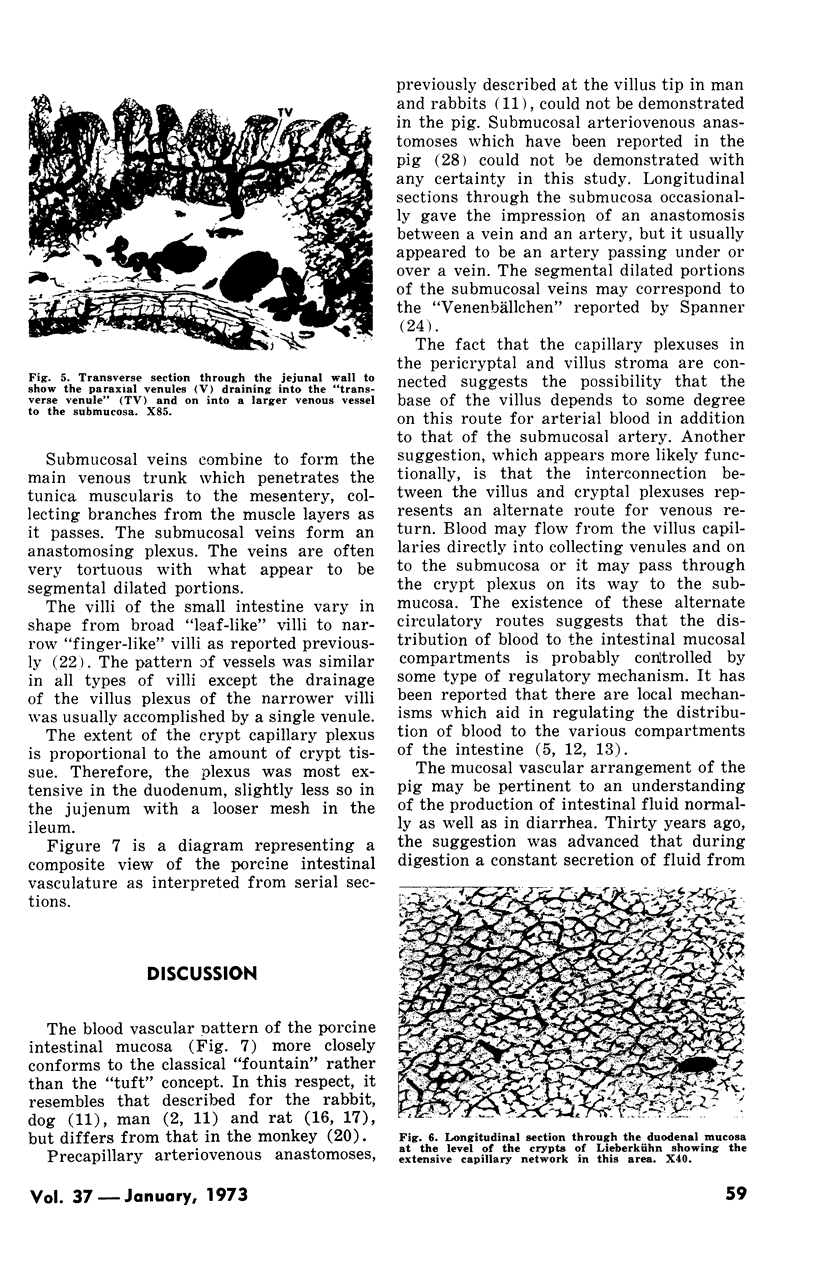
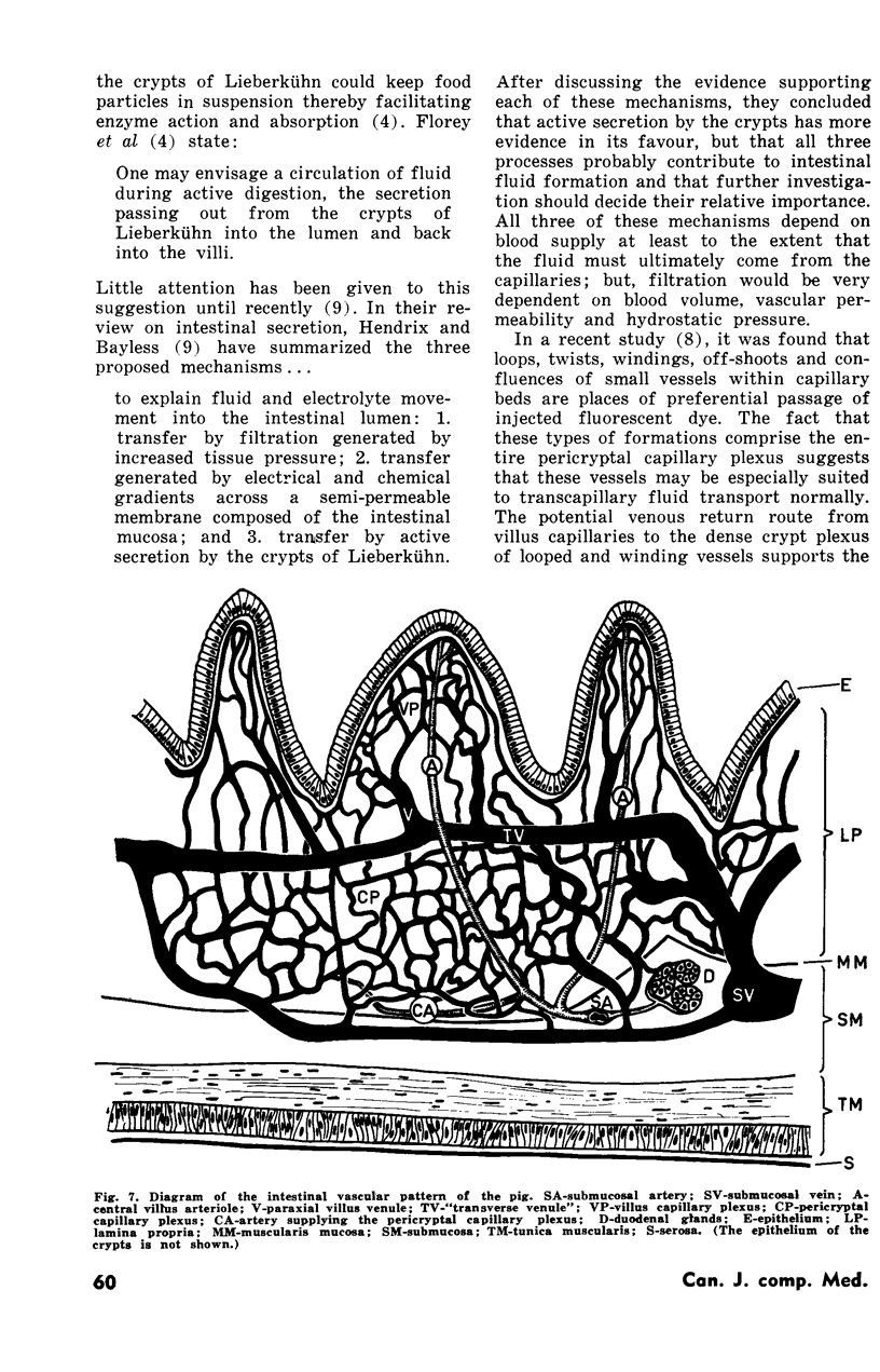
The vascular anatomy of the porcine small intestine was studied by injection of intestinal vessels with India Ink. Examination of transverse and longitudinal serial sections of the injected intestine facilitated a three-dimensional interpretation of the vascular pattern. An artery from the mesentery penetrated the tunica muscularis, supplied muscular branches and passed on to the submucosa where it formed an arterial rete. From the submucosal arteries, arterioles arose and followed a direct axial course to the tips of villi where they ramified into a subepithelial capillary plexus. Some of the capillaries, at the midpoint of the villus, fused into paraxial venules, which emptied into a “transverse venule” at the base of the villus. Other villus capillaries were continuous with those of the crypts. The pericryptal capillary plexus received a few arterial branches from the submucosal arteries. The transverse venule and the pericryptal capillary plexus emptied into large, segmentally dilated veins in the submucosa. The submucosal veins formed an extensive anastomosing network drained by large venous trunks which passed through the muscle layers to the mesentery.
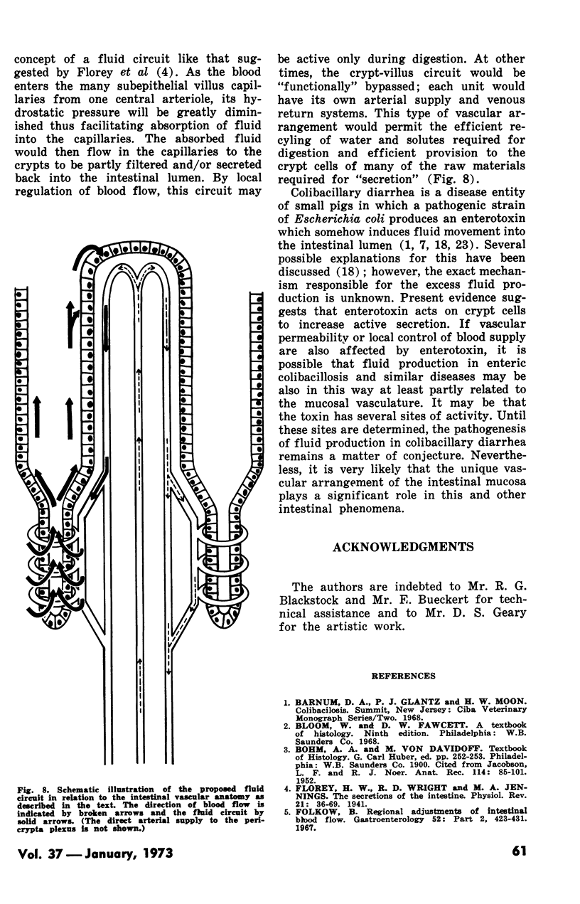
The observations suggest possible relationships between the vascular pattern and intestinal fluid movement.
Full text
PDF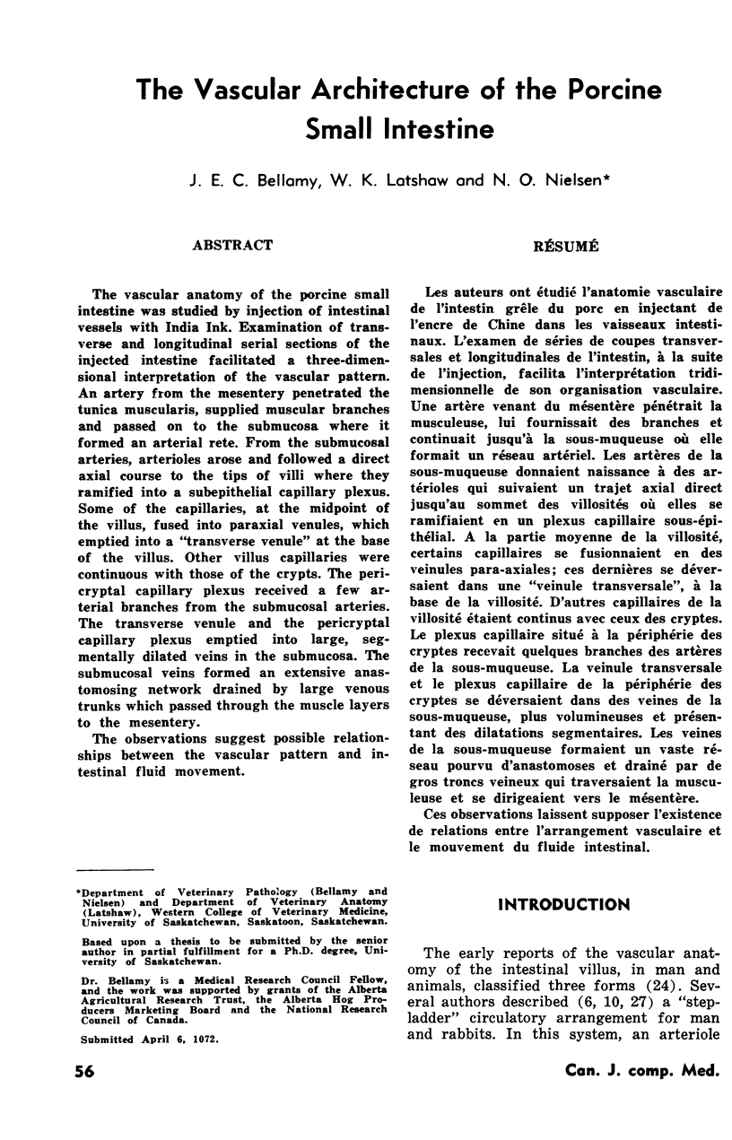
Images in this article

Selected References
These references are in PubMed. This may not be the complete list of references from this article.
- Folkow B. Regional adjustments of intestinal blood flow. Gastroenterology. 1967 Feb;52(2):423–432. [PubMed] [Google Scholar]
- Gyles C. L., Barnum D. A. A heat-labile enterotoxin from strains of Eschericha coli enteropathogenic for pigs. J Infect Dis. 1969 Oct;120(4):419–426. doi: 10.1093/infdis/120.4.419. [DOI] [PubMed] [Google Scholar]
- Hauck G. Luminescence-microscopic evidence for the existence of a gradient of vascular permeability in the mesentery capillary bed. Bibl Anat. 1969;10:221–224. [PubMed] [Google Scholar]
- Hendrix T. R., Bayless T. M. Digestion: intestinal secretion. Annu Rev Physiol. 1970;32:139–164. doi: 10.1146/annurev.ph.32.030170.001035. [DOI] [PubMed] [Google Scholar]
- JACOBSON L. F., NOER R. J. The vascular pattern of the intestinal villi in various laboratory animals and man. Anat Rec. 1952 Sep;114(1):85–101. doi: 10.1002/ar.1091140107. [DOI] [PubMed] [Google Scholar]
- JACOBSON L. F., NOER R. J. The vascular pattern of the intestinal villi in various laboratory animals and man. Anat Rec. 1952 Sep;114(1):85–101. doi: 10.1002/ar.1091140107. [DOI] [PubMed] [Google Scholar]
- JACOBSON L. F., NOER R. J. The vascular pattern of the intestinal villi in various laboratory animals and man. Anat Rec. 1952 Sep;114(1):85–101. doi: 10.1002/ar.1091140107. [DOI] [PubMed] [Google Scholar]
- JACOBSON L. F., NOER R. J. The vascular pattern of the intestinal villi in various laboratory animals and man. Anat Rec. 1952 Sep;114(1):85–101. doi: 10.1002/ar.1091140107. [DOI] [PubMed] [Google Scholar]
- JACOBSON L. F., NOER R. J. The vascular pattern of the intestinal villi in various laboratory animals and man. Anat Rec. 1952 Sep;114(1):85–101. doi: 10.1002/ar.1091140107. [DOI] [PubMed] [Google Scholar]
- JACOBSON L. F., NOER R. J. The vascular pattern of the intestinal villi in various laboratory animals and man. Anat Rec. 1952 Sep;114(1):85–101. doi: 10.1002/ar.1091140107. [DOI] [PubMed] [Google Scholar]
- JACOBSON L. F., NOER R. J. The vascular pattern of the intestinal villi in various laboratory animals and man. Anat Rec. 1952 Sep;114(1):85–101. doi: 10.1002/ar.1091140107. [DOI] [PubMed] [Google Scholar]
- Johnson P. C. Autoregulation of blood flow in the intestine. Gastroenterology. 1967 Feb;52(2):435–441. [PubMed] [Google Scholar]
- Lundgren O. Studies on blood flow distribution and countercurrent exchange in the small intestine. Acta Physiol Scand Suppl. 1967;303:1–42. doi: 10.1111/j.1748-1716.1967.tb03885.x. [DOI] [PubMed] [Google Scholar]
- Miller D. S., Rahman M. A., Tanner R., Mathan V. I., Baker S. J. The vascular architecture of the different forms of small intestinal villi in the rat (Rattus norvegicus). Scand J Gastroenterol. 1969;4(6):477–482. doi: 10.3109/00365526909180637. [DOI] [PubMed] [Google Scholar]
- Mohiuddin A. Blood and lymph vessels in the jejunal villi of the white rat. Anat Rec. 1966 Sep;156(1):83–89. doi: 10.1002/ar.1091560110. [DOI] [PubMed] [Google Scholar]
- Nielsen N. O., Moon H. W., Roe W. E. Enteric colibacillosis in swine. J Am Vet Med Assoc. 1968 Dec 15;153(12):1590–1606. [PubMed] [Google Scholar]
- Reynolds D. G., Brim J., Sheehy T. W. The vascular architecture of the small intestinal mucosa of the monkey (Macaca mulatta). Anat Rec. 1967 Oct;159(2):211–218. doi: 10.1002/ar.1091590210. [DOI] [PubMed] [Google Scholar]
- SLOSS M. W. The microscopic anatomy of the digestive tract of Sus scrofa domestica. Am J Vet Res. 1954 Oct;15(57):578–593. [PubMed] [Google Scholar]
- Smith H. W., Halls S. Studies on Escherichia coli enterotoxin. J Pathol Bacteriol. 1967 Apr;93(2):531–543. doi: 10.1002/path.1700930212. [DOI] [PubMed] [Google Scholar]