Abstract
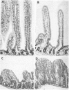
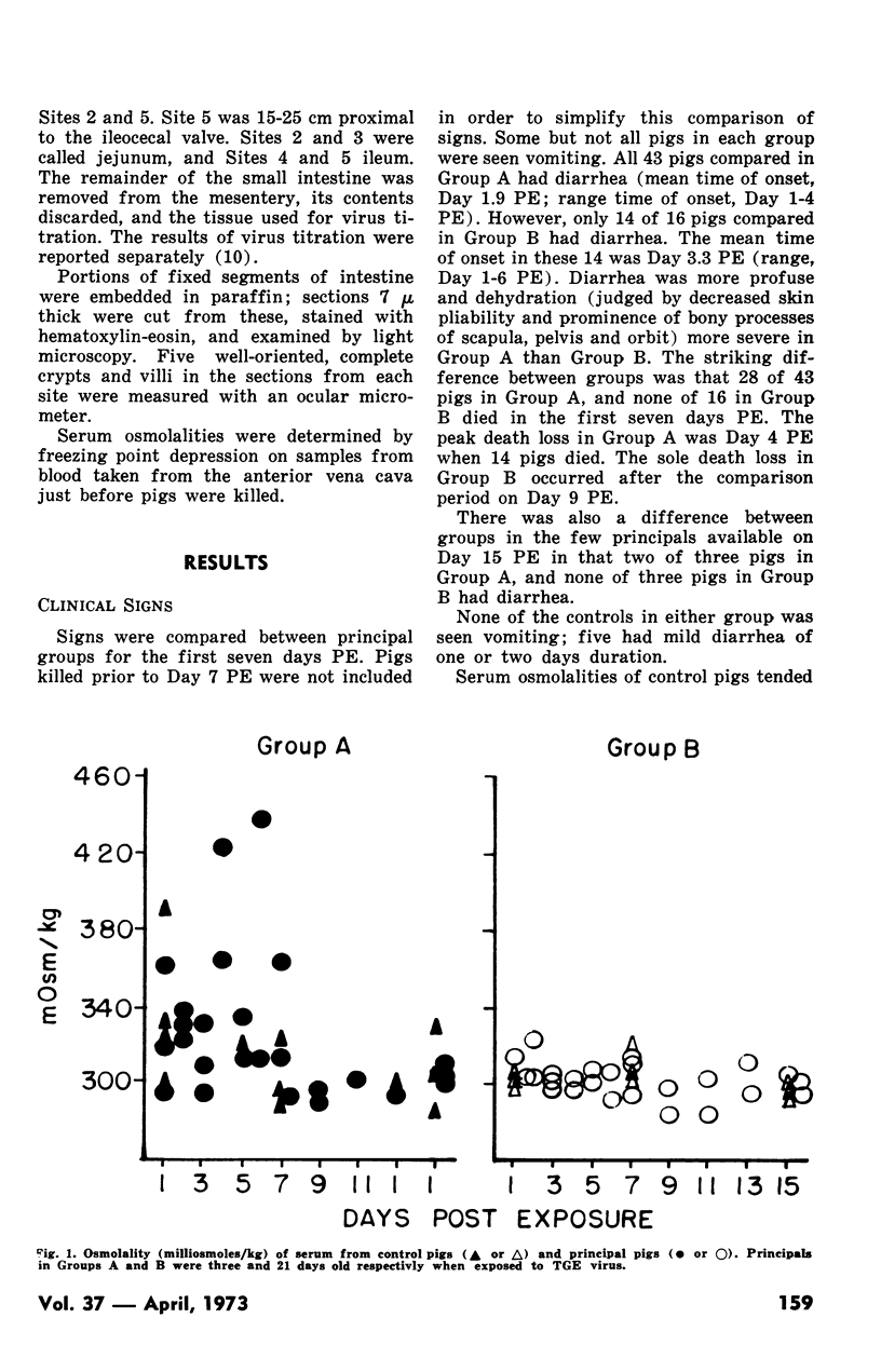
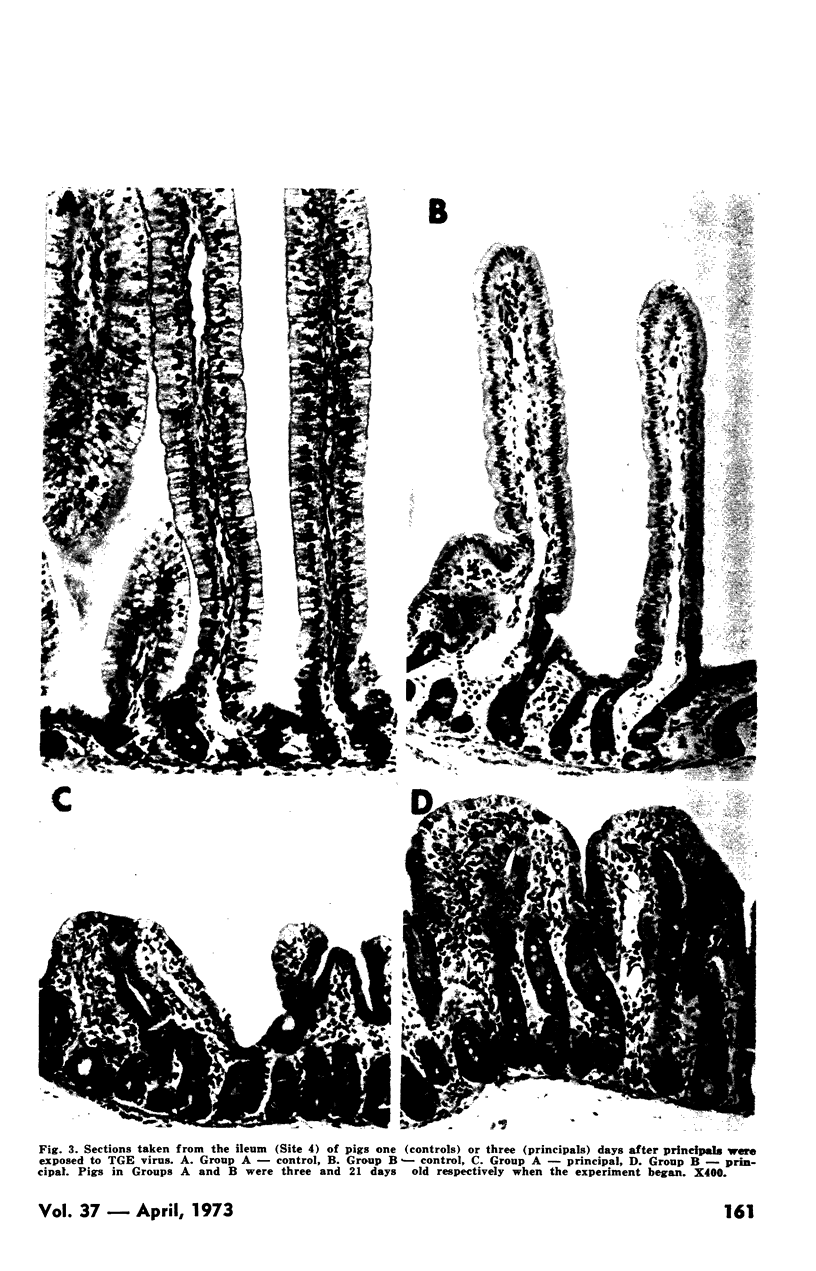
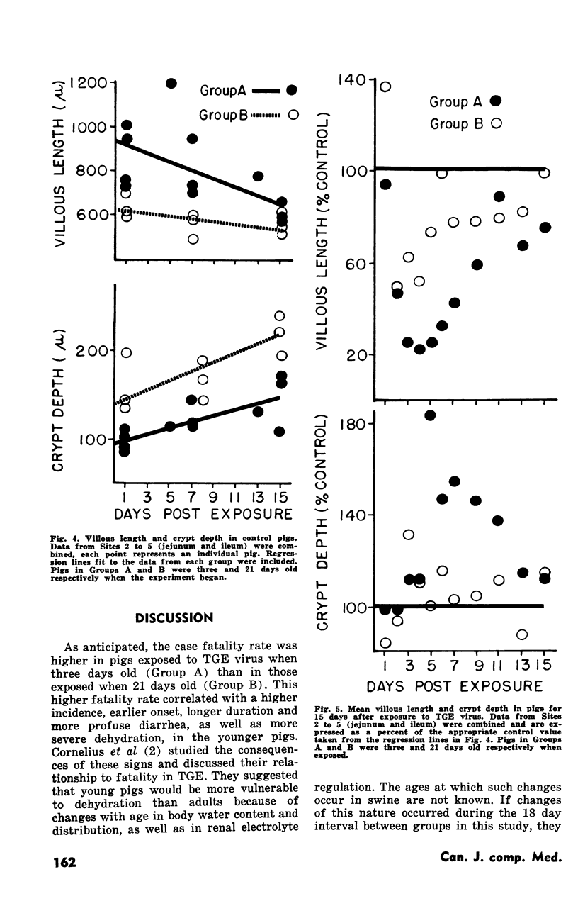
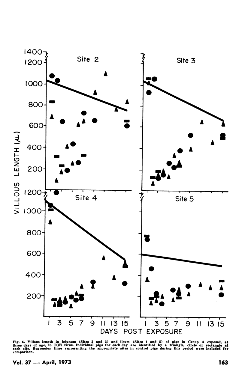
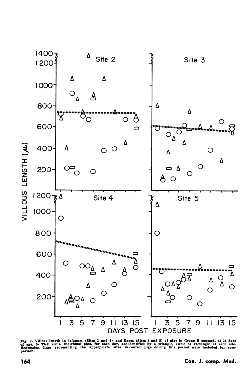
Pigs were exposed to transmissible gastroenteritis (TGE) virus when three days old or when 21 days old. Diarrhea was earliest in onset, most frequent, most profuse and most prolonged in the youngest group. Pigs exposed when three days old also had a higher case fatality rate than those exposed when 21 days old. The histological response of both groups to exposure was atrophy of villi and hyperplasia of crypts in jejunum and ileum. However, from days three to seven post-exposure, when most fatalities occurred in the younger group, atrophy of villi was both more intensive and extensive in the younger group. Hyperplasia of crypts was also greater and more prolonged in the younger group. Regeneration of atrophic villi was more rapid in jejunum than ileum in both groups. Results were interpreted to indicate two populations, with different rates of regeneration, in the 21-day old group. Based on this interpretation, regeneration of villi was more rapid in one population from the 21-day old group than in the three-day old group.
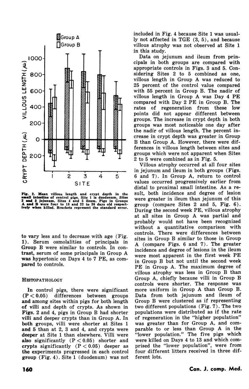
The length of villi and depth of crypts in control pigs varied longitudinally (i.e. from site to site) in the intestine, within each age group. Length of villi and depth of crypts in control pigs also varied with age.
Full text
PDF
Images in this article
Selected References
These references are in PubMed. This may not be the complete list of references from this article.
- Bohl E. H., Gupta R. K., McCloskey L. W., Saif L. Immunology of transmissible gastroenteritis. J Am Vet Med Assoc. 1972 Feb 15;160(4):543–549. [PubMed] [Google Scholar]
- Cross R. F., Bohl E. H. Some criteria for the field diagnosis of porcine transmissible gastroenteritis. J Am Vet Med Assoc. 1969 Feb 1;154(3):266–272. [PubMed] [Google Scholar]
- Haelterman E. O. On the pathogenesis of transmissible gastroenteritis of swine. J Am Vet Med Assoc. 1972 Feb 15;160(4):534–540. [PubMed] [Google Scholar]
- Moon H. W. Epithelial cell migration in the alimentary mucosa of the suckling pig. Proc Soc Exp Biol Med. 1971 May;137(1):151–154. doi: 10.3181/00379727-137-35533. [DOI] [PubMed] [Google Scholar]
- Mouwen J. M. White scours in piglets. I. Stereomicroscopy of the mucosa of the small intestine. Vet Pathol. 1971;8(4):364–380. doi: 10.1177/030098587100800407. [DOI] [PubMed] [Google Scholar]
- Norman J. O., Lambert G., Moon H. W., Stark S. L. Age dependent resistance to transmissible gastroenteritis of swine (TGE). II. Coronavirus titer in tissues of pigs after exposure. Can J Comp Med. 1973 Apr;37(2):167–170. [PMC free article] [PubMed] [Google Scholar]
- Olson L. D. Induction of transmissible gastroenteritis in feeder swine. Am J Vet Res. 1971 Mar;32(3):411–417. [PubMed] [Google Scholar]
- Pensaert M., Haelterman E. O., Burnstein T. Transmissible gastroenteritis of swine: virus-intestinal cell interactions. I. Immunofluorescence, histopathology and virus production in the small intestine through the course of infection. Arch Gesamte Virusforsch. 1970;31(3):321–334. doi: 10.1007/BF01253767. [DOI] [PubMed] [Google Scholar]
- Tamoglia T. W. Present status of products available for use against transmissible gastroenteritis. J Am Vet Med Assoc. 1972 Feb 15;160(4):554–558. [PubMed] [Google Scholar]
- Thake D. C. Jejunal epithelium in transmissible gastroenteritis of swine. An electron microscopic and histochemical study. Am J Pathol. 1968 Jul;53(1):149–168. [PMC free article] [PubMed] [Google Scholar]
- YOUNG G. A., HINZ R. W., UNDERDAHL N. R. Some characteristics of transmissible gastroenteritis (TGE) in disease-free antibody-devoid pigs. Am J Vet Res. 1955 Oct;16(61 Pt 1):529–535. [PubMed] [Google Scholar]


