Abstract
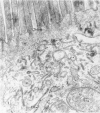
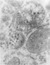
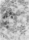
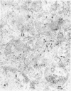
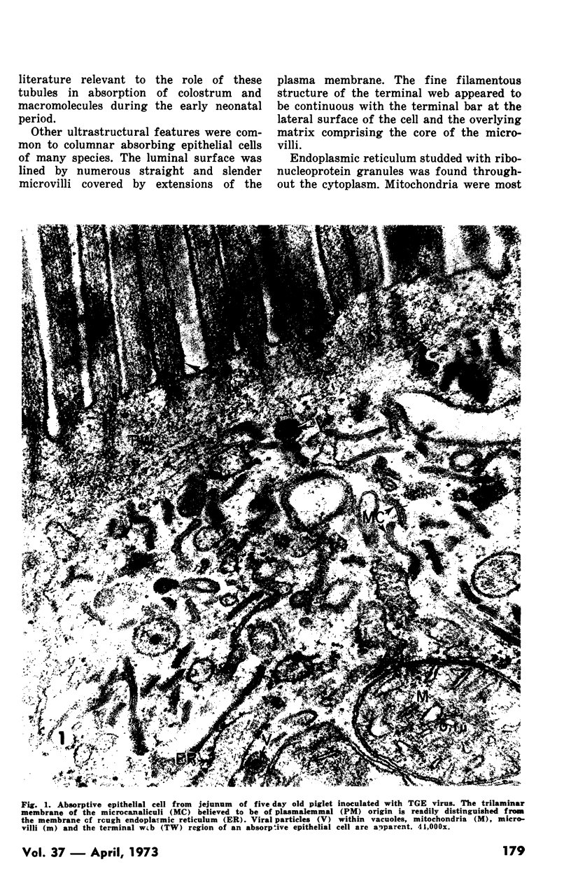
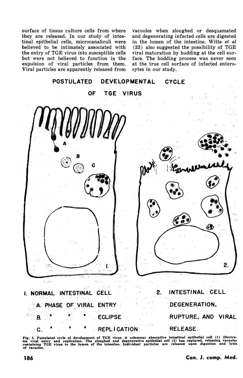
An electron microscopic study of intestinal epithelial cells of neonatal piglets infected with transmissible gastroenteritis (TGE) virus revealed a unique parasite-host cell interaction. Entry of the TGE virus into intestinal epithelial cells of newborn piglets is mediated through a network of cytoplasmic tubules of plasmalemma origin. the tubules, called microcanaliculi, are morphologically distinct from endoplasmic reticulum and Golgi. In uninfected animals similar tubules appear to be responsible for the indiscriminate uptake of large quantities of macromolecules from colostrum during the first few days of life.
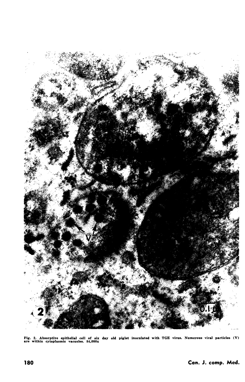
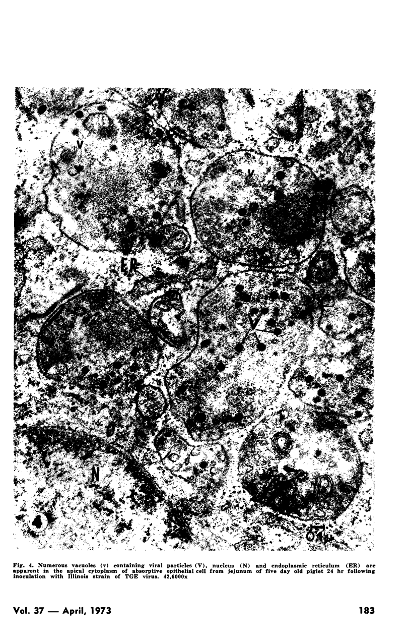
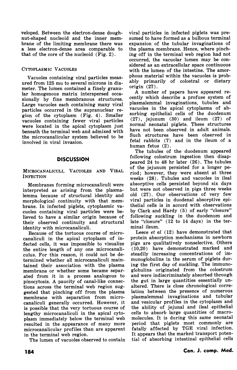
Thin-section profiles of plasmalemma invaginations resembled tubules or canals and frequently contained viral particles. TGE viral particles developed and accumulated within cytoplasmic vacuoles. Initially the vacuoles were continuous with the microcanaliculi formed by deep plasmalemma invagination. Mature viral particles were 60 to 85 mu in diameter with an electron dense doughnut-shaped nucleoid surrounded by a trilaminar membrane which resembled the vacuolar wall. Abundant evidence of viral effects was observed in absorbtive epithelial cells of the jejunum and ileum but not of the duodenum.
The ability of absorbtive intestinal epithelial cells to form deep cytoplasmic tubular invaginations is temporally related to the pathogenesis of TGE and may explain in part why pigs usually are fatally affected by TGE only during the neonatal period.
Full text
PDF
Images in this article
Selected References
These references are in PubMed. This may not be the complete list of references from this article.
- Adams W. R., Kraft L. M. Electron-Microscopic Study of the Intestinal Epithelium of Mice Infected with the Agent of Epizootic Diarrhea of Infant Mice (EDIM Virus). Am J Pathol. 1967 Jul;51(1):39–60. [PMC free article] [PubMed] [Google Scholar]
- BIERRING F., ANDERSEN H., EGEBERG J., BRO-RASMUSSEN F., MATTHIESSEN M. ON THE NATURE OF THE MECONIUM CORPUSCLES IN HUMAN FOETAL INTESTINAL EPITHELIUM. 1. ELECTRON MICROSCOPIC STUDIES. Acta Pathol Microbiol Scand. 1964;61:365–376. doi: 10.1111/apm.1964.61.3.365. [DOI] [PubMed] [Google Scholar]
- Becker W. B., McIntosh K., Dees J. H., Chanock R. M. Morphogenesis of avian infectious bronchitis virus and a related human virus (strain 229E). J Virol. 1967 Oct;1(5):1019–1027. doi: 10.1128/jvi.1.5.1019-1027.1967. [DOI] [PMC free article] [PubMed] [Google Scholar]
- CLARK S. L., Jr Cellular differentiation in the kidneys of newborn mice studies with the electron microscope. J Biophys Biochem Cytol. 1957 May 25;3(3):349–362. doi: 10.1083/jcb.3.3.349. [DOI] [PMC free article] [PubMed] [Google Scholar]
- Chandler R. L., Derbyshire J. B., Smith K. Observations on the experimental infection and cellular pathology of transmissible gastroenteritis in piglets. Res Vet Sci. 1969 Sep;10(5):435–439. [PubMed] [Google Scholar]
- Clarke R. M., Hardy R. N. Histological changes in the small intestine of the young pig and their relation to macromolecular uptake. J Anat. 1971 Jan;108(Pt 1):63–77. [PMC free article] [PubMed] [Google Scholar]
- Deren J. J., Strauss E. W., Wilson T. H. The development of structure and transport systems of the fetal rabbit intestine. Dev Biol. 1965 Dec;12(3):467–486. doi: 10.1016/0012-1606(65)90010-2. [DOI] [PubMed] [Google Scholar]
- Hooper B. E., Haelterman E. O. Growth of transmissible gastroenteritis virus in young pigs. Am J Vet Res. 1966 Jan;27(116):286–291. [PubMed] [Google Scholar]
- Karlsson B. W. Immunochemical studies on changes in blood serum proteins in piglets after colostrum ingestion and during neonatal and juvenile development. Acta Pathol Microbiol Scand. 1966;67(2):237–256. doi: 10.1111/apm.1966.67.2.237. [DOI] [PubMed] [Google Scholar]
- LUFT J. H. Improvements in epoxy resin embedding methods. J Biophys Biochem Cytol. 1961 Feb;9:409–414. doi: 10.1083/jcb.9.2.409. [DOI] [PMC free article] [PubMed] [Google Scholar]
- Okaniwa A., Harada K., Kaji T. Electron microscopy of tissue culture cells infected with swine transmissible gastroenteritis virus. Natl Inst Anim Health Q (Tokyo) 1968 Fall;8(3):148–163. [PubMed] [Google Scholar]
- Okaniwa A., Harada K., Park D. K. Structure of swine transmissible gastroenteritis virus examined by negative staining. Natl Inst Anim Health Q (Tokyo) 1968 Winter;8(4):175–181. [PubMed] [Google Scholar]
- Okaniwa A., Maeda M., Harada K., Kaji T. Electron microscopy of swine transmissible gastroenteritis (TGE) virus in tissue culture cells. Natl Inst Anim Health Q (Tokyo) 1966 Summer;6(2):119–120. [PubMed] [Google Scholar]
- PAYNE L. C., MARSH C. L. Absorption of gamma globulin by the small intestine. Fed Proc. 1962 Nov-Dec;21:909–912. [PubMed] [Google Scholar]
- Phillip J. I., Cartwright S. F., Scott A. C. The size and morphology of T.G.E. and vomiting and wasting disease viruses of pigs. Vet Rec. 1971 Mar 20;88(12):311–312. doi: 10.1136/vr.88.12.311. [DOI] [PubMed] [Google Scholar]
- REYNOLDS E. S. The use of lead citrate at high pH as an electron-opaque stain in electron microscopy. J Cell Biol. 1963 Apr;17:208–212. doi: 10.1083/jcb.17.1.208. [DOI] [PMC free article] [PubMed] [Google Scholar]
- RISTIC M., SIBINOVIC S., ALBERTS J. O. ELECTRON MICROSCOPY AND ETHER SENSITIVITY OF TRANSMISSIBLE GASTROENTERITIS VIRUS OF SWINE. Am J Vet Res. 1965 May;26:609–616. [PubMed] [Google Scholar]
- SABATINI D. D., BENSCH K., BARRNETT R. J. Cytochemistry and electron microscopy. The preservation of cellular ultrastructure and enzymatic activity by aldehyde fixation. J Cell Biol. 1963 Apr;17:19–58. doi: 10.1083/jcb.17.1.19. [DOI] [PMC free article] [PubMed] [Google Scholar]
- Staley T. E., Jones E. W., Corley L. D. Fine structure of duodenal absorptive cells in the newborn pig before and after feeding of colostrum. Am J Vet Res. 1969 Apr;30(4):567–581. [PubMed] [Google Scholar]
- Staley T. E., Jones E. W., Marshall A. E. The jejunal absorptive cell of the newborn pig: an electron microscopic study. Anat Rec. 1968 Aug;161(4):497–515. doi: 10.1002/ar.1091610412. [DOI] [PubMed] [Google Scholar]
- Tajima M. Morphology of transmissible gastroenteritis virus of pigs. A possible member of coronaviruses. Brief report. Arch Gesamte Virusforsch. 1970;29(1):105–108. doi: 10.1007/BF01253886. [DOI] [PMC free article] [PubMed] [Google Scholar]
- Thake D. C. Jejunal epithelium in transmissible gastroenteritis of swine. An electron microscopic and histochemical study. Am J Pathol. 1968 Jul;53(1):149–168. [PMC free article] [PubMed] [Google Scholar]
- VENABLE J. H., COGGESHALL R. A SIMPLIFIED LEAD CITRATE STAIN FOR USE IN ELECTRON MICROSCOPY. J Cell Biol. 1965 May;25:407–408. doi: 10.1083/jcb.25.2.407. [DOI] [PMC free article] [PubMed] [Google Scholar]
- Witte K. H., Tajima M., Easterday B. C. Morphologic characteristics and nucleic acid type of transmissible gastroenteritis virus of pigs. Arch Gesamte Virusforsch. 1968;23(1):53–70. doi: 10.1007/BF01242114. [DOI] [PubMed] [Google Scholar]


