

Abstract
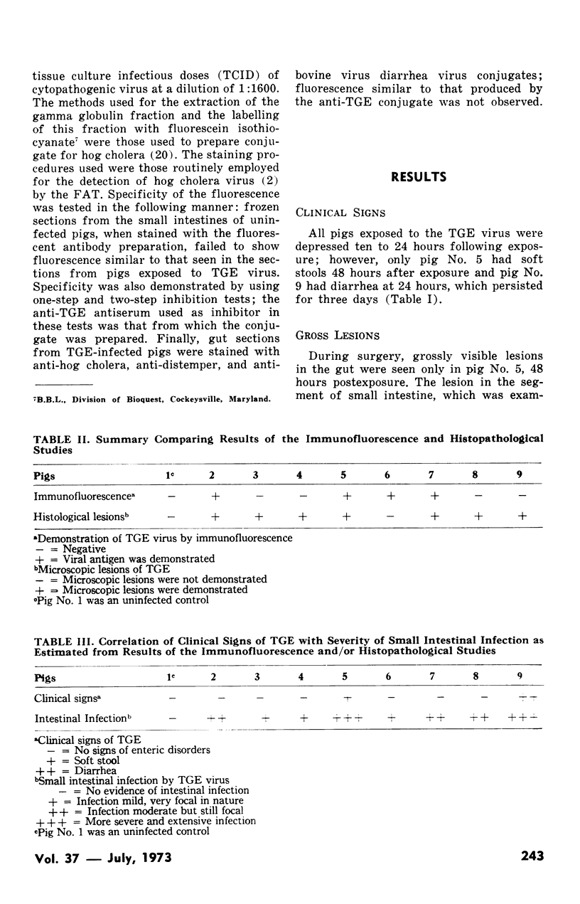
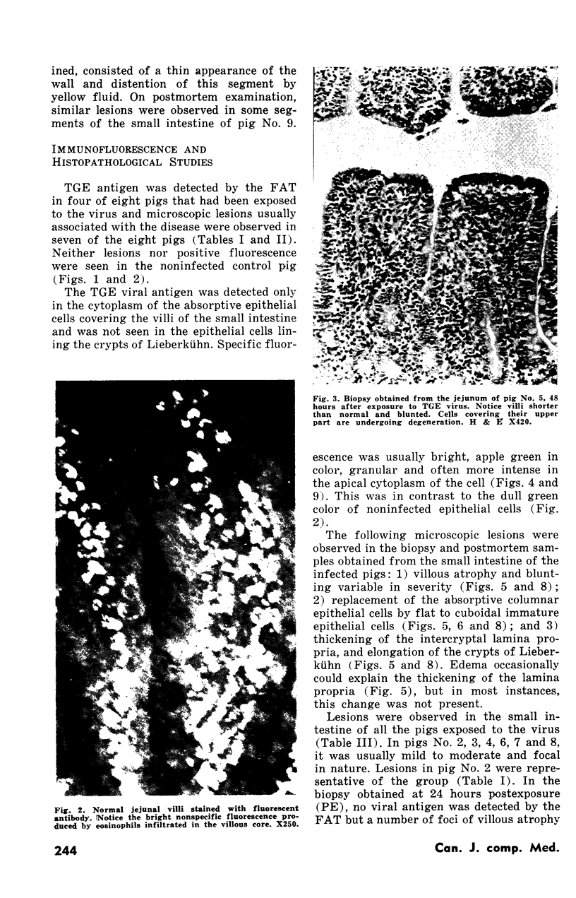
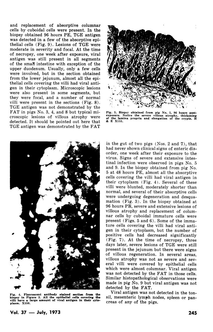
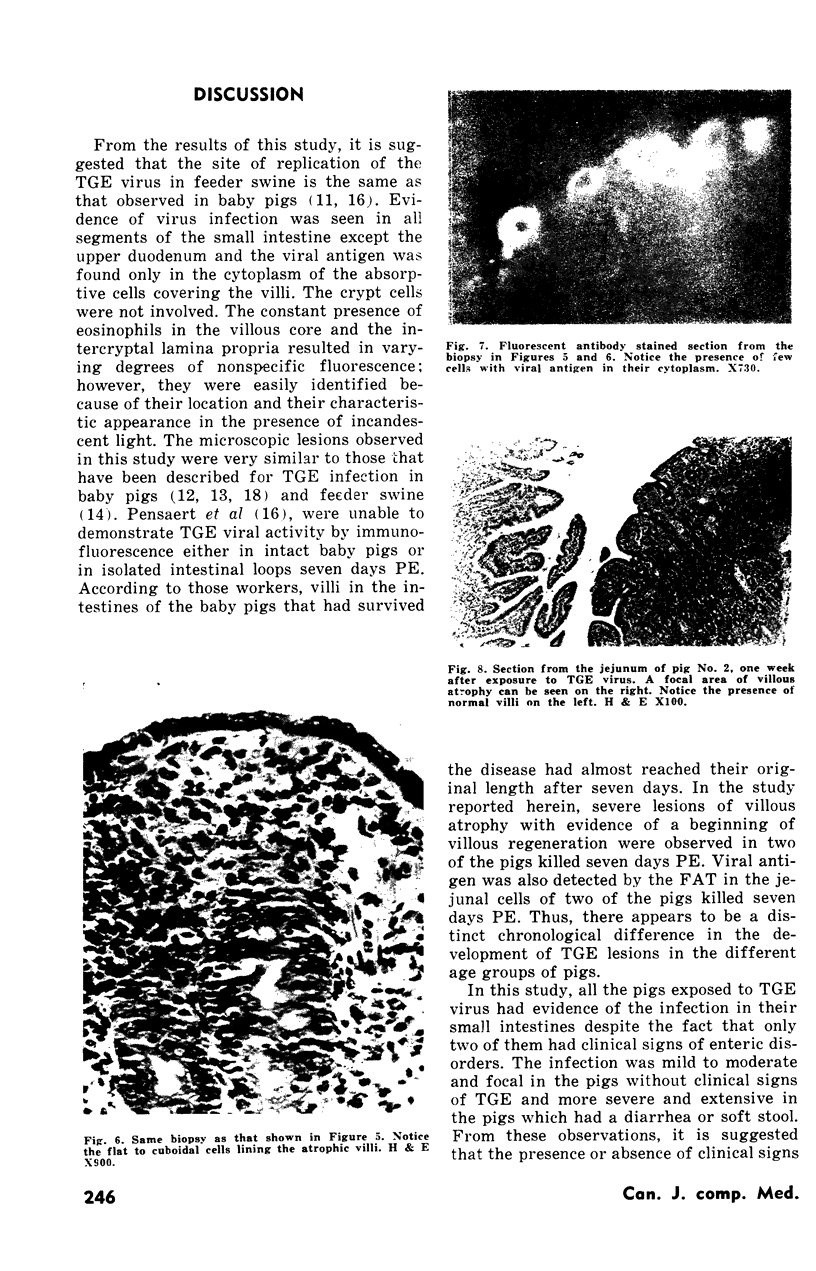
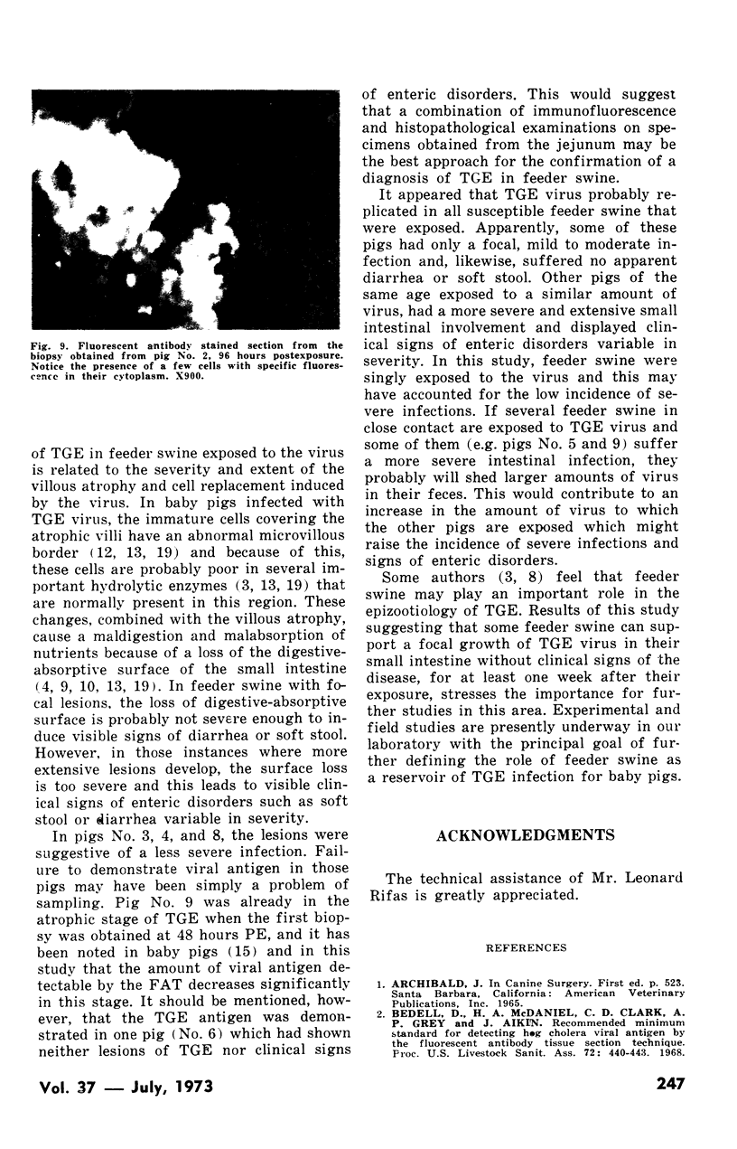
Eight feeder swine (four to six months of age) were inoculated orally with 200,000 to 500,000 pig infectious doses (PID) of the Purdue strain of transmissible gastroenteritis (TGE) virus. Biopsies obtained from their small intestines were examined histopathologically and by fluorescent antibody tissue section technique at intervals that included 24, 48, 72 and 96 hours postexposure, and similar examinations were carried out at necropsy 168 hours postexposure. Evidence of virus infection was demonstrated in all segments of the small intestine except the upper duodenum and the viral antigen was found only in the cytoplasm of the absorptive cells covering the villi. Although six of the eight pigs failed to show clinical signs of TGE, typical microscopic lesions of villous atrophy with replacement of columnar absorptive cells by cuboidal cells were observed in seven pigs, and TGE virus antigen was demonstrated in the intestinal cells of four of eight pigs during the first week postexposure. The infection was usually mild to moderate and focal in the pigs without clinical signs of the disease and more severe and extensive in the pigs with clinical signs of the disease variable in severity. It was concluded that TGE virus probably replicated in all feeder swine exposed, and that the presence or absence of clinical signs of TGE in these pigs was related to the severity and extent of the villous atrophy and columnar cell replacement induced in their small intestines.
Full text
PDF
Images in this article
Selected References
These references are in PubMed. This may not be the complete list of references from this article.
- Aelterman E. O., Hooper B. E. Transmissible gastroenteritis of swine as a model for the study of enteric disease. Gastroenterology. 1967 Jul;53(1):109–113. [PubMed] [Google Scholar]
- Badell D., McDaniel H. A., Grey A. P., Aikin J. Recommended minimun standards for detecting hog cholera viral antigen by the fluorescet antibody tissue section technique. Proc Annu Meet U S Anim Health Assoc. 1968;72:440–443. [PubMed] [Google Scholar]
- Ferris D. H. Epizootiologic features of transmissible swine gastroenteritis. J Am Vet Med Assoc. 1971 Jul 15;159(2):184–194. [PubMed] [Google Scholar]
- GOODWIN R. F., JENNINGS A. R. Infectious gastro-enteritis of pigs. I. The disease in the field. J Comp Pathol. 1959 Jan;69(1):87–97. doi: 10.1016/s0368-1742(59)80008-4. [DOI] [PubMed] [Google Scholar]
- Hooper B. E., Haelterman E. O. Growth of transmissible gastroenteritis virus in young pigs. Am J Vet Res. 1966 Jan;27(116):286–291. [PubMed] [Google Scholar]
- Hooper B. E., Haelterman E. O. Lesions of the gastrointestinal tract of pigs infected with transmissible gastroenteritis. Can J Comp Med. 1969 Jan;33(1):29–36. [PMC free article] [PubMed] [Google Scholar]
- Maronpot R. R., Whitehair C. K. Experimental sprue-like small intestinal lesions in pigs. Can J Comp Med Vet Sci. 1967 Dec;31(12):309–316. [PMC free article] [PubMed] [Google Scholar]
- Olson L. D. Induction of transmissible gastroenteritis in feeder swine. Am J Vet Res. 1971 Mar;32(3):411–417. [PubMed] [Google Scholar]
- Pensaert M. B., Haelterman E. O., Burnstein T. Diagnosis of transmissible gastroenteritis in pigs by means of immunofluorescence. Can J Comp Med. 1968 Oct;32(4):555–561. [PMC free article] [PubMed] [Google Scholar]
- Pensaert M., Haelterman E. O., Burnstein T. Transmissible gastroenteritis of swine: virus-intestinal cell interactions. I. Immunofluorescence, histopathology and virus production in the small intestine through the course of infection. Arch Gesamte Virusforsch. 1970;31(3):321–334. doi: 10.1007/BF01253767. [DOI] [PubMed] [Google Scholar]
- Pensaert M., Haelterman E. O., Hinsman E. J. Transmissible gastroenteritis of swine: virus-intestinal cell interactions. II. Electron microscopy of the epithelium in isolated jejunal loops. Arch Gesamte Virusforsch. 1970;31(3):335–351. doi: 10.1007/BF01253768. [DOI] [PubMed] [Google Scholar]
- Thake D. C. Jejunal epithelium in transmissible gastroenteritis of swine. An electron microscopic and histochemical study. Am J Pathol. 1968 Jul;53(1):149–168. [PMC free article] [PubMed] [Google Scholar]
- Trapp A. L., Sanger V. L., Stalnaker E. Lesions of the small intestinal mucosa in transmissible gastroenteritis-infected germfree pigs. Am J Vet Res. 1966 Nov;27(121):1695–1702. [PubMed] [Google Scholar]