Abstract
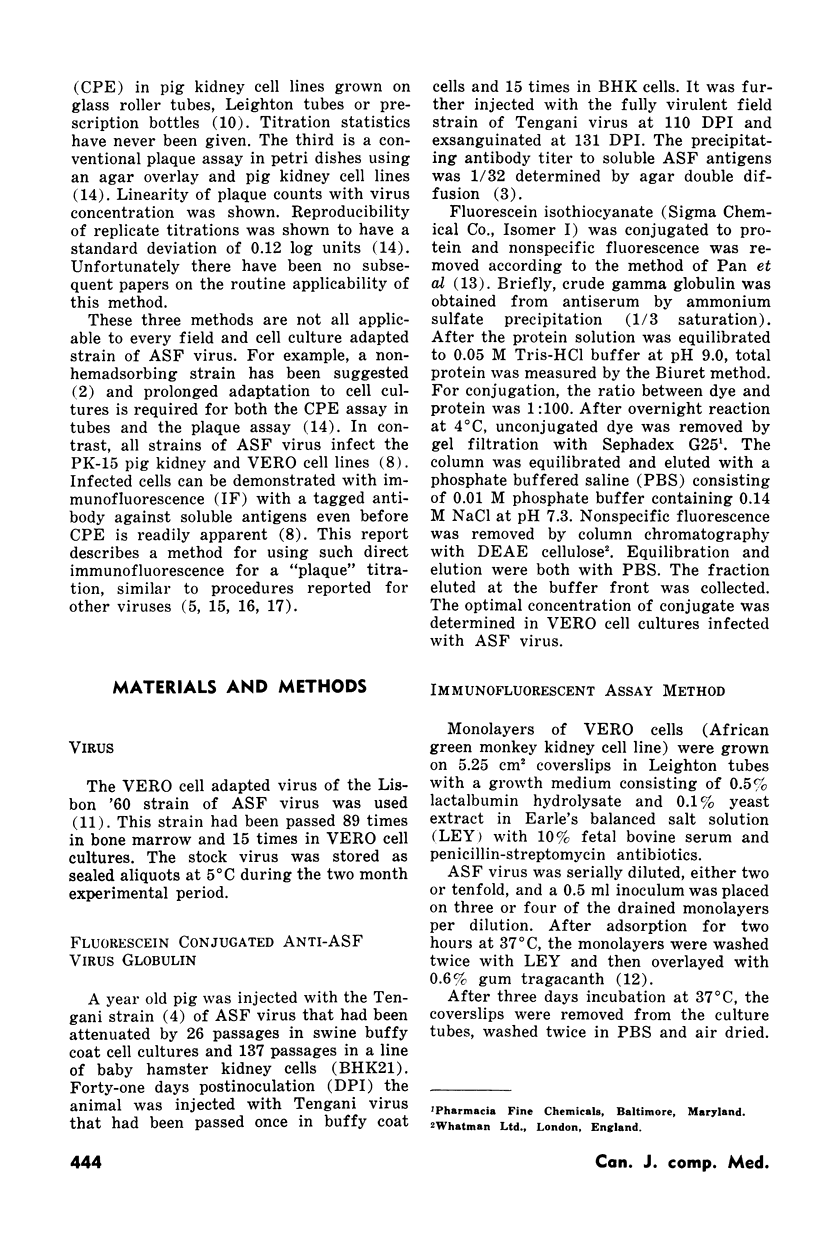
Suitably diluted cell culture adapted African swine fever virus preparations were inoculated on VERO cell monolayers and grown on coverslips. Gum tragacanth was used as an overlay. After three days incubation at 37°C the infected cultures were fixed with acetone and stained with fluorescent antibody conjugate. Fluorescing plaques consisted of 20-30 infected cells.
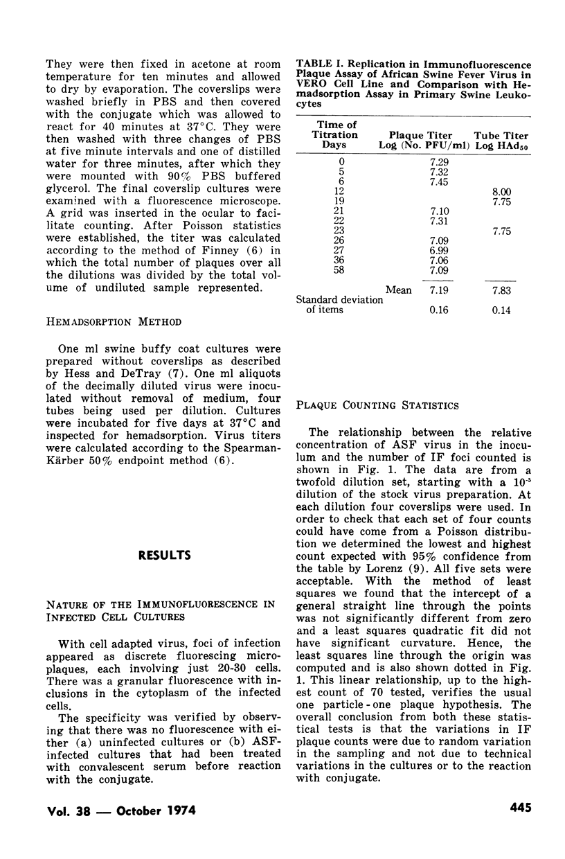
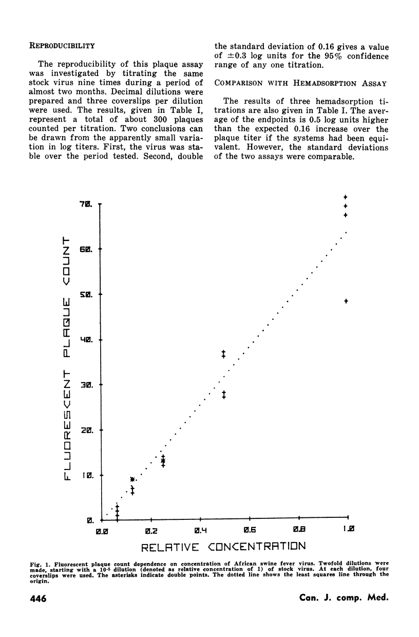
Three statistical criteria for a quantitatively reliable assay were met: the Poisson distribution for plaque counts, linearity of the relationship between the concentration of virus and the plaque count and reproducibility of replicate titrations. The method is suitable for counts up to at least 70 plaques per 5 cm2 coverslip and computed titers are reproducible within 0.16 log units with a total of 300 plaques enumerated.
Full text
PDF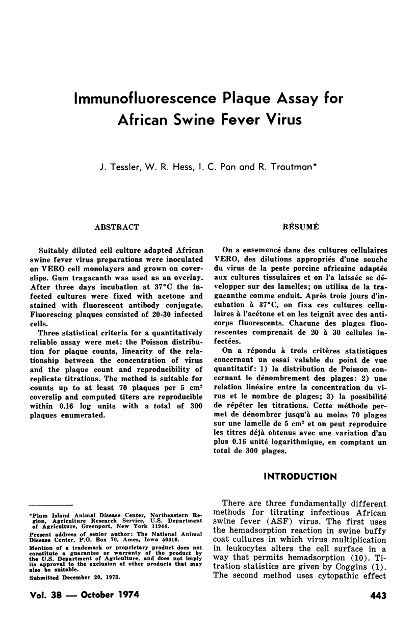
Selected References
These references are in PubMed. This may not be the complete list of references from this article.
- Coggins L., Heuschele W. P. Use of agar diffusion precipitation test in the diagnosis of African swine fever. Am J Vet Res. 1966 Mar;27(117):485–488. [PubMed] [Google Scholar]
- Coggins L. Segregation of a nonhemadsorbing African swine fever virus in tissue culture. Cornell Vet. 1968 Jan;58(1):12–20. [PubMed] [Google Scholar]
- DEIBEL R., HOTCHIN J. E. Quantitative applications of fluorescent antibody technique to influenza-virus-infected cell cultures. Virology. 1959 Jul;8(3):367–380. doi: 10.1016/0042-6822(59)90036-4. [DOI] [PubMed] [Google Scholar]
- Heuschele W. P., Hess W. R. Diagnosis of African swine fever by immunofluorescence. Trop Anim Health Prod. 1973 Aug;5(3):181–186. doi: 10.1007/BF02251387. [DOI] [PubMed] [Google Scholar]
- LORNEZ R. J. [On statistics of the plaque test]. Arch Gesamte Virusforsch. 1962;12:108–137. [PubMed] [Google Scholar]
- MALMQUIST W. A., HAY D. Hemadsorption and cytopathic effect produced by African Swine Fever virus in swine bone marrow and buffy coat cultures. Am J Vet Res. 1960 Jan;21:104–108. [PubMed] [Google Scholar]
- Mirchamsy H., Rapp F. A new overlay for plaquing animal viruses. Proc Soc Exp Biol Med. 1968 Oct;129(1):13–17. doi: 10.3181/00379727-129-33237. [DOI] [PubMed] [Google Scholar]
- PHILIPSON L. Adenovirus assay by the fluorescent cell-counting procedure. Virology. 1961 Nov;15:263–268. doi: 10.1016/0042-6822(61)90357-9. [DOI] [PubMed] [Google Scholar]
- Pan I. C., Tsai K. S., Karstad L. Glomerulonephritis in Aleutian disease of mink: histological and immunofluorescence studies. J Pathol. 1970 Jun;101(2):119–127. doi: 10.1002/path.1711010207. [DOI] [PubMed] [Google Scholar]
- Parker J., Plowright W. Plaque formation by African swine fever virus. Nature. 1968 Aug 3;219(5153):524–525. doi: 10.1038/219524a0. [DOI] [PubMed] [Google Scholar]
- RAPP F., RASMUSSEN L. E., BENYESH-MELNICK M. THE IMMUNOFLUORESCENT FOCUS TECHNIQUE IN STUDYING THE REPLICATION OF CYTOMEGALOVIRUS. J Immunol. 1963 Nov;91:709–719. [PubMed] [Google Scholar]
- WHEELOCK E. F., TAMM I. Enumeration of cell-infecting particles of Newcastle disease virus by the fluorescent antibody technique. J Exp Med. 1961 Feb 1;113:301–316. doi: 10.1084/jem.113.2.301. [DOI] [PMC free article] [PubMed] [Google Scholar]


