Abstract
Context: Although isotonic and isokinetic exercises are commonly used in sports medicine and rehabilitation, studies comparing their effects on the neuromuscular system have provided conflicting results.
Objective: To compare responses of the neuromuscular system to isotonic and isokinetic contractions by controlling the total external amount of work performed and the mean angular movement velocity.
Design: A familiarization session was followed by isotonic and isokinetic sessions of tests performed on an isokinetic dynamometer. Each subject participated in 3 sessions.
Setting: A sport sciences research laboratory.
Patients or Other Participants: Nine healthy adult males with no history of knee injury.
Intervention(s): The isotonic session consisted of 3 sets of 8 knee extensions at 80% of each subject's maximal voluntary isotonic contraction. The isokinetic session involved 3 sets of n knee extensions at a preset velocity equivalent to the mean velocity measured during the corresponding isotonic sets; n represented the number of repetitions subjects had to achieve to equalize the total external amount of work performed during the corresponding isotonic sets.
Main Outcome Measure(s): We recorded mechanical parameters, n, and surface electromyographic signals from the vastus medialis, rectus femoris, and vastus lateralis muscles. Then root mean square, mean power frequency, and neuromuscular efficiency values were calculated for each repetition.
Results: As expected, the total external amount of work and mean angular velocity were similar between the isotonic and isokinetic sessions. The number of repetitions performed was equivalent in both sessions. In addition, although no “shift” of mean power frequency occurred, mean neuromuscular efficiency decreased linearly with repetitions for both modes in no differentiated way.
Conclusions: Standardization of isotonic and isokinetic contractions based on total external amount of work and movement velocity is possible. This method can be applied by future investigators aiming to compare chronic effects of these 2 contraction modes on the neuromuscular system.
Keywords: isotonic, isokinetic, surface electromyography, external work, knee extensors
Isotonic and isokinetic contractions present different biomechanical characteristics. In the isotonic mode, the neuromuscular system has to overcome an initial resistance (constant throughout the movement) to move the lever arm. Consequently, isotonic contraction is supposed to maximally load the neuromuscular system only at the weakest mechanical points of the range of motion, whereas the rest is worked at less than maximal capacity.1,2 By contrast, the isokinetic mode implies an accommodating resistance, which allows a constant angular velocity once the preset velocity is reached. Therefore, the isokinetic movement is expected to maximally load the neuromuscular system through the overall range of motion.1,3 Thus, the isokinetic contraction theoretically allows the muscle to perform a greater amount of work than the isotonic contraction over the same range of motion.3 Then, isotonic and isokinetic training may induce specific changes on torque-angle and torque-velocity relationships.
Some authors1,2,4,5 have already attempted to compare the effectiveness of isotonic versus isokinetic training on strength development. Some researchers1,4 reported that isokinetic training was more effective than isotonic training in improving maximal strength and explosive muscular properties, whereas others2,5 observed that isotonic training resulted in greater increases in maximal strength and muscle mass than isokinetic training. These apparent discrepancies seem to originate from the protocols set up to compare these 2 training modes. Indeed, none of these investigators have quantified the total external amount of work performed during isotonic and isokinetic training sessions. Although movement velocity has been reported to affect strength gains,6,7 only 1 study5 attempted to control angular velocity. Some authors1,4,5 also used different training and/or testing devices, which might have been inappropriate because of ergometer-type specific effects.8 Consequently, it appears necessary to build a standardized protocol allowing comparison of both isotonic and isokinetic modes in order to analyze the specific effects of each training mode on the neuromuscular system.
Our main purpose was to investigate a method of comparing isotonic and isokinetic knee extensions with the same ergometer by standardizing the total external amount of work performed by subjects and controlling the mean angular velocity of movement. We also aimed to provide some insight concerning the mechanical and electrophysiologic behaviors of the neuromuscular system during both isotonic and isokinetic contractions. We hypothesized that because isokinetic contractions theoretically allowed the muscle group to perform a greater amount of external work over the same range of motion than isotonic contractions, more isotonic contractions would be necessary to achieve the same total external amount of work.
METHODS
Subjects
Nine healthy adult males without any previous history of knee injury volunteered for this study. All subjects were informed of the nature and the aim of this study before they signed an informed consent form. This study was conducted according to the Helsinki Statement (1964). The mean age, height, mass, and body fat percentage of the subjects were 23.3 ± 2.1 years, 177.8 ± 8.2 cm, 71.9 ± 7.6 kg, and 14.6 ± 3.6%, respectively. Body fat percentage was estimated from 4 skinfold thicknesses (biceps, triceps, subscapular, and suprailliac) according to the equation of Siri.9
Dynamometry
Isotonic and isokinetic tests were performed on a Biodex System 3 Pro dynamometer (Biodex Medical Systems, Shirley, NY). In isotonic mode, the subject had to overcome a preloaded resistance (ie, a level of torque) before the actuator arm initiated movement. Then, “any increase in applied force by the subject would be absorbed by the dynamometer and returned as a directly proportional increase in velocity.”2 Therefore, the isotonic mode simulated by the dynamometer differs somewhat from the conventional isotonic method, which consists of lifting a constant load through the overall range of motion. In isokinetic mode, the dynamometer compared the velocity of the lever arm to the preset velocity through the range of motion. If the preset velocity was not reached, no resistance was encountered by the subject. But when the preset velocity was reached, the dynamometer accommodated resistance in order to keep angular velocity constant. This device also allowed us to end the exercises when a predetermined number of repetitions was reached or when a preset amount of external work was achieved through the use of visual feedback. During the tests, adjustable seat belts were firmly attached across the subject's chest and hips to prevent hip joint movements during concentric knee extensions. The dominant lower leg (the one used to kick a ball) was attached to the mobile part of the dynamometer just above the ankle joint. Ergometer settings and the seat position were saved to be reproduced during all test sessions. During contractions, subjects were asked to cross their arms over their chests. Mechanical signals were recorded at a sampling frequency of 100 Hz. All recorded torques were gravity corrected through the overall range of motion using Biodex software.
Electromyography
Bipolar electromyographic signals (SEMG) were recorded from surface electrodes (4-mm diameter Ag-AgCl, In Vivo Metric, Healdsburg, CA) on the vastus medialis, rectus femoris, and vastus lateralis muscles with a 13-mm interelectrode distance. Electrode-skin impedance was reduced below 55 kΩ10 using standard skin-preparation procedures.11 According to the “Surface Electromyography for the Non-Invasive Assessment of Muscles” recommendations,12 we placed surface electrodes between the distal tendon and the innervation zone with respect to the direction of the assumed fibers. Moreover, surface electrodes were placed for the knee joint set at the angle of peak torque, ie, an individualized biomechanical position. Three reference electrodes were placed over the lateral and medial epicondyles. Sensor locations were marked with indelible ink during the first test session so that we could exactly replace the electrodes during the subsequent test sessions. Then, EMG signals were preamplified (gain = 600) and sampled at 1024 Hz with a 12-bit A/D converter (Myodata, Electronique du Mazet, Le Mazet Saint Voy, France; input impedance = 10 GΩ, common mode-rejection ratio at 50/60 Hz = 100 dB, sampling frequency = 0 to 400 Hz). Data were stored in a flash memory card (20 MB) and transferred to a computer hard disk for further analysis.
Experimental Design
Each subject participated in 3 test sessions. The first session allowed the subjects to be familiarized with isotonic and isokinetic contraction modes. The second and third sessions consisted of isotonic and isokinetic tests, respectively, and were completed at the same time of the day with 1 day of rest in between.
During the first session, the subject's anthropometric characteristics (height, mass, body fat percentage) were determined. After a 5-minute general warm-up on a cycle ergometer (100 W), the subject was seated on the dynamometer so that the hip was flexed to 85° (0° = full hip extension) with the leg in horizontal position. The motor rotation axis was visually aligned with the anatomical axis of the knee. The knee range of motion was 90° to 30° (0° = leg in horizontal position) to ensure that all subjects could perform the movement through the overall range of motion, especially in isotonic mode, as determined during preliminary studies. Then, after a specific warm-up on the dynamometer (6 knee extensions at 50 Nm in isotonic mode), we assessed the maximal voluntary isotonic contraction of the knee extensors. Maximal voluntary isotonic contraction was defined as the maximal level of torque the subject could overcome. As a familiarization sequence, the subject performed 8 isotonic knee extensions at 80% of maximal voluntary isotonic contraction. Then the subject completed 8 maximal isokinetic knee extensions at a preset velocity similar to the mean velocity measured during the isotonic set. The isokinetic set also allowed us to determine the individual angle for peak torque in order to apply surface electrodes at this angle.
In the second and third test sessions, the subject warmed up as in the first session. During the second session, the subject performed 3 sets of 8 isotonic repetitions at 80% of maximal voluntary isotonic contraction. The subject had to reach this preset level of torque to initiate the movement, and then the dynamometer controlled this value and kept it constant through the range of motion. Each subject performed the 3 sets of 8 knee extensions through the overall range of motion. The third test session consisted of 3 sets of n maximal isokinetic repetitions at a preset velocity similar to the mean velocity measured during the corresponding isotonic sets; n represented the number of maximal isokinetic repetitions the subject had to achieve in order to equalize the external amount of work performed during the corresponding isotonic set. Each isokinetic set was automatically stopped when this amount of work was reached. Therefore, the order of the second and the third test sessions was not randomized. No verbal encouragement was given during any test session, but subjects were required to perform ballistic movements. A 2-minute rest period13 between sets was respected in both the isotonic and isokinetic sessions. The SEMG of the vastus lateralis, rectus femoris, and vastus medialis muscles was recorded during the second and third sessions from the sites defined during the first session.
Data Analysis
Mechanical measurements (position, torque, and velocity) and SEMG data were collected during the second and third test sessions. Data processing was conducted using a custom program (Protags, Labview, National Instruments, Austin, TX) for mechanical data, the Biodex software for the quantification of external amount of work, and the Myodata software for SEMG signals.
For each knee extension, the total external amount of work (W) was calculated at each time interval (t) from the torque (T) and the angular position (α), as expressed by the following equation:
| W = Σ [(T(t) − T(t − 1)) × (α(t) − α(t − 1))] |
In isotonic mode, we calculated the mean angular velocity of each knee extension between the beginning (90°) and end (30°) of the movement. Then, the mean angular velocity of each isotonic set was calculated in order to preset velocity at an equivalent value during the corresponding isokinetic set. Maximal external torque was determined during each repetition of both isotonic and isokinetic sessions. The SEMG analyses were performed in both time and frequency domains. For each muscle, root mean square (RMS) values were calculated every 20 ms14 from SEMG bursts corresponding to repetitions 2 through 8. Then, for each repetition and for each muscle, a mean RMS value was determined by dividing the sum of the RMS values by the burst duration, ie, the amount of time required to complete the 60° range of motion. The SEMG bursts lasted about 420 ms. A mean RMS value of knee extensors was calculated for each repetition by summing the mean RMS values of the vastus lateralis, rectus femoris, and vastus medialis muscles. Neuromuscular efficiency (NME) was then calculated for each repetition by dividing maximal torque by the RMS of the knee extensors.15 Also, after filtering SEMG signals with a bandwidth of 6 to 400 Hz, we performed a Fast Fourier Transformation to calculate mean power frequency (MPF) values of the second and last bursts of each set. The first repetition of the set was not taken into account in case it was not representative of the rest of the set.16
Statistical Analysis
Before each comparison procedure, normality of data was verified with the Kolmogorov-Smirnov test. Thus, we used a Student paired t test to analyze potential changes between the isotonic and isokinetic modes in the total external amount of work (for set 3), the mean angular velocity, and the number of repetitions performed. A Wilcoxon signed rank test was used to compare the total external work achieved between the isotonic and isokinetic modes for sets 1 and 2. Mean maximal external torque, mean NME, and mean RMS were linearly regressed against repetitions 2 to 8 for each set in both contraction modes. When regressions were significant, we used slope comparisons to determine potential differences between sets 1 and 3 for both modes and between isotonic and isokinetic sets. Possible differences in MPF between the beginning and end of the sets were tested for each muscle using a Student paired t test for sets 1 and 2 and a Wilcoxon signed rank test for set 3. For all tests, the significance level was fixed at P < .05.
RESULTS
Controlled Parameters
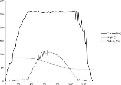
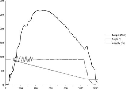
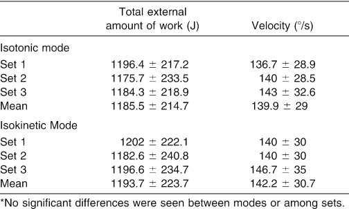
Figures 1 and 2 show typical raw mechanical data obtained during isotonic and isokinetic contractions, respectively, for 1 repetition. The mean total external amount of work performed during the isotonic session (1185.5 ± 214.7 J) was not significantly different from that realized during the isokinetic session (1193.7 ± 223.7 J) for set 1 (P = .2), set 2 (P = .07), or set 3 (P = .41) (Table 1). Mean velocity measured in isotonic mode (139.9 ± 29°/s) was not significantly different from mean preset velocity fixed in isokinetic mode (142.2 ± 30.7°/s) for set 1 (P = .19), set 2 (P = .99), or set 3 (P = .35) (Table 1).
Figure 1. Typical raw mechanical data obtained during isotonic contraction for 1 repetition. Subjects have to overcome a preloaded amount of torque before the actuation arm initiates the movement. In this example, the level of torque is preset at 250 N·m.
Figure 2. Typical raw mechanical data obtained during isokinetic contraction for 1 repetition. The dynamometer accommodates resistance to the subject's effort to guarantee a constant angular velocity. In this example, angular velocity is preset at 90°/s.
Table 1. Mean Total External Amount of Work Performed and Mean Velocity of Movement During Sets of Isotonic and Isokinetic Modes*.
Number of Repetitions
We found no significant difference in number of repetitions required by set to perform the same total external amount of work between the isokinetic and isotonic modes: 7.8 ± 1.0 versus 8 for set 1 (P = .65), set 2 (P = .64), and set 3 (P = .57).
Torque-Repetitions Relationships
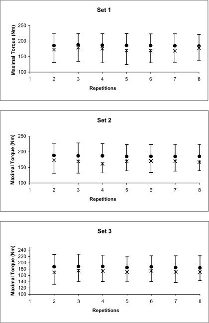
The mean maximal external torque values remained steady in both isotonic and isokinetic modes (Figure 3). Percentages of changes in mean torque between repetitions 2 and 8 were −0.9%, −1.5%, and −2.1% for sets 1, 2, and 3, respectively, in isotonic mode and +2.6%, −3.2%, and +0.7% for sets 1, 2, and 3, respectively, in isokinetic mode.
Figure 3. Mean maximal external torque values in isotonic and isokinetic modes. •, isotonic mode; ×, isokinetic mode.
Root Mean Square-Repetitions Relationships
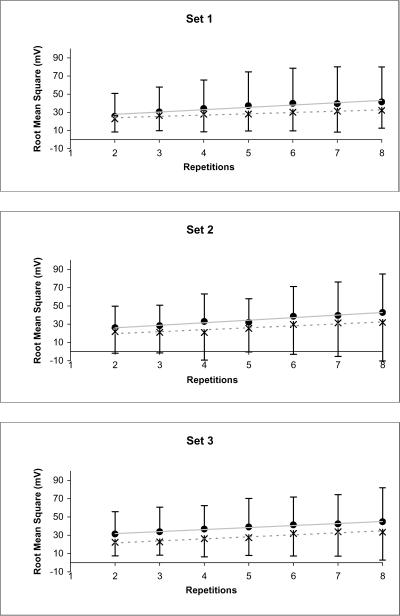
The mean RMS-repetitions relationships showed a linear increase for all sets in the isotonic (0.92 < r2 < 0.99) and isokinetic (0.87 < r2 < 0.94) modes (Figure 4). In isotonic mode, the mean RMS increased 62.1%, 63.8%, and 42.7% for sets 1, 2, and 3, respectively, whereas in isokinetic mode, it increased 41.2%, 45.9%, and 50.1% for sets 1, 2, and 3, respectively. The slopes for set 1 were not significantly different from those for set 3 in either mode. Isotonic slopes were not significantly different from isokinetic slopes for all 3 sets.
Figure 4. Mean root mean square-repetitions relationships in isotonic and isokinetic modes. •, isotonic mode; ×, isokinetic mode. Slope of set 1 is not significantly different from slope of set 3 for both modes. Isotonic slopes are not significantly different from isokinetic slopes in all sets.
Neuromuscular Efficiency-Repetitions Relationships
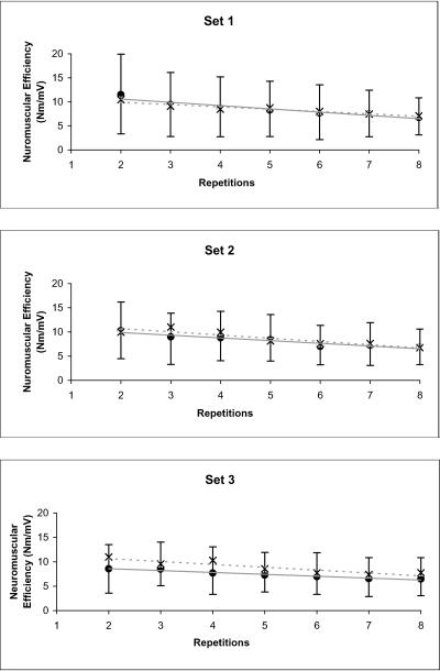
The mean NME-repetitions relationships showed a linear decrease for all sets in the isotonic (0.89 < r2 < 0.96) and isokinetic (0.83 < r2 < 0.90) modes (Figure 5). The decreases in mean NME were −41.1%, −33.9%, and −24.4%, for sets 1, 2, and 3, respectively, in isotonic mode, and −32%, −32.3%, and −29.3% for sets 1, 2, and 3, respectively, in isokinetic mode. No significant difference was noted between the slopes of set 1 and set 3 for either mode or between the isotonic and isokinetic slopes for all 3 sets.
Figure 5. Mean neuromuscular efficiency-repetitions relationships in isotonic and isokinetic modes. •, isotonic mode; ×, isokinetic mode. Slope of set 1 is not significantly different from slope of set 3 for both modes. Isotonic slopes are not significantly different from isokinetic slopes in all sets.
Mean Power Frequency Values
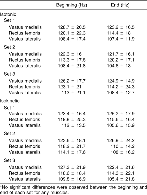
For the vastus lateralis, rectus femoris, and vastus medialis muscles, we found no significant difference in the levels of MPF between the beginning and end of the set for set 1 (P = .65), set 2 (P = .63), or set 3 (P = .32) in isotonic mode, or for set 1 (P = .19), set 2 (P = .59), or set 3 (P = .81) in isokinetic mode (Table 2).
Table 2. Mean Power Frequency Values at the Beginning (Second Repetition) and the End (Last Repetition) of Each Isotonic and Isokinetic Set for Each Muscle*.
DISCUSSION
Our main purpose was to investigate a methodologic approach allowing comparison of isotonic and isokinetic concentric knee extensions on the same ergometer by equalizing the total external amount of work performed and the mean angular velocity of movement. Some authors interested in such a comparison have discussed several points. For instance, Smith and Melton1 suggested that equalization of the external amount of work performed was a critical question and that, ideally, a computerized work integrator should be connected to both isotonic and isokinetic devices. O'Hagan et al5 attempted to evaluate the effectiveness of these 2 contraction modes by taking into account training stimuli (ie, intensity of contraction, number of contractions, and time required to complete a training session). Compared with isokinetic training sessions, a possible lack of intensity in the isotonic training sessions was compensated for by an increased number of muscular actions. However, the total amount of work was not quantified. These authors also endeavored to keep the velocity of isotonic movement as close as possible to the preset isokinetic velocity by using a metronome. However, they noted that the last repetitions became slower than the metronome's speed because of fatigue, particularly in the isotonic mode. Similarly, Schmitz and Westwood3 tried to equalize movement velocity. They compared 10 isotonic (at 50% of maximal voluntary isometric contraction) and 10 isokinetic knee extensions by setting isokinetic velocity at 180°/s for all subjects. In our study, the mean isokinetic velocity was close to that used by Schmitz and Westwood3 (140°/s versus 180°/s), but it was individually fixed from the movement velocity measured during the isotonic session. Finally, our results, as expected, indicate that the total external amount of work and angular velocity of movement measured during each set were not different between the contraction modes. Therefore, it could be assumed that the isotonic and isokinetic sessions were standardized for these 2 parameters.
Because an isokinetic contraction theoretically allows the muscle to perform more work over a same given range of motion than an isotonic contraction,3 we hypothesized that more isotonic contractions would be required to perform the same total external amount of work, yet we found no significant difference between the number of repetitions in isotonic mode and isokinetic mode. Consequently, our results do not support this hypothesis. Kovaleski et al2 also disagreed with this hypothesis, but in a different way. These authors assumed that isokinetic training would develop muscle strength to a greater extent than isotonic training. However, they found greater strength improvements with isotonic training than with isokinetic training on isometric tests at 10°, 30°, 50°, 70°, and 90° of knee flexion. Moreover, the muscle group trained in isotonic mode produced greater isotonic and isokinetic power than the isokinetic-trained muscle group. In our study, the mean maximal isotonic torque was higher than the mean maximal isokinetic torque (186.2 versus 171.4 Nm, P < .001). Antagonist muscle coactivation has been shown to occur during both static and dynamic contractions.17,18 For instance, in isokinetic mode (180°/s), Miller et al18 evaluated the amount of medial hamstring coactivation at 15.2% and biceps femoris coactivation at 39% of their agonist activity at the same movement velocity. However, to our knowledge, such data are not available for the isotonic mode. It could be suggested that greater coactivation of hamstring muscles may have occurred in isokinetic than in isotonic mode, resulting therefore in a weaker work : repetition ratio in isokinetic mode. This could lead to a greater number of isokinetic contractions than expected and could explain the equivalent number of repetitions found in both modes in this study.
Our results indicate that no significant “shift” of MPF occurred for any considered muscles, no matter the contraction mode. A shift of MPF from high frequency toward low frequency has been reported by several authors as a mark of neuromuscular fatigue during isometric19 and dynamic contractions.20 Indeed, this spectrum shift has been attributed mainly to fatigue of peripheral origin.21 In contrast, Svantesson et al22 did not show any decrease in MPF after approximately 48 isokinetic concentric contractions, despite a significant decrease in the total external amount of work performed. These authors concluded that repeated dynamic muscle actions at high intensity were not necessarily associated with a reduction in MPF. Thus, the lack of change in MPF values in our study does not necessarily indicate that no muscular fatigue occurs.
Indeed, our results show that NME, defined as “the responsiveness of muscle to neural excitation,”15 decreases linearly with the repetitions of a set for both modes. Hermann and Barnes23 also reported a reduction in NME during 50 maximal isokinetic concentric or eccentric contractions of trunk extensors. Because NME is the torque : SEMG ratio, a decrease in maximal external torque and/or an increase in SEMG activity could explain the decline in NME during a set of contractions. In this way, Hermann and Barnes23 essentially described this reduction in NME by decrements in maximal concentric and eccentric torque (−30% and −24%, respectively) because no change in SEMG activity occurred. On the contrary, our results showed that the decrease in NME (−27% to −38%) may be explained mainly by an increase in SEMG activity (+36% to +65%), because maximal external torque seems to remain steady in the course of repetitions during both contraction modes. Increased EMG activity during submaximal contractions has often been associated with a central process aiming to progressively fully activate motor units of a muscle in order to maintain the same level of force.24,25 This hypothesis could explain the increase in RMS during submaximal isotonic contractions, but it seems less evident in isokinetic mode because isokinetic contractions are supposed to be maximal. Nevertheless, Wretling et al20 showed that during 150 maximum isokinetic knee extensions at 90°/s, the RMS values of the vastus lateralis muscle increased during the first 7 contractions before reaching a plateau. In their study, the increase in RMS was associated with an increase in peak torque. These authors consequently concluded that subjects were unable to maximally recruit their muscles during the initial isokinetic knee extensions. This finding is somewhat different from our results, because we found that in isokinetic mode, RMS increased, whereas maximal external torque remained steady in the course of repetitions. It could be suggested that hamstring coactivation, by opposing force to the developed external torque, may have induced a progressive increase in SEMG activity of the quadriceps muscles26 without changes in the external extension torque. Finally, our results indicate that neuromuscular responses to standardized isotonic and isokinetic concentric contractions occur in an undifferentiated way: the slopes of the RMS-repetitions relationships and the NME-repetitions relationships show no significant difference between isotonic and isokinetic modes.
In conclusion, this study showed that equalizing total external amount of work and movement velocity to allow comparison of the isotonic and isokinetic contraction modes is conceivable. Both contraction modes seem to involve the same number of repetitions to achieve the same external amount of work, an unexpected finding given the biomechanical characteristics of each of these contraction modes. This result could be partly explained by differences in the degree of hamstring coactivation. Future investigators should seek to clarify this last point. Our findings also suggest that this method could be applied by future authors aiming to study the specific effects of isotonic versus isokinetic training on the neuromuscular system. Such research should potentially interest both clinicians, in order to optimize their rehabilitation programs, and athletic coaches, in order to adapt the contraction mode to the specific requirements of the activity.
Acknowledgments
This study was supported by grants from the DRDJS (Direction Régionale et Départementale de la Jeunesse et des Sports), Pays de la Loire, and Loire-Atlantique (France).
Footnotes
Anthony Remaud, MS, contributed to acquisition and analysis and interpretation of the data and drafting and final approval of the article. Christophe Cornu, PhD, and Arnaud Guével, PhD, contributed to conception and design, analysis and interpretation of the data, and critical revision and final approval of the article.
Address correspondence to Christophe Cornu, PhD, Laboratoire “Motricité, Interactions, Performance” (J.E. 2438), UFR STAPS, Université de Nantes, 25 bis boulevard Guy Mollet, BP 72206, 44322 Nantes Cedex 3, France. Address e-mail to christophe.cornu@univ-nantes.fr.
REFERENCES
- Smith MJ, Melton P. Isokinetic versus isotonic variable-resistance training. Am J Sports Med. 1981;9:275–279. doi: 10.1177/036354658100900420. [DOI] [PubMed] [Google Scholar]
- Kovaleski JE, Heitman RH, Trundle TL, Gilley WF. Isotonic preload versus isokinetic knee extension resistance training. Med Sci Sports Exerc. 1995;27:895–899. [PubMed] [Google Scholar]
- Schmitz RJ, Westwood KC. Knee extensor electromyographic activity-to-work ratio is greater with isotonic than isokinetic contractions. J Athl Train. 2001;36:384–387. [PMC free article] [PubMed] [Google Scholar]
- Pipes TV, Wilmore JH. Isokinetic vs. isotonic strength training in adult men. Med Sci Sports. 1975;7:262–274. [PubMed] [Google Scholar]
- O'Hagan FT, Sale DG, MacDougall JD, Garner SH. Comparative effectiveness of accomodating and weight resistance training modes. Med Sci Sports Exerc. 1995;27:1210–1219. [PubMed] [Google Scholar]
- Aagaard P, Simonsen EB, Trolle M, Bangsbo J, Klausen K. Specificity of training velocity and training load on gains in isokinetic knee joint strength. Acta Physiol Scand. 1996;156:123–129. doi: 10.1046/j.1365-201X.1996.438162000.x. [DOI] [PubMed] [Google Scholar]
- Pereira MI, Gomes PS. Movement velocity in resistance training. Sports Med. 2003;33:427–438. doi: 10.2165/00007256-200333060-00004. [DOI] [PubMed] [Google Scholar]
- Abernethy P, Wilson G, Logan P. Strength and power assessment: issues, controversies and challenges. Sports Med. 1995;19:401–417. doi: 10.2165/00007256-199519060-00004. [DOI] [PubMed] [Google Scholar]
- Siri WE. The gross composition of the body. Adv Biol Med Phys. 1956;4:239–280. doi: 10.1016/b978-1-4832-3110-5.50011-x. [DOI] [PubMed] [Google Scholar]
- Hewson DJ, Hogrel JY, Langeron Y, Duchêne J. Evolution in impedance at the electrode-skin interface of two types of surface EMG electrodes during long-term recordings. J Eletromyogr Kinesiol. 2003;13:273–279. doi: 10.1016/s1050-6411(02)00097-4. [DOI] [PubMed] [Google Scholar]
- Maisetti O, Guével A, Legros P, Hogrel JY. SEMG power spectrum changes during a sustained 50% maximum voluntary isometric torque do not depend upon the prior knowledge of the exercise duration. J Electromyogr Kinesiol. 2002;12:103–109. doi: 10.1016/s1050-6411(02)00010-x. [DOI] [PubMed] [Google Scholar]
- Hermens HJ, Freriks B, Disselhorst-Klug C, Rau G. Development of recommendations for SEMG sensors and sensor placement procedures. J Eletromyogr Kinesiol. 2000;10:361–374. doi: 10.1016/s1050-6411(00)00027-4. [DOI] [PubMed] [Google Scholar]
- Kraemer WJ, Adams K, Cafarelli E. American College of Sports Medicine position stand: progression models in resistance training for healthy adults. Med Sci Sports Exerc. 2002;34:364–380. doi: 10.1097/00005768-200202000-00027. et al. [DOI] [PubMed] [Google Scholar]
- Colson S, Pousson M, Martin A, Van Hoecke J. Isokinetic elbow flexion and coactivation following eccentric training. J Electromyogr Kinesiol. 1999;9:13–20. doi: 10.1016/s1050-6411(98)00025-x. [DOI] [PubMed] [Google Scholar]
- Deschenes MR, Giles JA, McCoy RW, Volek JS, Gomez AL, Kraemer WJ. Neural factors account for strength decrements observed after short-term muscle unloading. Am J Physiol Regul Integr Comp Physiol. 2002;282:R578–R583. doi: 10.1152/ajpregu.00386.2001. [DOI] [PubMed] [Google Scholar]
- Kellis E, Kellis S. Effects of agonist and antagonist muscle fatigue on muscle coactivation around the knee in pubertal boys. J Electromyogr Kinesiol. 2001;11:307–318. doi: 10.1016/s1050-6411(01)00014-1. [DOI] [PubMed] [Google Scholar]
- Psek JA, Cafarelli E. Behavior of coactive muscles during fatigue. J Appl Physiol. 1993;74:170–175. doi: 10.1152/jappl.1993.74.1.170. [DOI] [PubMed] [Google Scholar]
- Miller JP, Croce RV, Hutchins R. Reciprocal coactivation patterns of the medial and lateral quadriceps and hamstrings during slow, medium and high speed isokinetic movements. J Electromyogr Kinesiol. 2000;10:233–239. doi: 10.1016/s1050-6411(00)00012-2. [DOI] [PubMed] [Google Scholar]
- Van Dieen JH, Oude Vrielink HHE, Housheer AF, Lotters FB, Toussaint HM. Trunk extensor endurance and its relationship to electromyogram parameters. Eur J Appl Physiol Occup Physiol. 1993;66:388–396. doi: 10.1007/BF00599610. [DOI] [PubMed] [Google Scholar]
- Wretling ML, Henriksson-Larsen K, Gerdle B. Inter-relationship between muscle morphology, mechanical output and electromyographic activity during fatiguing dynamic knee-extensions in untrained females. Eur J Appl Physiol Occup Physiol. 1997;76:483–490. doi: 10.1007/s004210050279. [DOI] [PubMed] [Google Scholar]
- De Luca CJ. Myoelectrical manifestations of localized muscular fatigue in humans. Crit Rev Biomed Eng. 1984;11:251–279. [PubMed] [Google Scholar]
- Svantesson U, Österberg U, Thomeé R, Peeters M, Grimby G. Fatigue during repeated eccentric-concentric and pure concentric muscle actions of the plantar flexors. Clin Biomech (Bristol, Avon) 1998;13:336–343. doi: 10.1016/s0268-0033(98)00099-0. [DOI] [PubMed] [Google Scholar]
- Hermann KM, Barnes WS. Effects of eccentric exercise on trunk extensor torque and lumbar paraspinal EMG. Med Sci Sports Exerc. 2001;33:971–977. doi: 10.1097/00005768-200106000-00017. [DOI] [PubMed] [Google Scholar]
- Ebenbichler G, Kollmitzer J, Quittan M, Uhl F, Kirtley C, Fialka V. EMG fatigue patterns accompanying isometric fatiguing knee-extensions are different in mono- and bi-articular muscles. Electroencephalogr Clin Neurophysiol. 1998;109:256–262. doi: 10.1016/s0924-980x(98)00015-0. [DOI] [PubMed] [Google Scholar]
- Bigland-Ritchie B, Furbush F, Woods JJ. Fatigue of intermittent submaximal voluntary contractions: central and peripheral factors. J Appl Physiol. 1986;61:421–429. doi: 10.1152/jappl.1986.61.2.421. [DOI] [PubMed] [Google Scholar]
- Weir JP, Keefe DA, Eaton JF, Augustine RT, Tobin DM. Effect of fatigue on hamstring coactivation during isokinetic knee extensions. Eur J Appl Physiol Occup Physiol. 1998;78:555–559. doi: 10.1007/s004210050460. [DOI] [PubMed] [Google Scholar]


