Abstract
Context: Cervical resistance training has been purported to aid in reducing the severity of brain injuries in athletes.
Objective: To determine the effect of an 8-week resistance-training program on head-neck segment dynamic stabilization in male and female collegiate soccer players.
Design: Pretest and posttest control group design.
Setting: University research laboratory and fitness center.
Patients or Other Participants: Thirty-six National Collegiate Athletic Association Division I collegiate soccer players (17 men, 19 women).
Intervention(s): The resistance training group underwent an 8-week cervical resistance training program that consisted of 3 sets of 10 repetitions of neck flexion and extension at 55% to 70% of their 10-repetition maximum 2 times a week. Participants in the control group performed no cervical resistance exercises.
Main Outcome Measure(s): Head-neck segment kinematics and stiffness, electromyographic activity of the upper trapezius and sternocleidomastoid muscles during force application to the head, and neck flexor and extensor isometric strength.
Results: No kinematic, electromyographic, or stiffness training effects were seen. The posttest resistance training group isometric neck flexor strength was 15% greater than the pretest measurement. Isometric neck extensor strength in the female resistance training group was 22.5% greater at the posttest than at the pretest. Women's neck girth increased 3.4% over time regardless of training group level. Women exhibited 7% less head-neck segment length and 26% less head-neck segment mass than men.
Conclusions: Despite increases in isometric strength and girth, the 8-week isotonic cervical resistance training did not enhance head-neck segment dynamic stabilization during force application in collegiate soccer players. Future researchers should examine the effect of head-neck segment training protocols that include traditional and neuromuscular activities (eg, plyometrics) with the focus of reducing head acceleration on force application.
Keywords: cervical musculature, concussion, head acceleration
It has been estimated that soccer players head the soccer ball 2000 times during games in their careers,1 with the total number of headers likely higher when practice is factored in.2 The long-term cumulative cognitive effects of the relatively low loads during soccer heading are not known. Some authors have reported no neurocognitive effect with soccer participation3,4 whereas others have shown deficits in neural function with participation.5–8 As with acute brain injury,9 any long-term cognitive deficits may be related to the amount of head acceleration experienced at impact.
It is estimated that 5% to 22% of high school and collegiate soccer injuries each year are concussions.10–13 The true incidence of concussion may be higher, however, because soccer players do not always seek medical attention due to failure to recognize the symptoms of concussion as well as other factors.14 Males are reported to have a higher risk of concussion than females,10,11,15 but this finding is based on studies with methodologic limitations, including single-team cohort studies and limited longitudinal assessments. Powell and Barber-Foss13 reported on a 3-year high school injury surveillance system funded by the National Athletic Trainers' Association and determined that women's soccer had a higher percentage of concussions (6.2%) than men's soccer (5.7%). Covassin et al12 examined data from 3 years of the National Collegiate Athletic Association's Injury Surveillance System and concluded that concussions accounted for 11.4% and 2.4% of game and practice injuries in women's soccer and 7% and 1.7% of game and practice injuries in men's soccer, respectively.
The concussion disparity between the sexes in soccer may be caused by anatomical and biomechanical differences. Tierney et al16 reported that physically active females have greater head-neck segment acceleration than males when their heads are subjected to the same load. These differences are attributed to females' having less head mass and neck girth, leading to less head-neck segment stiffness and strength than males. Such findings are consistent with those of other studies in which males had stronger neck flexors and extensors17,18 and greater neck girth than females.19 Accordingly, if neck girth and strength were increased in females, stiffness values would likely increase, potentially resulting in decreased head-neck segment acceleration and, ostensibly, a decreased risk of concussion.
It is reasonable that the head-neck segment dynamic restraint system would provide protective properties similar to those demonstrated in the ankle, knee, and shoulder.20,21 Dynamic restraint relies on both feed-forward and feedback motor control to anticipate and react to segmental loads and movement.22 The feed-forward mechanism incorporates previous experience in the production of a motor response and is thought to be responsible for preparatory muscle activity.22 Preparatory activity of the sternocleidomastoid and trapezius muscles has been reported to increase resistance to head motion.16,23 The feedback mechanism is associated with reactive muscle activity and uses reflex pathways to regulate motor control.22 Reflex responses to control head and neck movements are elicited by vestibular, visual, and mechanoreceptor signals.24,25 Progressive resistance training has been reported to increase fiber size,26 but more importantly, it may improve neuromuscular control, which increases the rate and amount of muscle force development.20,27
Several groups19,26,28–31 have demonstrated increased cervical muscle strength and girth as a result of various resistance training programs. Cervical strength training has also been purported to aid in reducing the incidence or severity of concussion injury.32 No researchers have reported on the effect of resistance training on head-neck segment kinematics and muscle activity in response to an external force application. Our purpose was to determine the effect of an 8-week cervical resistance training program on head-neck dynamic stabilization in male and female collegiate soccer players.
METHODS
Research Design
A pretest and posttest control group design was used to assess the following independent and dependent variables. The independent variables were sex, training group (resistance training group [RTG] versus control group [CG]), session (pretest versus posttest), force application knowledge (known versus unknown), and force direction (forced flexion [eccentric tension of upper trapezius muscle] versus forced extension [eccentric tension of sternocleidomastoid]). The dependent variables were kinematic measurements of angular acceleration and displacement of the head-neck segment; electromyography (EMG) measurements of peak amplitude, area, and latency of the upper trapezius and sternocleidomastoid muscles; head-neck segment stiffness; and isometric neck flexor and extensor strength.
Subjects
Thirty-six National Collegiate Athletic Association Division I collegiate soccer players (17 men [age = 19.21 ± 0.918 years, mass = 74.33 ± 5.11 kg, and height = 69.87 ± 2.75 cm] and 19 women [age = 19.16 ± 0.898 years, mass = 62.15 ± 6.36 kg, and height = 64.93 ± 2.40 cm]) volunteered and participated in this study. One male participant dropped out of the study after pretesting for personal reasons, and his data were not used in the analysis. The players were participating in an offseason training session during which they were completing a resistance training and conditioning program in preparation for the upcoming spring season. All participants denied participation in a cervical resistance training program and any history of concussion or neck injury within 6 months of the study and were free of other neurologic disorders (eg, epilepsy, seizures). If injured previously, participants had been cleared by a physician to return to play before study participation. A university institutional review board approved the study.
Instrumentation
Anthropometric Assessments
Participant height, weight, head-neck segment length, and neck girth were assessed. Participant height in cm was measured using a metric tape measure (Medco Sports Medicine, Towanda, NY). Weight was assessed in lbs using a scale (Detecto Scales, Brooklyn, NY) and converted to mass in kg. Head-neck segment mass was calculated according to the method of Plagenhoef et al33 by multiplying a predetermined head-neck segment value (for men, 8.26%; for women, 8.20%) by the total body mass. Head-neck segment length and neck girth were assessed using a metric tape measure while the participant sat straight and looked at an object placed at eye level. Head-neck segment length was measured from the seventh cervical vertebrae to the most superior region of the head observed in the frontal plane. Neck girth was measured just above the thyroid cartilage. The primary investigator (J.M.) performed all measurements with a reliability of .99 as determined by intraclass correlation coefficient (model 2,1).
External Force Applicator
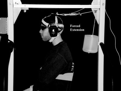
We used an external force applicator, designed and previously described by Tierney et al,16 to apply the external force to the head-neck segment. The applicator consisted of an outer metal frame, headgear, 2 cords with plastic stoppers, and 2 pulleys (Figure 1). Cords from the front and back of the headgear (Strength Systems Inc, Jefferson, LA) wrapped around the pulleys and connected to plastic stoppers at the end of the cords opposite the headgear. A load of 50 N, which was confirmed during testing with a tension force load cell (model ELFS-T3; Entran Devices, Inc, Fairfield, NJ), was created when a 1-kg weight was dropped from a height of 15 cm into the plastic stopper. With the participant seated, the pulleys were arranged at a 90° angle to the head-neck segment, which was verified by tester visual inspection. The participant's chair was positioned so that when located in front of the participant, the pulley caused forced flexion, whereas when the pulley was located in back of the participant, it caused forced extension.
Figure 1. Forced extension trial setup with external force applicator.
PEAK Motus Motion Analysis System
The PEAK Motus Motion Analysis System (Peak Performance Technologies Inc, Englewood, CO) was used to gather 2-dimensional kinematic data. All trials were recorded with a color video camera (model AG456 Proline; Panasonic, Secaucus, NJ) and collected at 60 Hz with a shutter speed of 1/500 s. Four reflective markers aided in the digitizing process by creating a head-neck segment and a torso segment. The markers were placed at the most superior aspect of the participant's head in the frontal plane, which corresponded with the most superior portion of the headgear; the fourth cervical vertebra spinous process; the acromion process; and the tenth rib.
Raw video data were autodigitized, filtered (fourth-order, zero-lag Butterworth filter with a 6-Hz cutoff), and analyzed using the PEAK Motus software, version 6.1 (Peak Performance Technologies Inc). Data collected with this system were used to determine peak head-neck segment angular acceleration and displacement values. Peak head-neck segment angular acceleration was calculated as the greatest acceleration within 1 trial. Total head-neck segment angular displacement was calculated from the onset of force application until motion stopped in the direction of the applied force.
Noraxon Telemyo System
We used the Noraxon Telemyo System (Noraxon USA, Scottsdale, AZ) to assess the EMG activity of the right sternocleidomastoid and upper trapezius muscles. These muscles were chosen to enable direct comparison with previous findings.16 Skin over the right upper trapezius and sternocleidomastoid muscles was shaved, abraded, and cleaned with 70% alcohol. The subject was instructed to perform an isometric contraction to allow identification of the approximate middle of the muscle, and we placed self-adhesive silver/silver-chloride bipolar surface electrodes measuring 10 mm in diameter on the skin 10 mm apart and parallel to the muscle fiber direction. Resistance between the electrodes was measured using a digital multimeter (model 982017; Sears, Roebuck & Co, Hoffman Estates, IL). If resistance was greater than 2 kΩ, the skin-preparation process was repeated until the criterion value was met.
Signals from the electrodes were passed to a battery-operated, 9-channel FM transmitter worn around the participant's waist. The signal was amplified (gain 1000) with a single-ended amplifier (impedance > 10 MΩ) and filtered with a fourth-order Butterworth filter (10–500 Hz) and common mode rejection ratio of 130 db at direct current (minimum 85 db across the entire frequency of 10 to 500 Hz). An antenna receiver (Antennex, Inc, Glendale, IL) with a sixth-order filter (gain 2, total gain 2000) further amplified the signal. This signal was converted to a digital signal using an analog-to-digital converter card (model KPCMCIA 12A1-C; Keithley Instruments, Inc, Cleveland, OH). The raw digital signal was sampled at a rate of 960 Hz, rectified, and smoothed using a root mean square algorithm over a 20-milisecond moving window. Data were stored in the MyoResearch software (version 2.02; Noraxon USA).
Data collected were used to determine peak muscle amplitude, muscle amplitude area, and muscle onset latency. Peak maximal voluntary isometric contraction values were assessed for each participant, and the peak muscle amplitude and muscle amplitude area were normalized to those values. Peak muscle amplitude was defined as the highest amplitude during 1 trial. Muscle amplitude area was defined as the sum of the amplitudes of activity over the total time of the trial. Muscle onset latency was defined as the time between force application and the first upswing of myoelectric activity from baseline34 and measured only during the unknown force-application trials.
Head-Neck Segment Stiffness Assessment
We evaluated head-neck segment stiffness using the tension force load cell and the PEAK Motus Motion Analysis System. Tension force was assessed throughout the trial by the in-line load cell, and an analog signal (range, −2.5 to 2.5 V) was amplified by the load cell's PS 30A amplifier. The analog signal was converted to a digital signal by the analog-to-digital converter card and stored in the MyoResearch software.
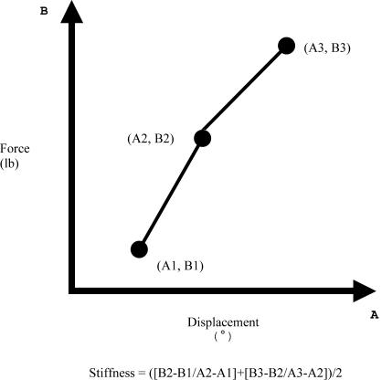
Head-neck segment stiffness was defined as a change in force over a change in length or displacement.35 The end of a stiffness assessment during a trial was marked by the peak force. Head-neck segment stiffness was determined by the average slope of the line on a force-displacement curve (Figure 2).
Figure 2. Stiffness calculation example. This force-displacement line was created from a trial with 3 force (B1, B2, and B3) and 3 displacement (A1, A2, and A3) data points. These points create 2 lines, and stiffness was determined from the average slope of the lines.25.
Head-Neck Segment Isometric Strength Assessment
The Microfet Hand-Held Dynamometer (Hoggan Health Industries, Inc, West Draper, UT) was used to quantify isometric neck flexor and extensor muscle strength. The participant was seated in the chair and stabilized at the thorax with a Velcro (Velcro USA Inc, Manchester, NH) strap and the arms crossed over the chest. Neck flexor strength was assessed with the dynamometer placed in the center of the participant's forehead. Neck extensor strength was evaluated with the dynamometer placed just above the participant's occipital protuberance. The participant was asked to apply maximal force against the dynamometer for 3 seconds during each of 3 trials and rested for 30 seconds between trials. The peak force was recorded for each trial, and the average of the 3 trials was reported. The primary investigator performed all measurements with a reliability of .96 (intraclass correlation coefficient model 2,1).
Procedures
Potential participants read and signed informed consent and consent to videotape forms and completed a health history questionnaire. Subjects who met the inclusion criteria and did not have any exclusionary factors proceeded with the testing.
Pretest
Participants completed 2 test sessions (1 pretest and 1 posttest) separated by at least 9 but no more than 11 weeks. To begin testing, participants performed a neck warm-up consisting of 15 seconds of clockwise neck rotations, 15 seconds of counterclockwise neck rotations, and 2 repetitions each of 15 seconds of neck flexion and 15 seconds of neck extension stretching. The skin was then prepared for electrode placement over the right upper trapezius and sternocleidomastoid muscles. A ground electrode was positioned over the right clavicle. Subjects were fitted with the headgear, which served as the attachment for the pulleys during the trials. Reflective markers were placed as previously described.
Participants were seated in a chair placed inside the external force applicator and stabilized as noted above. They performed 3 maximal voluntary isometric contractions for both flexion and extension against manual pressure with the dynamometer. For the head-neck segment dynamic stabilization tests, the effects of visual feedback were eliminated by having subjects wear modified goggles covered with opaque black athletic tape. The effects of auditory feedback were minimized for each participant with headphones over the ears.
Before the force-application trials started, the pulley from the external force applicator was secured to the back of the headgear, and the 1-kg weight was placed inside the plastic stopper to allow the participant to become accustomed to the weight. The first 3 trials performed were forced neck extension with the participant's knowing the timing of the force application, followed by 3 trials without the participant's knowing the timing. Force-application order was not randomized, so direct comparisons could be made with previous research.16 Upon collection of the extension testing data, the subject's chair was turned 180°, the pulley was attached to the front of the headgear, and the reflective markers were moved to the right side of the body. Three forced-flexion trials were then performed with the participant's knowing the force-application timing, followed by 3 trials without the participant's knowing the timing. During trials with knowledge, subjects were instructed to preactivate their cervical muscles and resist the load that would be dropped after a 3-second countdown. During trials without knowledge, participants were instructed to relax their muscles and were informed that, within the next 30 seconds, they would feel a tug resulting from a force of the same magnitude as in the knowledge condition. They were instructed to resist the tug at its onset. If muscle preactivation occurred, the trial was not included.
Resistance Training Program
Participants were randomly assigned to either the RTG or the CG by the primary investigator using a Latin square table. All subjects participated in offseason team upper and lower extremity weight lifting and conditioning sessions 2 days per week. In addition, the RTG participants underwent a cervical resistance training program twice a week for 8 weeks. The CG subjects performed no cervical resistance exercises during the 8-week period. Both groups were instructed not to perform any exercise aside from team lifting and practices during the 8 weeks. No participant missed more than 1 session during the 8-week training period.
The RTG participants were instructed on how to properly perform neck extension and flexion exercises on the isotonic neck resistance training machine (Trotter Strength Manufacturing Facility, Owatonna, MN). Seat height and back pad position were adjusted to each subject's height and recorded for use in all training sessions. The axis of rotation of the cervical isotonic resistance training machine was positioned such that resistance was applied over a full range of motion for both flexion and extension. The primary investigator monitored the neck-strengthening exercises performed by the RTG to ensure compliance and proper setup and execution. The exercises were performed at the end of the team weight-lifting session.
The initial weight for resistance training was determined by having each subject perform a 10-repetition maximum test on the same isotonic neck machine used for the resistance training program. Fifty-five percent of each participant's 10-repetition maximum was used in the first 2 weeks of training (4 sessions). For example, if the maximum weight a participant could lift 10 times was 20 pounds (9.07 kg), 55% of that weight (11 pounds [4.99 kg]) was used as the starting weight. The weight was incrementally increased by 5% of the 10-repetition maximum every 2 weeks to a 70% effort. The resistance-training program consisted of 3 sets of 10 repetitions through the full range of motion in neck flexion, followed by 3 sets of 10 repetitions through the full range of motion in neck extension. Participants rested for 90 seconds between sets.
Posttest
The posttest measurements were taken no more than 1 week after completion of the resistance training program. The posttests were performed in the same manner as the pretests.
Data Analysis
A power analysis was performed a priori to determine appropriate sample size. An effect size of 1.0 was calculated for head-neck segment acceleration from previous research using similar methods.16 For this effect size and an alpha level of .05, the sample size needed to be at least 13 to achieve a power of .80.
Data analysis consisted of descriptive and inferential statistics. Statistical tests included multiple multivariate and univariate analyses of variance with appropriate follow-up univariate analyses of variance and post hoc t tests (α ≤ .05). Forced flexion and extension were analyzed using separate statistical tests because previous researchers16–19 have reported clear differences between neck flexor and extensor strength and stabilization ability.
Specifically, head-neck segment length and mass were analyzed with a 2 (sex) × 2 (group) multivariate analysis of variance (MANOVA). Neck girth was analyzed with a 2 (sex) × 2 (group) × 2 (session) analysis of variance (ANOVA) with repeated measures on the last factor. Head-neck segment peak angular acceleration and displacement were analyzed with a 2 (sex) × 2 (group) × 2 (session) × 2 (knowledge) MANOVA with repeated measures on the last 2 factors. The EMG dependent variables consisted of the sternocleidomastoid and trapezius peak muscle activity, muscle activity area, and muscle onset latency. Peak muscle activity and muscle activity area were analyzed with a 2 (sex) × 2 (group) × 2 (session) × 2 (knowledge) MANOVA with repeated measures on the last 2 factors. Muscle onset latency was analyzed with a 2 (sex) × 2 (group) × 2 (session) ANOVA with repeated measures on the last factor. Head-neck segment stiffness was analyzed with a 2 (sex) × 2 (group) × 2 (session) × 2 (knowledge) ANOVA with repeated measures on the last 2 factors. Neck strength was analyzed with a 2 (sex) × 2 (group) × 2 (session) ANOVA with repeated measures on the last factor. Neck girth was analyzed with a 2 (sex) × 2 (group) × 2 (session) ANOVA with repeated measures on the last factor.
Potential covariates in the kinematic, EMG, head-neck segment stiffness, and isometric strength analyses were head-neck segment mass (kg) and length (cm) as well as neck girth (cm). Covariates were correlated a priori with the appropriate dependent variables. A correlation value of r ≥ .60 was used as the criterion for inclusion as a covariate.36 Neck girth and head mass were correlated (r > .60) with head-neck segment isometric flexion and extension strength. They were not statistically significant (P > .05) in the analysis of covariance model, however, and were therefore not included in the analysis. None of the potential covariates met this criterion for any other dependent variable, and they were not used in any analysis. We used the SPSS for Windows statistical program (version 11.5; SPSS Inc, Chicago, IL) for data analysis.
RESULTS
Head-Neck Segment Anthropometric Measurements
A 2 (sex) × 2 (group) MANOVA for head-neck segment length and mass revealed a significant main effect for sex (Table 1). No other significant differences were noted. Follow-up ANOVAs revealed a significant sex difference in head-neck segment length (F1,32 = 5.10, P = .031) and mass (F1,32 = .62.91, P < .001). Women exhibited 7% less head-neck segment length and 20% less head-neck segment mass than men.
Table 1. Participant Anthropometric Measurements.
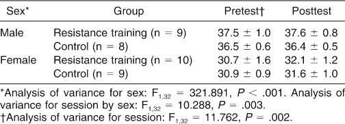
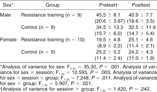
A 2 (sex) × 2 (group) × 2 (session) ANOVA demonstrated a significant time × sex interaction effect for neck girth (Table 2). No other significant differences were shown. Post hoc t tests revealed that pretest to posttest neck girth was significantly different for women (t = −4.70, P < .001) but not for men (t = −0.190, P = .855) (Table 2). Specifically, the women's neck girth increased 3.4% over time regardless of group level.
Table 2. Participant Neck Girth (cm) by Session.
Head-Neck Segment Kinematics
The 2 (sex) × 2 (group) × 2 (knowledge) × 2 (session) MANOVA for forced flexion revealed a significant main effect for force-application knowledge and session (Table 3). No other significant differences for forced flexion were seen. The follow-up ANOVAs for force-application knowledge revealed a significant main effect for displacement (F1,31 = 19.30, P < .001) but not for acceleration (F1,31 = 0.210, P = .650, power = .073). Specifically, participants exhibited 23% more displacement during the unknown versus the known force-application trials. The follow-up ANOVAs for session revealed a significant main effect for acceleration (F1,31 = 8.489, P = .007) but not for displacement (F1,31 = 0.243, P = .626, power = .077). Specifically, subjects exhibited 40% greater head-neck segment acceleration during posttesting than during pretesting.
Table 3. Forced-Flexion Head-Neck Segment Kinematic Data*.
The 2 × 2 × 2 × 2 MANOVA for forced extension also demonstrated a significant main effect for force-application knowledge (Table 4). No other significant differences for forced extension were noted. The follow-up ANOVAs were statistically significant for displacement (F1,32 = 19.60, P < .001) but not for acceleration (F1,32 = 2.90, P = .098, power = .379). Specifically, participants exhibited 25% more displacement during the unknown versus the known force-application trials.
Table 4. Forced-Extension Head-Neck Segment Kinematic Data*.
Peak Muscle Activity and Muscle Activity Area
The 2 (sex) × 2 (group) × 2 (knowledge) × 2 (session) MANOVA revealed no significant differences for upper trapezius muscle activity (Table 5). The 2 × 2 × 2 × 2 MANOVA for sternocleidomastoid muscle activity revealed a significant main effect for force-application knowledge (Table 6). No other significant differences existed. The follow-up ANOVAs revealed a significant difference for peak muscle activity (F1,32 = 6.76, P = .014) but not for muscle activity area (F1,32 = 0.071, P = .791, power = .058). Specifically, subjects exhibited 18% greater peak muscle activity in the unknown force-application condition than in the known condition.
Table 5. Upper Trapezius Peak and Area Muscle Activity*.
Table 6. Sternocleidomastoid Peak and Area Muscle Activity*.
Muscle Onset Latency
The 2 (sex) × 2 (group) × 2 (session) ANOVA for upper trapezius onset latency demonstrated a significant main effect for session (Table 7). No other significant differences were seen. Trapezius onset latency was 40% slower in posttesting versus pretesting. The 2 × 2 × 2 ANOVA for sternocleidomastoid onset latency revealed a significant sex × group interaction (Table 8). No other reportable significant differences were shown. Post hoc t tests indicated a significant difference (t = 3.49, P = .003) for the male RTG but not for the female RTG (t = −0.644, P = .528). Specifically, sternocleidomastoid onset latency was 64% faster in the male CG than in the male RTG regardless of test session.
Table 7. Upper Trapezius Muscle Onset Latency (ms).
Table 8. Sternocleidomastoid Muscle Onset Latency (ms).
Head-Neck Segment Stiffness
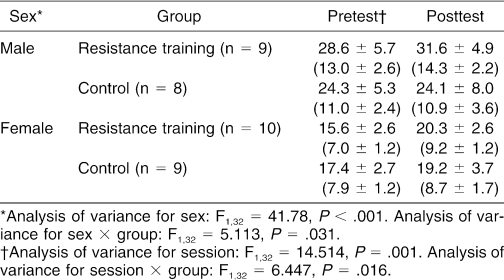
The 2 (sex) × 2 (group) × 2 (knowledge) × 2 (session) ANOVA for forced flexion head-neck segment stiffness revealed a significant main effect for group (Table 9). No other significant differences existed. Specifically, the RTG exhibited 43% more stiffness than the CG.
Table 9. Forced Flexion Head-Neck Segment Stiffness in lb/° (kg/°).
A 2 × 2 × 2 × 2 ANOVA for forced extension head-neck segment stiffness revealed a sex × knowledge × session × training group interaction effect (Table 10). Post hoc 2 (group) × 2 (knowledge) × 2 (session) ANOVAs within the sexes showed a significant knowledge × session × group interaction effect for males (F1,15 = 5.81, P = .029) but not for females (F1,15 = 2.29, P = .151, power = .271). However, 2 (knowledge) × 2 (time) further ANOVAs within the male CG and RTG did not indicate where the significant differences existed.
Table 10. Forced Extension Head-Neck Segment Stiffness in lb/° (kg/°).
Isometric Head-Neck Segment Muscle Strength
The 2 (sex) × 2 (group) × 2 (session) ANOVA for isometric neck flexor strength revealed significant session × group and sex × training group interaction effects (Table 11). For the session × group interaction, post hoc t tests revealed a significant difference (t = −5.95, P < .001) for the RTG between pretest and posttest but not for the CG (t = −0.783, P = .445). Specifically, RTG posttest isometric neck flexor strength was 15% greater than pretest strength. For the sex × group interaction effect, post hoc t tests revealed a significant difference (t = −2.17, P = .015) between male groups but not between female groups (t = 0.346, P = .733). Isometric neck flexor strength was 20% greater in the male RTG than in the male CG.
Table 11. Neck Flexor Isometric Strength in lb (kg).
The 2 × 2 × 2 ANOVA for isometric neck extensor strength revealed a significant sex × session × group interaction (Table 12). Follow-up 2 (group) × 2 (session) ANOVAs within the sexes revealed a significant interaction effect for women (F1,17 = 7.31, P = .015) but not for men (F1,15 = 0.191, P = .292, power = .180). Post hoc t tests revealed a significant difference (t = −3.22, P = .010) in the female RTG over time but not in the female CG (t = 0.603, P = .563). Specifically, isometric neck extensor strength in the female RTG was 22.5% greater posttest versus pretest.
Table 12. Neck Extensor Isometric Strength in lb (kg).
DISCUSSION
Our results revealed that 8 weeks of isotonic cervical resistance training did not enhance head-neck segment dynamic restraint in collegiate soccer players. Although cervical resistance training increased neck girth (women only) and isometric strength (male neck flexors only and female neck flexors and extensors), no training effect was noted for the kinematic, EMG, or stiffness values on force application. We also found no sex differences in kinematic, EMG, or stiffness values despite greater neck girth, head-neck segment mass, and isometric neck strength in male versus female soccer players.
Resistance Training Effect
Isometric neck flexor strength increased in the training groups by 15% from pretest to posttest, and isometric neck extensor strength and neck girth increased in women by 22.5% and 4.5% from pretest to posttest, respectively. These findings are similar to earlier results in which 8 weeks of cervical resistance training resulted in increased strength19,30,37 and girth19,30 of the neck musculature. We saw no reduction, however, in head-neck segment acceleration on force application after training. These findings suggest that although a traditional cervical resistance training program changes muscle structure, the neuromuscular plasticity needed to enhance dynamic restraint and reduce head acceleration on force application is not evident.
Isometric flexor strength increased significantly in the male training group, yet there were no changes in neck girth, reactive muscle activity, or head kinematics on force application. These results may suggest the need for neck muscle training tasks that elicit feed-forward and feedback motor control mechanisms to better use the dynamic stabilizers for both protection and performance enhancement. The traditional resistance training program was selected because isotonic training is currently the most accepted method for training the head-neck segment. Ballistic activities (eg, plyometrics) were not included in this study but have been reported to enhance neuromuscular control and dynamic stabilization at other joints.20,38 Although plyometric training of the head-neck segment has the potential for being similar to heading (low accelerative head movements), consideration should be given for its inclusion in cervical resistance training programs because of its benefits to motor control.20,38 This recommendation is predicated on safeguards being taken to protect the head-neck segment from direct and indirect injury.
Sex Differences
No sex differences existed in kinematic, EMG, or stiffness values despite greater neck girth, head-neck segment length and mass, and isometric strength in males. Our kinematic (ie, acceleration) findings for sex are similar to those of Morris and Popper,39 but differ from those of Tierney et al.16 The differences can be attributed to subject populations. Tierney et al16 tested physically active male and female subjects who had no experience with force being applied to their heads. Morris and Popper39 tested male and female volunteers who were accustomed to head accelerative forces because they had undergone training. Our subjects were also trained to resist low accelerative forces because of heading activities during soccer practices and games. This type of experience for subjects in the latter 2 studies may have enhanced head-neck segment dynamic restraint through neuromuscular adaptations that led to greater preparatory muscle activity in both male and female subjects. Soccer players are also accustomed to absorbing loads (eg, 180 N) greater than loads used in this study.40 This is illustrated by the fact that the 50-N load used in both our study and Tierney et al16 was insufficient to elicit a sex effect for head acceleration in soccer players but did in physically active subjects.
Force-Application Knowledge
We found no reduction in head acceleration for either sex with awareness of force application. Tierney et al16 reported that force-application awareness and muscle preactivation enabled physically active male but not female subjects to significantly reduce their head acceleration. Both groups used the same load (50 N), method of force application (pulley system), and order of force application (known trials followed by unknown trials) but different subject populations. In comparing the 2 studies, male and female soccer players exhibited lower pretest mean head angular accelerations (male, 984°/s2; female, 1080°/s2) than male and female physically active participants (male, 1218°/s2; female, 1790°/s2). This would suggest greater dynamic restraint in the soccer players. The cervical dynamic restraint system in soccer players is trained by absorbing impact forces of more than 180 N during heading activities.40 Because of the relatively low load applied during testing, male and female soccer players were able to absorb the external forces equally, resulting in no force-application knowledge-by-sex interaction.
Our study revealed greater head-neck segment displacement and muscle peak activity during unknown force-application trials. Greater segment displacement indicates a greater distance over which the muscles can provide stabilization. During the unknown trials, the muscles were not preactivated and head-neck segment stabilization was primarily controlled with reactive reflex muscle firing generated from increased strain on static stabilizers and mechanoreceptors. These findings are similar to those of previous researchers,16,23 in that head-neck segment kinematics in participants were greater when the timing of force application was unknown. Although we did not measure muscle preactivation, these findings indicate its importance in segment stabilization and warrant future study in head-neck segment research.
CONCLUSIONS
Despite increases in isometric strength and girth, the 8-week isotonic cervical resistance training did not enhance head-neck segment dynamic restraint during force application to the head in male and female collegiate soccer players. This finding is clinically relevant because resistance training is purported to aid in protecting athletes from head injury32 but may not elicit the desired outcome. Future investigators should examine whether higher-intensity isotonic training and other types of training (eg, plyometrics) stimulate the neuromuscular changes necessary to enhance head-neck segment dynamic restraint in soccer players and other populations. Future researchers should also include more ballistic testing and examine the effects of neck resistance training on mild head injury occurrence and long-term neurologic outcomes.
Acknowledgments
We thank the members of the Temple University soccer teams for their participation in this study. We also thank Jack Reed for his help in the previous design and construction of the external force applicator.
Footnotes
Jamie Mansell, MEd, ATC; Ryan T. Tierney, PhD, ATC; Michael R. Sitler, EdD, ATC; and Kathleen A. Swanik, PhD, ATC, contributed to conception and design; acquisition and analysis and interpretation of the data; and drafting, critical revision, and final approval of the article. David Stearne, MS, ATC, contributed to conception and design, acquisition of the data, and drafting and final approval of the article.
Address correspondence to Ryan T. Tierney, PhD, ATC, 127 Pearson Hall, Temple University, Philadelphia, PA 19122. Address e-mail to rtierney@temple.edu.
REFERENCES
- Tysvaer A, Storli O. Association football injuries to the brain: a preliminary report. Br J Sports Med. 1981;15:163–166. doi: 10.1136/bjsm.15.3.163. [DOI] [PMC free article] [PubMed] [Google Scholar]
- Broglio SP, Yu YY, Broglio MD, Sell TC. The efficacy of soccer headgear. J Athl Train. 2003;38:220–224. [PMC free article] [PubMed] [Google Scholar]
- Guskiewicz KM. No evidence of impaired neurocognitive performance in collegiate soccer players. Am J Sports Med. 2002;30:630. [PubMed] [Google Scholar]
- Broglio SP, Guskiewicz KM, Sell TC, Lephart SM. No acute changes in postural control after soccer heading. Br J Sports Med. 2004;38:561–567. doi: 10.1136/bjsm.2003.004887. [DOI] [PMC free article] [PubMed] [Google Scholar]
- Matser JT, Kessels AG, Jordan BD, Lezak MD, Troost J. Chronic traumatic brain injury in professional soccer players. Neurology. 1998;51:791–796. doi: 10.1212/wnl.51.3.791. [DOI] [PubMed] [Google Scholar]
- Matser EJ, Kessels AG, Lezak MD, Jordan BD, Troost J. Neuropsychological impairment in amateur soccer players. JAMA. 1999;282:971–973. doi: 10.1001/jama.282.10.971. [DOI] [PubMed] [Google Scholar]
- Tysvaer AT, Storli OV. Soccer injuries to the brain: a neurologic and electroencephalographic study of active football players. Am J Sports Med. 1989;17:573–578. doi: 10.1177/036354658901700421. [DOI] [PubMed] [Google Scholar]
- Tysvaer AT, Lochen EA. Soccer injuries to the brain: a neuropsychologic study of former soccer players. Am J Sports Med. 1991;19:56–60. doi: 10.1177/036354659101900109. [DOI] [PubMed] [Google Scholar]
- Gennarelli TA, Seggawa H, Wald U, Czernicki Z, Marsh K, Thompson C. Physiological response to angular acceleration of the head. In: Grossman RG, Gildenberg PL, eds. Head Injury: Basic and Clinical Aspects. New York, NY: Raven Press; 1982:129–140.
- Barnes BC, Cooper L, Kirkendall DT, McDermott TP, Jordan BD, Garrett WE., Jr. Concussion history in elite male and female soccer players. Am J Sports Med. 1998;26:433–438. doi: 10.1177/03635465980260031601. [DOI] [PubMed] [Google Scholar]
- Boden BP, Kirkendall DT, Garrett WE., Jr. Concussion incidence in elite college soccer players. Am J Sports Med. 1998;26:238–241. doi: 10.1177/03635465980260021301. [DOI] [PubMed] [Google Scholar]
- Covassin T, Swanik CB, Sachs ML. Epidemiological considerations of concussions among intercollegiate athletes. Appl Neuropsychol. 2003;10:12–22. doi: 10.1207/S15324826AN1001_3. [DOI] [PubMed] [Google Scholar]
- Powell JW, Barber-Foss KD. Traumatic brain injury in high school athletes. JAMA. 1999;282:958–963. doi: 10.1001/jama.282.10.958. [DOI] [PubMed] [Google Scholar]
- Delaney JS, Lacroix VJ, Leclerc S, Johnston KM. Concussions among university football and soccer players. Clin J Sport Med. 2002;12:331–338. doi: 10.1097/00042752-200211000-00003. [DOI] [PubMed] [Google Scholar]
- Dick RW. A summary of head and neck injuries in collegiate athletics using the NCAA Injury Surveillance System. In: Hoerner EF, ed. Head and Neck Injuries in Sports. Philadelphia, PA: American Society for Testing and Materials; 1994:13–19.
- Tierney RT, Sitler MR, Swanik CB, Swanik KA, Higgins M, Torg JS. Gender differences in head-neck dynamic stabilization during head acceleration. Med Sci Sports Exerc. 2005;37:272–279. doi: 10.1249/01.mss.0000152734.47516.aa. [DOI] [PubMed] [Google Scholar]
- Garces GL, Medina D, Milutinovic L, Garavote P, Guerado E. Normative database of isometric cervical strength in a healthy population. Med Sci Sports Exerc. 2002;34:464–470. doi: 10.1097/00005768-200203000-00013. [DOI] [PubMed] [Google Scholar]
- Jordan A, Mehlsen J, Bulow PM, Ostergaard K, Danneskiold-Samsoe B. Maximal isometric strength of the cervical musculature in 100 healthy volunteers. Spine. 1999;24:1343–1348. doi: 10.1097/00007632-199907010-00012. [DOI] [PubMed] [Google Scholar]
- Maeda A, Nakashima T, Shibayama H. Effect of training on the strength of cervical muscle. Ann Physiol Anthropol. 1994;13:59–67. doi: 10.2114/ahs1983.13.59. [DOI] [PubMed] [Google Scholar]
- Swanik KA, Lephart SM, Swanik CB, Lephart SP, Stone DA, Fu FH. The effects of shoulder plyometric training on proprioception and muscle performance characteristics. J Shoulder Elbow Surg. 2002;11:579–586. doi: 10.1067/mse.2002.127303. [DOI] [PubMed] [Google Scholar]
- Hewett TE, Stroupe AL, Nance TA, Noyes FR. Plyometric training in female athletes: decreased impact forces and increased hamstring torques. Am J Sports Med. 1996;24:765–773. doi: 10.1177/036354659602400611. [DOI] [PubMed] [Google Scholar]
- Swanik CB, Lephart SM, Giannantonio FP, Fu FH. Reestablishing proprioception and neuromuscular control in the ACL-injured athlete. J Sport Rehabil. 1997;6:182–206. [Google Scholar]
- Kumar S, Narayan Y, Amell T. Role of awareness in head-neck acceleration in low velocity rear-end impacts. Accid Anal Prev. 2000;32:233–241. doi: 10.1016/s0001-4575(99)00114-1. [DOI] [PubMed] [Google Scholar]
- Ito Y, Corna S, vo Brevern MV, Bronstein A, Gresty M. The functional effectiveness of neck muscle reflexes for head-righting in response to sudden fall. Exp Brain Res. 1997;117:266–272. doi: 10.1007/s002210050221. [DOI] [PubMed] [Google Scholar]
- Schor RH, Kearney RE, Dieringer N. Reflex stabilization of the head. In: Peterson B, Richmond F, eds. Control of Head Movement. New York, NY: Oxford University Press;1988;141–166.
- Heck RW, McKeever KH, Alway SE. Resistance training-induced increases in muscle mass and performance in ponies. Med Sci Sports Exerc. 1996;28:877–883. doi: 10.1097/00005768-199607000-00015. et al. [DOI] [PubMed] [Google Scholar]
- Brooks GA, Fahey TD, White TP, Baldwin KM. Exercise Physiology: Human Bioenergetics and Its Applications. 3rd ed. New York, NY: McGraw Hill; 2000.
- Bland JH. Conditioning provides protection: helping athletes avoid neck injuries. J Musculoskelet Med. 1996;13:30–38. [Google Scholar]
- Goodman R, Frew L. Effectiveness of progressive strength resistance training for whiplash: a pilot study. Physiother Can. 2000;52:211–214. [Google Scholar]
- Stump J, Rash G, Semon J, Christian W, Miller K. A comparison of two modes of cervical exercise in adolescent male athletes. J Manipulative Physiol Ther. 1993;16:155–160. [PubMed] [Google Scholar]
- Pollock ML, Graves JE, Bamman MM. Frequency and volume of resistance training: effect on cervical extension strength. Arch Phys Med Rehabil. 1993;74:1080–1086. doi: 10.1016/0003-9993(93)90065-i. et al. [DOI] [PubMed] [Google Scholar]
- Cross KM, Serenelli C. Training and equipment to prevent athletic head and neck injuries. Clin Sports Med. 2003;22:639–657. doi: 10.1016/s0278-5919(02)00099-6. [DOI] [PubMed] [Google Scholar]
- Plagenhoef S, Evans FG, Abdelnour T. Anatomical data for analyzing human motion. Res Q Exerc Sport. 1983;54:169–178. [Google Scholar]
- Brunt D, Williams J, Rice RR. Analysis of EMG activity and temporal components of gait during recovery from perturbation. Arch Phys Med Rehabil. 1990;71:473–477. [PubMed] [Google Scholar]
- McNair PJ, Hewson DJ, Dombroski E, Stanley SN. Stiffness and passive peak force changes at the ankle joint: the effect of different joint angular velocities. Clin Biomech (Bristol, Avon) 2002;17:536–540. doi: 10.1016/s0268-0033(02)00062-1. [DOI] [PubMed] [Google Scholar]
- Portney LG, Watkins MP. Foundations of Clinical Research: Applications to Practice. 2nd ed. Upper Saddle River, NJ: Prentice-Hall; 2000.
- Randlov A, Ostergaard M, Manniche C. Intensive dynamic training for females with chronic neck/shoulder pain: a randomized controlled trial. Clin Rehabil. 1998;12:200–210. doi: 10.1191/026921598666881319. et al. [DOI] [PubMed] [Google Scholar]
- Chimera NJ, Swanik KA, Swanik CB, Straub SJ. Effects of plyometric training on muscle-activation strategies and performance in female athletes. J Athl Train. 2004;39:24–31. [PMC free article] [PubMed] [Google Scholar]
- Morris CE, Popper SE. Gender and effect of impact acceleration on neck motion. Aviat Space Environ Med. 1999;70:851–856. [PubMed] [Google Scholar]
- Bauer JA, Thomas TS, Cauraugh JH, Kaminski TW, Hass CJ. Impact forces and neck muscle activity in heading by collegiate female soccer players. J Sports Sci. 2001;19:171–179. doi: 10.1080/026404101750095312. [DOI] [PubMed] [Google Scholar]


