

Abstract
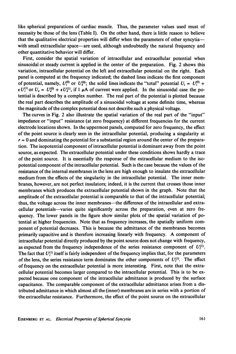
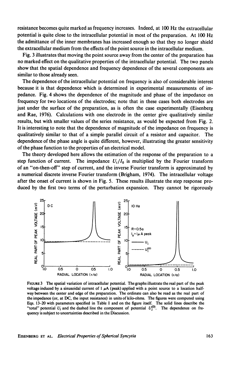
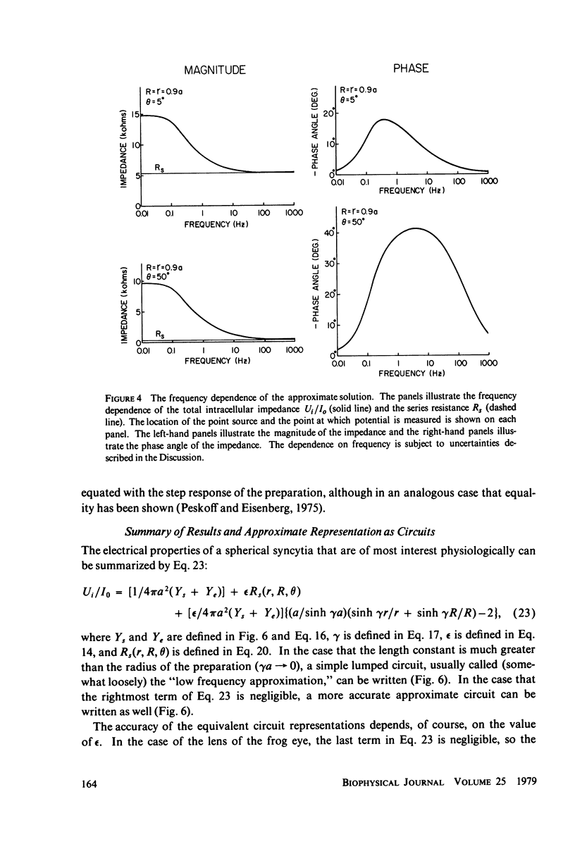
Syncytial tissues consist of many cells whose intracellular spaces are electrically coupled one to another. Such tissues typically include narrow, tortuous extracellular space and often have specialized membranes at their outer surface. We derive differential equations to describe the potentials induced when a sinusoidal or steady current is applied to the intracellular space with a microelectrode. We derive solutions for spherical preparations with isotropic properties or with a particular anisotropy in effective extracellular and intracellular resistivities. Solutions are presented in an approximate form with a simple physical interpretation. The leading term in the intracellular potential describes an "isopotential" cell in which there is no spatial variation of intracellular potential. The leading term in the extracellular potential, and thus the potential across the inner membranes, varies with radial position, even at zero frequency. The next term of the potentials describes the direct effects of the point source of current and, for the parameters given here, acts as a series resistance producing a large local potential drop essentially independent of frequency. A lumped equivalent circuit describes the "low frequency" behavior of the syncytium, and a distributed circuit gives a reasonably accurate general description. Graphs of the spatial variation and frequency dependence of intracellular, extracellular, and transmembrane potential are given, the response to sinusoidal currents is used to calculate numerically the response to a step function of current.
Full text
PDF


Selected References
These references are in PubMed. This may not be the complete list of references from this article.
- Chandler W. K., Schneider M. F. Time-course of potential spread along a skeletal muscle fiber under voltage clamp. J Gen Physiol. 1976 Feb;67(2):165–184. doi: 10.1085/jgp.67.2.165. [DOI] [PMC free article] [PubMed] [Google Scholar]
- Dehaan R. L., Fozzard H. A. Membrane response to current pulses in spheroidal aggregates of embryonic heart cells. J Gen Physiol. 1975 Feb;65(2):207–222. doi: 10.1085/jgp.65.2.207. [DOI] [PMC free article] [PubMed] [Google Scholar]
- Eisenberg R. S., Mathias R. T., Rae J. S. Measurement, modeling, and analysis of the linear electrical properties of cells. Ann N Y Acad Sci. 1977 Dec 30;303:342–354. [PubMed] [Google Scholar]
- Eisenberg R. S., Rae J. L. Current-voltage relationships in the crystalline lens. J Physiol. 1976 Nov;262(2):285–300. doi: 10.1113/jphysiol.1976.sp011596. [DOI] [PMC free article] [PubMed] [Google Scholar]
- FALK G., FATT P. LINEAR ELECTRICAL PROPERTIES OF STRIATED MUSCLE FIBRES OBSERVED WITH INTRACELLULAR ELECTRODES. Proc R Soc Lond B Biol Sci. 1964 Apr 14;160:69–123. doi: 10.1098/rspb.1964.0030. [DOI] [PubMed] [Google Scholar]
- Frömter E. The route of passive ion movement through the epithelium of Necturus gallbladder. J Membr Biol. 1972;8(3):259–301. doi: 10.1007/BF01868106. [DOI] [PubMed] [Google Scholar]
- Haylett D. G., Jenkinson D. H. Effects of noradrenaline on potassium reflux, membrane potential and electrolyte levels in tissue slices prepared from guinea-pig liver. J Physiol. 1972 Sep;225(3):721–750. doi: 10.1113/jphysiol.1972.sp009966. [DOI] [PMC free article] [PubMed] [Google Scholar]
- Makowski L., Caspar D. L., Phillips W. C., Goodenough D. A. Gap junction structures. II. Analysis of the x-ray diffraction data. J Cell Biol. 1977 Aug;74(2):629–645. doi: 10.1083/jcb.74.2.629. [DOI] [PMC free article] [PubMed] [Google Scholar]
- Mathias R. T. An analysis of the electrical properties of a skeletal muscle fiber containing a helicoidal T system. Biophys J. 1978 Aug;23(2):277–284. doi: 10.1016/S0006-3495(78)85448-4. [DOI] [PMC free article] [PubMed] [Google Scholar]
- Mathias R. T., Eisenberg R. S., Valdiosera R. Electrical properties of frog skeletal muscle fibers interpreted with a mesh model of the tubular system. Biophys J. 1977 Jan;17(1):57–93. doi: 10.1016/S0006-3495(77)85627-0. [DOI] [PMC free article] [PubMed] [Google Scholar]
- Mathias R. T., Rae J. L., Eisenberg R. S. Electrical properties of structural components of the crystalline lens. Biophys J. 1979 Jan;25(1):181–201. doi: 10.1016/S0006-3495(79)85284-4. [DOI] [PMC free article] [PubMed] [Google Scholar]
- Matthews E. K., Sakamoto Y. Electrical characteristics of pancreatic islet cells. J Physiol. 1975 Mar;246(2):421–437. doi: 10.1113/jphysiol.1975.sp010897. [DOI] [PMC free article] [PubMed] [Google Scholar]
- Peskoff A., Eisenberg R. S. Interpretation of some microelectrode measurements of electrical properties of cells. Annu Rev Biophys Bioeng. 1973;2:65–79. doi: 10.1146/annurev.bb.02.060173.000433. [DOI] [PubMed] [Google Scholar]
- Petersen O. H. Electrophysiological studies on gland cells. Experientia. 1974 Feb 15;30(2):130–134. doi: 10.1007/BF01927689. [DOI] [PubMed] [Google Scholar]
- Purves R. D. Current flow and potential in a three-dimensional syncytium. J Theor Biol. 1976 Jul 21;60(01):147–162. doi: 10.1016/0022-5193(76)90160-0. [DOI] [PubMed] [Google Scholar]
- Schoenberg M., Dominguez G., Fozzard H. A. Effect of diameter on membrane capacity and conductance of sheep cardiac Purkinje fibers. J Gen Physiol. 1975 Apr;65(4):441–458. doi: 10.1085/jgp.65.4.441. [DOI] [PMC free article] [PubMed] [Google Scholar]
- Sheridan J. D. Electrical coupling between fat cells in newt fat body and mouse brown fat. J Cell Biol. 1971 Sep;50(3):795–803. doi: 10.1083/jcb.50.3.795. [DOI] [PMC free article] [PubMed] [Google Scholar]
- Weidmann S. The diffusion of radiopotassium across intercalated disks of mammalian cardiac muscle. J Physiol. 1966 Nov;187(2):323–342. doi: 10.1113/jphysiol.1966.sp008092. [DOI] [PMC free article] [PubMed] [Google Scholar]