Abstract
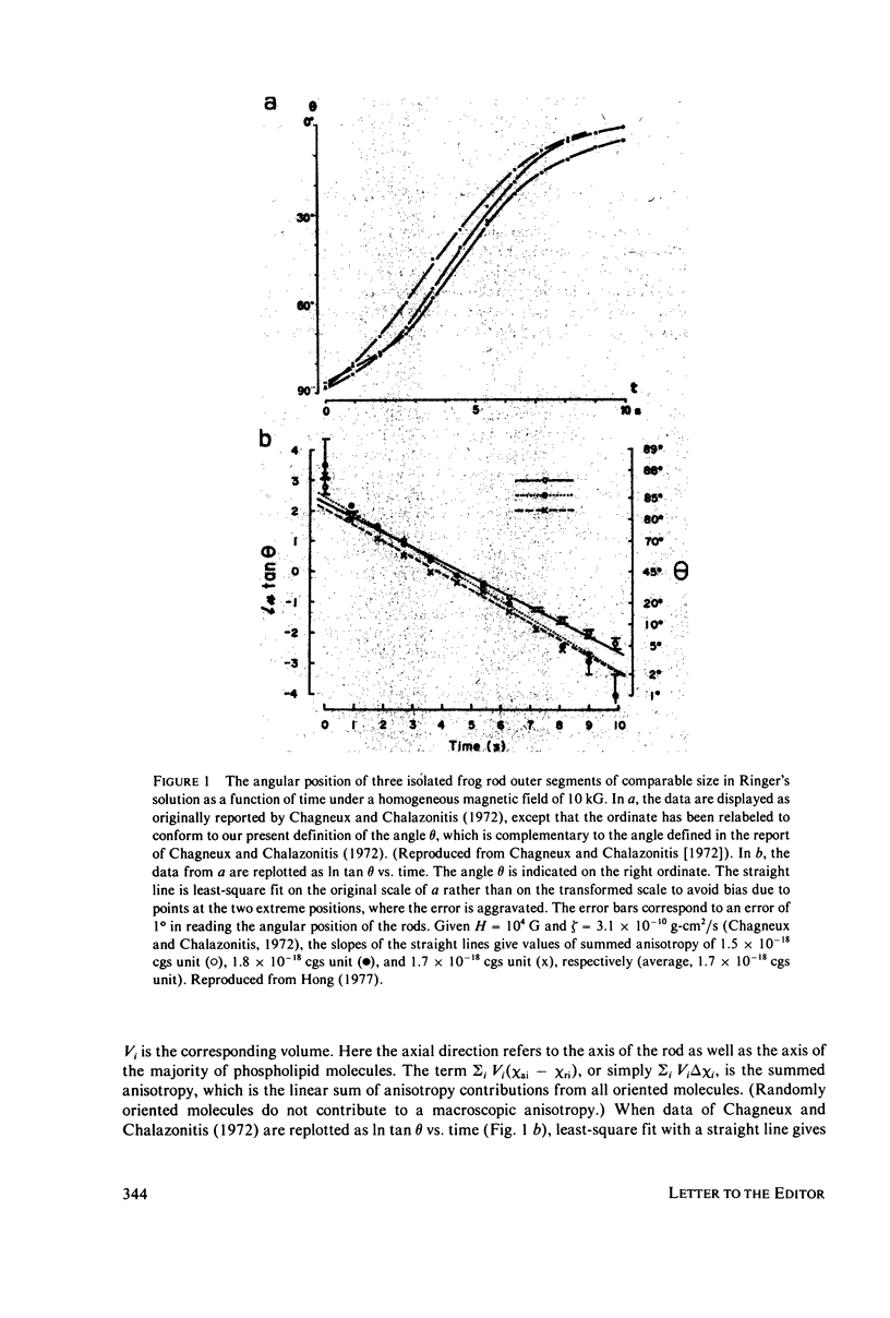
A new estimate of diamagnetic anisotropy of the frog rhodopsin is reported. The estimate is obtained by combining the data of magnetic field induced orientation of isolated frog rod outer segments as measured by Chagneux and Chalazonitis (1972) and the data of diamagnetic anisotropy of lecithin membranes as recently reported by Boroske and Helfrich (1978). The anisotropy of the volume susceptibilities of frog rhodopsin is calculated to be 4.4 X 10(-8) cgs unit/cm3, which corresponds to 1.5 X 10(-27) cgs unit/molecule, or 9.0 X 10(-4) cgs unit/mol.
Full text
PDF


Selected References
These references are in PubMed. This may not be the complete list of references from this article.
- Becker J. F., Trentacosti F., Geacintov N. E. A linear dichroism study of the orientation of aromatic protein residues in magnetically oriented bovine rod outer segments. Photochem Photobiol. 1978 Jan;27(1):51–54. doi: 10.1111/j.1751-1097.1978.tb07564.x. [DOI] [PubMed] [Google Scholar]
- Blasie J. K., Worthington C. R. Molecular localization of frog retinal receptor photopigment by electron microscopy and low-angle X-ray diffraction. J Mol Biol. 1969 Feb 14;39(3):407–416. doi: 10.1016/0022-2836(69)90135-1. [DOI] [PubMed] [Google Scholar]
- Boroske E., Helfrich W. Magnetic anisotropy of egg lecithin membranes. Biophys J. 1978 Dec;24(3):863–868. doi: 10.1016/S0006-3495(78)85425-3. [DOI] [PMC free article] [PubMed] [Google Scholar]
- Chabre M., Breton J. Orientation of aromatic residues in rhodopsin. Rotation of one tryptophan upon the meta I to meta II transition afer illumination. Photochem Photobiol. 1979 Aug;30(2):295–299. doi: 10.1111/j.1751-1097.1979.tb07150.x. [DOI] [PubMed] [Google Scholar]
- Chabre M. Diamagnetic anisotropy and orientation of alpha helix in frog rhodopsin and meta II intermediate. Proc Natl Acad Sci U S A. 1978 Nov;75(11):5471–5474. doi: 10.1073/pnas.75.11.5471. [DOI] [PMC free article] [PubMed] [Google Scholar]
- Chagneux R., Chalazonitis N. Evaluation de l'anisotropie magnétique des cellules multimembranaires dans un champ magnétique constant (segments externes des bâtonnets de la rétine de grenouille. C R Acad Sci Hebd Seances Acad Sci D. 1972 Jan 10;274(2):317–320. [PubMed] [Google Scholar]
- Chalazonitis N., Chagneux R., Arvanitaki A. Rotation des segments externes des photorécepeurs dans le champ magnétique constant. C R Acad Sci Hebd Seances Acad Sci D. 1970 Jul 6;271(1):130–133. [PubMed] [Google Scholar]
- Hong F. T., Mauzerall D., Mauro A. Magnetic anisotropy and the orientation of retinal rods in a homogeneous magnetic field. Proc Natl Acad Sci U S A. 1971 Jun;68(6):1283–1285. doi: 10.1073/pnas.68.6.1283. [DOI] [PMC free article] [PubMed] [Google Scholar]
- LIEBMAN P. A. In situ microspectrophotometric studies on the pigments of single retinal rods. Biophys J. 1962 Mar;2:161–178. doi: 10.1016/s0006-3495(62)86847-7. [DOI] [PMC free article] [PubMed] [Google Scholar]
- NILSSON S. E. THE ULTRASTRUCTURE OF THE RECEPTOR OUTER SEGMENTS IN THE RETINA OF THE LEOPARD FROG (RANA PIPIENS). J Ultrastruct Res. 1965 Feb;12:207–231. doi: 10.1016/s0022-5320(65)80016-8. [DOI] [PubMed] [Google Scholar]
- Worcester D. L. Structural origins of diamagnetic anisotropy in proteins. Proc Natl Acad Sci U S A. 1978 Nov;75(11):5475–5477. doi: 10.1073/pnas.75.11.5475. [DOI] [PMC free article] [PubMed] [Google Scholar]


