

Abstract
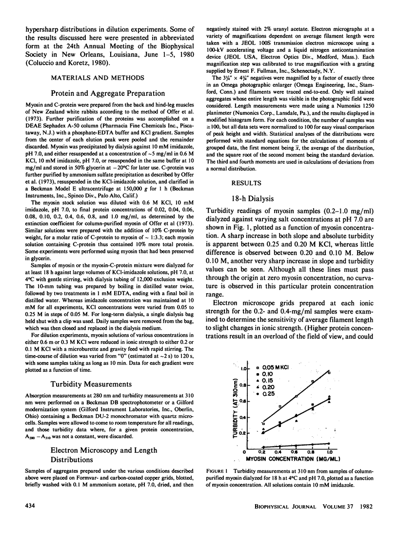
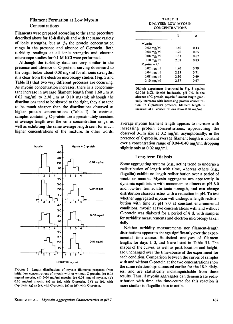
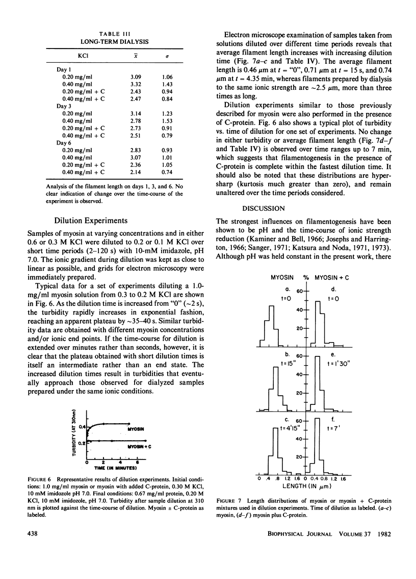
The aggregation properties of column-purified rabbit skeletal myosin at pH 7.0 were investigated as functions of ionic strength, protein concentration, and time. Filaments prepared by dialysis exhibited the same average length and population distribution at 0.10 and 0.15 M KCl at protein concentrations greater than 0.10 mg/ml; similar results were obtained at .0.20 M KCl, although average filament length was approximately 0.5 micrometer shorter. Once formed, these length distributions remained virtually unchanged over an 8-d period. At and below 0.10 mg/ml, average filament length decreased as a function of protein concentration; filaments prepared from an initial concentration of 0.02 mg/ml were half the length of those prepared at 0.2 mg/ml. Filaments prepared by dilution exhibited a sharp increase in average length as the time-course increased up to 40 s, then altered only slightly over a further period of 4 min. Addition of C-protein in a molar ratio of 1-3.3 myosin molecules affected most of these results. Average filament length was affected neither by ionic strength nor by initial protein concentration down to 0.04 mg/ml or over an 8-d period. Filaments formed by dilution in the presence of C-protein exhibited a constant average length and hypersharp length distribution over variable time courses up to 7 min. It is possible that C-protein acts to stabilize the antiparallel intermediate during filamentogenesis, and may also affect subunit addition to this nucleus.
Full text
PDF


Images in this article
Selected References
These references are in PubMed. This may not be the complete list of references from this article.
- Craig R., Offer G. The location of C-protein in rabbit skeletal muscle. Proc R Soc Lond B Biol Sci. 1976 Mar 16;192(1109):451–461. doi: 10.1098/rspb.1976.0023. [DOI] [PubMed] [Google Scholar]
- Godfrey J. E., Harrington W. F. Self-association in the myosin system at high ionic strength. I. Sensitivity of the interaction to pH and ionic environment. Biochemistry. 1970 Feb 17;9(4):886–893. doi: 10.1021/bi00806a025. [DOI] [PubMed] [Google Scholar]
- Godfrey J. E., Harrington W. F. Self-association in the myosin system at high ionic strength. II. Evidence for the presence of a monomer--dimer equilibrium. Biochemistry. 1970 Feb 17;9(4):894–908. doi: 10.1021/bi00806a026. [DOI] [PubMed] [Google Scholar]
- Hinssen H., D'Haese J., Small J. V., Sobieszek A. Mode of filament assembly of myosins from muscle and nonmuscle cells. J Ultrastruct Res. 1978 Sep;64(3):282–302. doi: 10.1016/s0022-5320(78)90037-0. [DOI] [PubMed] [Google Scholar]
- Huxley H. E., Brown W. The low-angle x-ray diagram of vertebrate striated muscle and its behaviour during contraction and rigor. J Mol Biol. 1967 Dec 14;30(2):383–434. doi: 10.1016/s0022-2836(67)80046-9. [DOI] [PubMed] [Google Scholar]
- Josephs R., Harrington W. F. Studies on the formation and physical chemical properties of synthetic myosin filaments. Biochemistry. 1966 Nov;5(11):3474–3487. doi: 10.1021/bi00875a013. [DOI] [PubMed] [Google Scholar]
- Kaminer B., Bell A. L. Myosin filamentogenesis: effects of pH and ionic concentration. J Mol Biol. 1966 Sep;20(2):391–401. doi: 10.1016/0022-2836(66)90070-2. [DOI] [PubMed] [Google Scholar]
- Kaminer B. Synthetic myosin filaments from vertebrate smooth muscle. J Mol Biol. 1969 Jan;39(2):257–264. doi: 10.1016/0022-2836(69)90315-5. [DOI] [PubMed] [Google Scholar]
- Katsura I., Noda H. Further studies on the formation of reconstituted myosin filaments. J Biochem. 1973 Feb;73(2):245–256. [PubMed] [Google Scholar]
- Katsura I., Noda H. Studies on the formation and physical chemical properties of synthetic myosin filaments. J Biochem. 1971 Jan;69(1):219–229. doi: 10.1093/oxfordjournals.jbchem.a129449. [DOI] [PubMed] [Google Scholar]
- Koretz J. F. Effects of C-protein on synthetic myosin filament structure. Biophys J. 1979 Sep;27(3):433–446. doi: 10.1016/S0006-3495(79)85227-3. [DOI] [PMC free article] [PubMed] [Google Scholar]
- Koretz J. F. Structural studies of synthetic filaments prepared from column-purified myosin. Biophys J. 1979 Sep;27(3):423–432. doi: 10.1016/S0006-3495(79)85226-1. [DOI] [PMC free article] [PubMed] [Google Scholar]
- Miyahara M., Noda H. Interaction of C-protein with myosin. J Biochem. 1980 May;87(5):1413–1420. doi: 10.1093/oxfordjournals.jbchem.a132882. [DOI] [PubMed] [Google Scholar]
- Moos C., Offer G., Starr R., Bennett P. Interaction of C-protein with myosin, myosin rod and light meromyosin. J Mol Biol. 1975 Sep 5;97(1):1–9. doi: 10.1016/s0022-2836(75)80017-9. [DOI] [PubMed] [Google Scholar]
- Morimoto K., Harrington W. F. Substructure of the thick filament of vertebrate striated muscle. J Mol Biol. 1974 Feb 15;83(1):83–97. doi: 10.1016/0022-2836(74)90425-2. [DOI] [PubMed] [Google Scholar]
- Offer G., Moos C., Starr R. A new protein of the thick filaments of vertebrate skeletal myofibrils. Extractions, purification and characterization. J Mol Biol. 1973 Mar 15;74(4):653–676. doi: 10.1016/0022-2836(73)90055-7. [DOI] [PubMed] [Google Scholar]
- Pepe F. A., Dowben P. The myosin filament. V. Intermediate voltage electron microscopy and optical diffraction studies of the substructure. J Mol Biol. 1977 Jun 15;113(1):199–218. doi: 10.1016/0022-2836(77)90050-x. [DOI] [PubMed] [Google Scholar]
- Pepe F. A., Drucker B. The myosin filament. III. C-protein. J Mol Biol. 1975 Dec 25;99(4):609–617. doi: 10.1016/s0022-2836(75)80175-6. [DOI] [PubMed] [Google Scholar]
- Pepe F. A., Drucker B. The myosin filament. VI. Myosin content. J Mol Biol. 1979 Jun 5;130(4):379–393. doi: 10.1016/0022-2836(79)90429-7. [DOI] [PubMed] [Google Scholar]
- Pepe F. A. The myosin filament. I. Structural organization from antibody staining observed in electron microscopy. J Mol Biol. 1967 Jul 28;27(2):203–225. doi: 10.1016/0022-2836(67)90016-2. [DOI] [PubMed] [Google Scholar]
- Potter J. D. The content of troponin, tropomyosin, actin, and myosin in rabbit skeletal muscle myofibrils. Arch Biochem Biophys. 1974 Jun;162(2):436–441. doi: 10.1016/0003-9861(74)90202-1. [DOI] [PubMed] [Google Scholar]
- Reisler E., Smith C., Seegan G. Myosin minifilaments. J Mol Biol. 1980 Oct 15;143(1):129–145. doi: 10.1016/0022-2836(80)90127-8. [DOI] [PubMed] [Google Scholar]
- Squire J. M. Symmetry and three-dimensional arrangement of filaments in vertebrate striated muscle. J Mol Biol. 1974 Nov 25;90(1):153–160. doi: 10.1016/0022-2836(74)90263-0. [DOI] [PubMed] [Google Scholar]
- Starr R., Offer G. Polypeptide chains of intermediate molecular weight in myosin preparations. FEBS Lett. 1971 Jun 2;15(1):40–44. doi: 10.1016/0014-5793(71)80075-3. [DOI] [PubMed] [Google Scholar]
- Starr R., Offer G. The interaction of C-protein with heavy meromyosin and subfragment-2. Biochem J. 1978 Jun 1;171(3):813–816. doi: 10.1042/bj1710813. [DOI] [PMC free article] [PubMed] [Google Scholar]
- Tregear R. T., Squire J. M. Myosin content and filament structure in smooth and striated muscle. J Mol Biol. 1973 Jun 25;77(2):279–290. doi: 10.1016/0022-2836(73)90336-7. [DOI] [PubMed] [Google Scholar]