Abstract
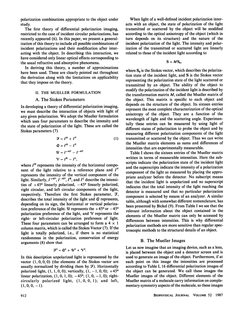
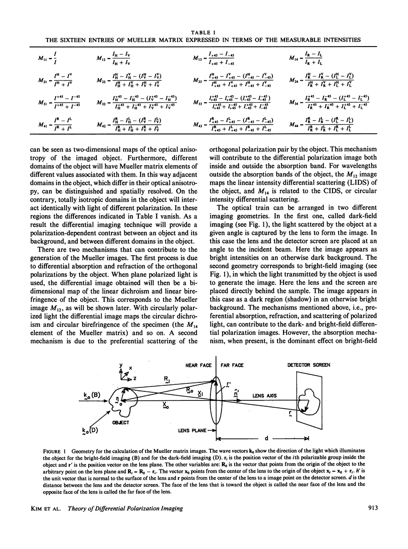
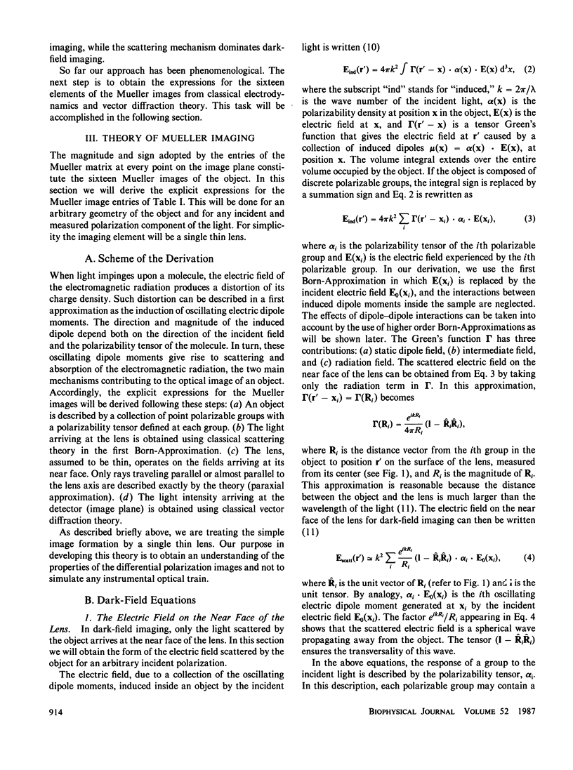
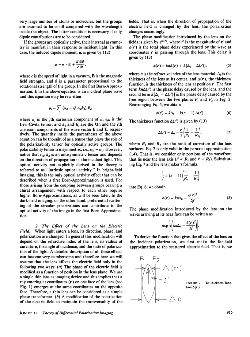
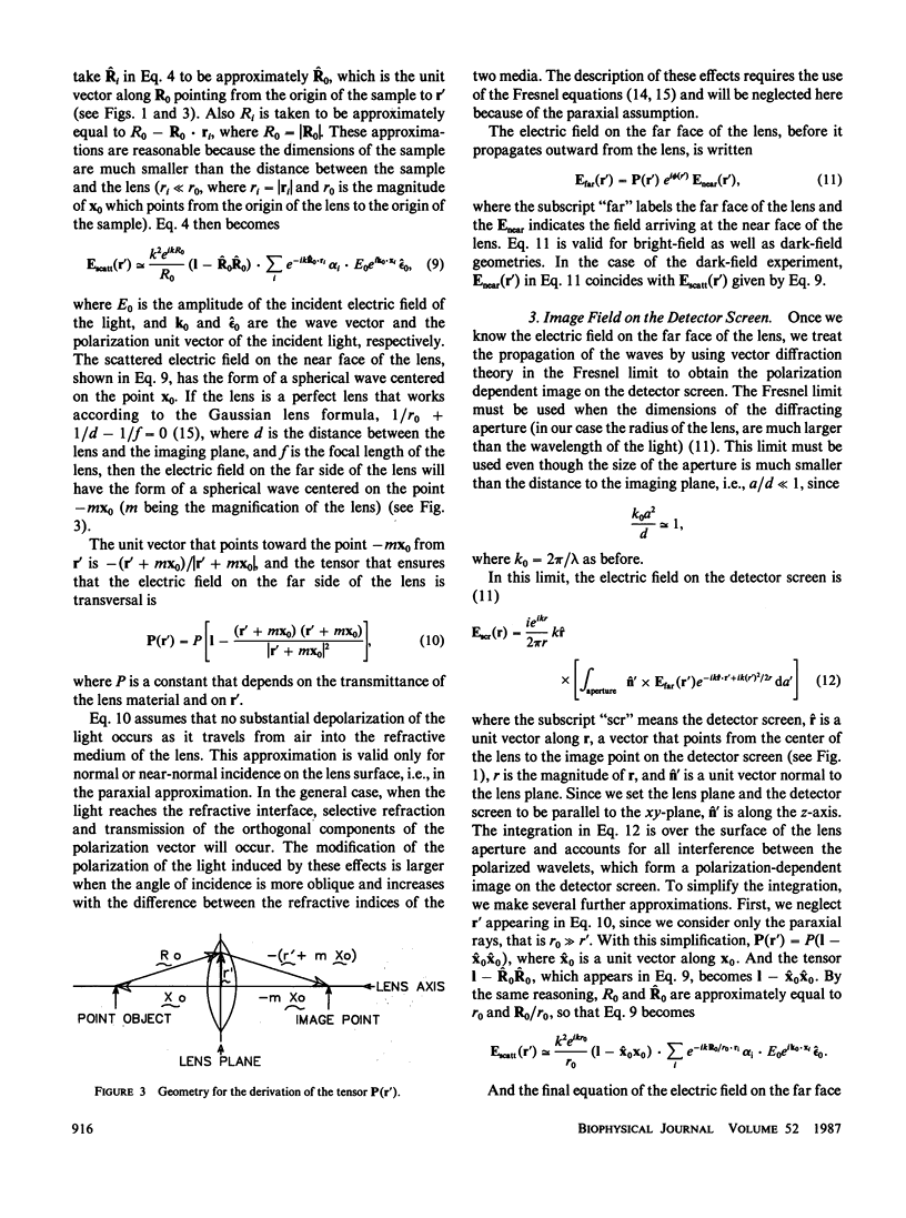
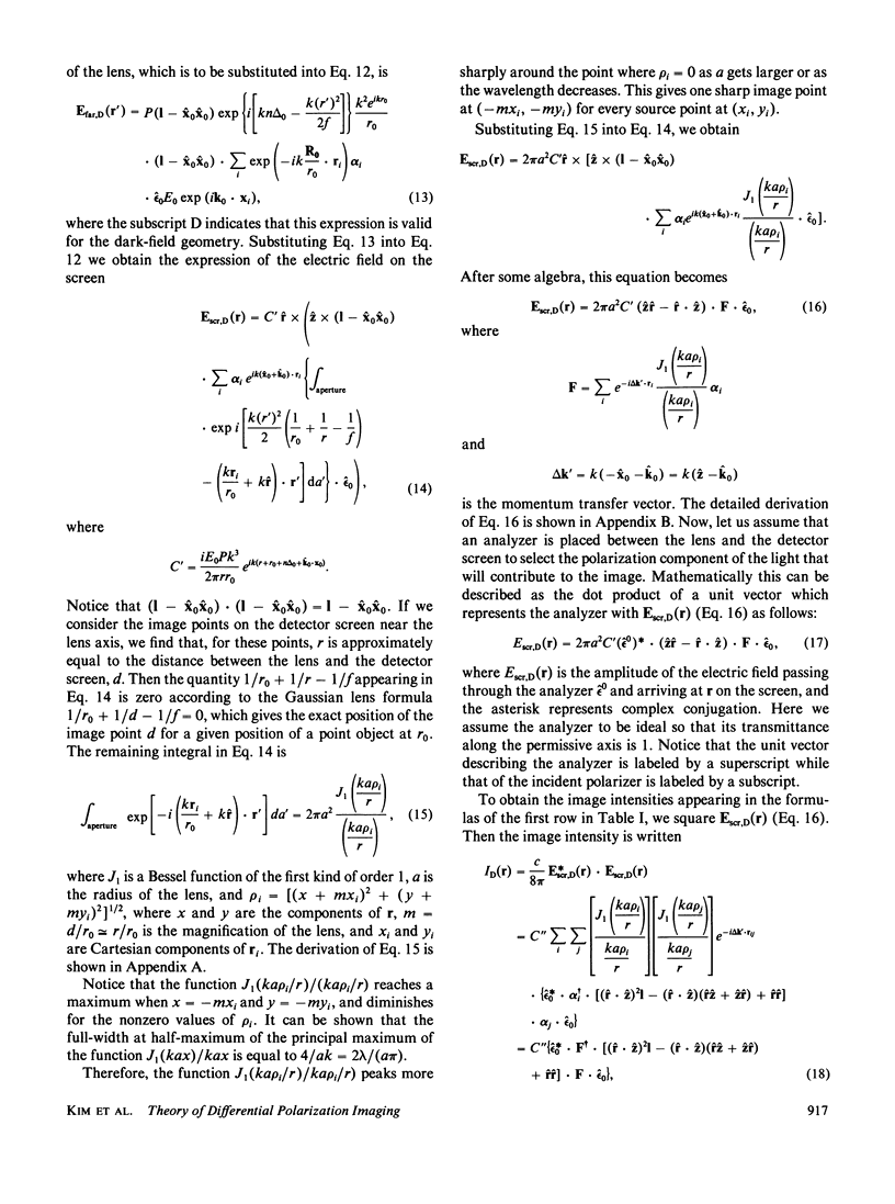
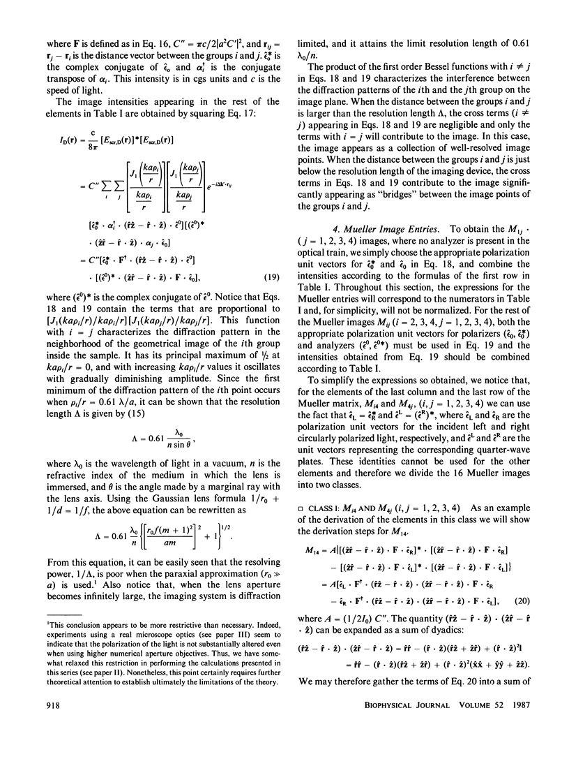
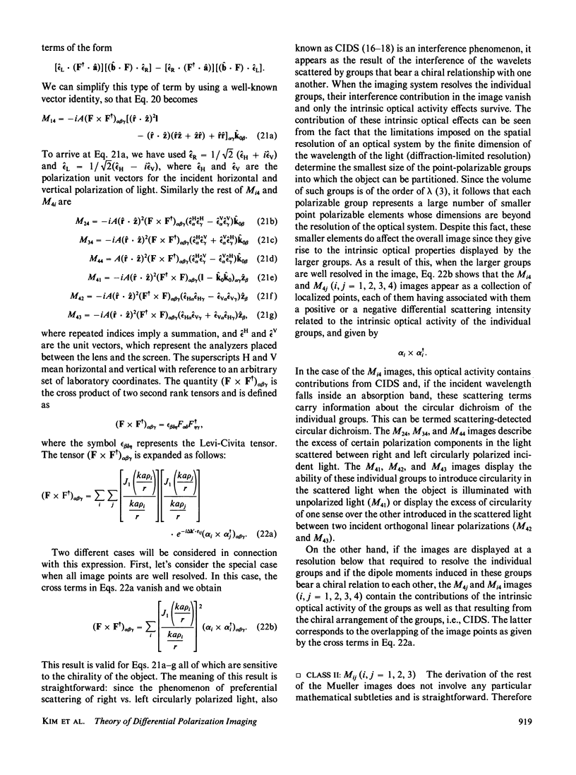
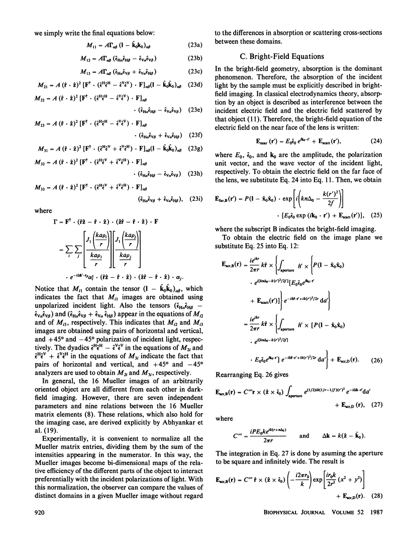
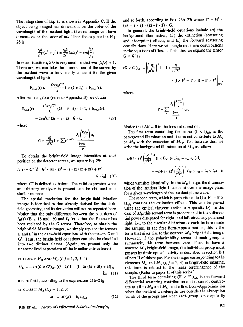
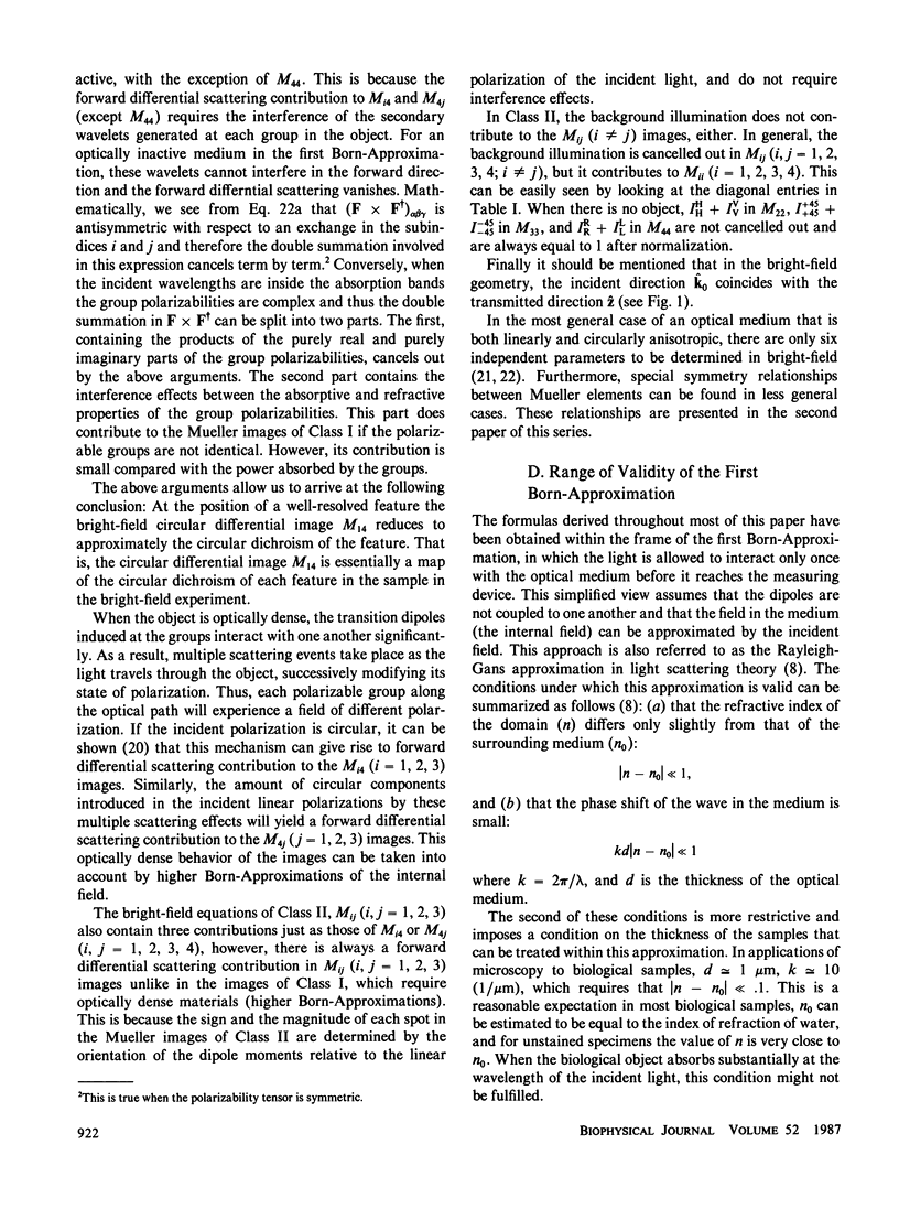
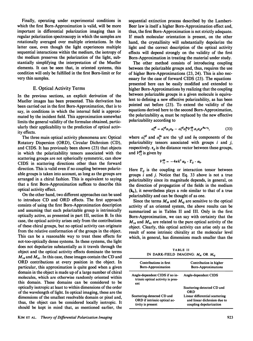
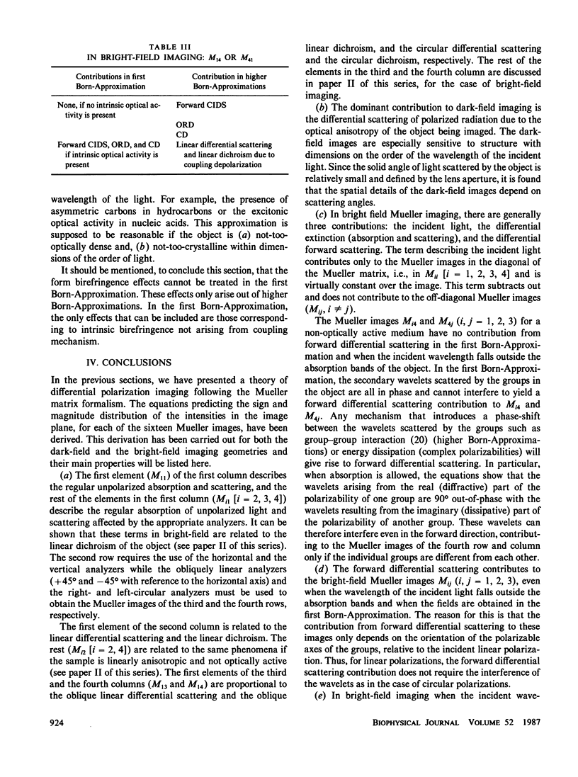
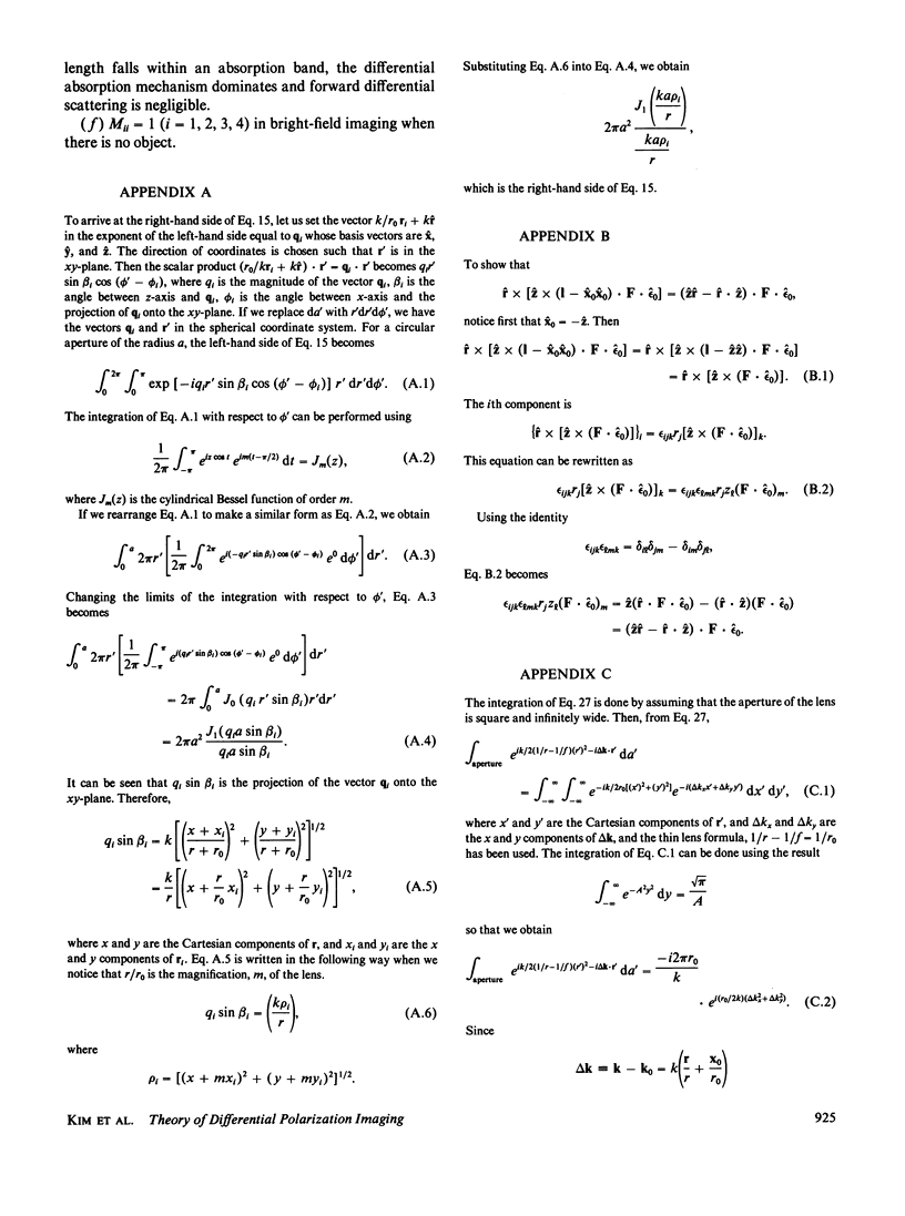
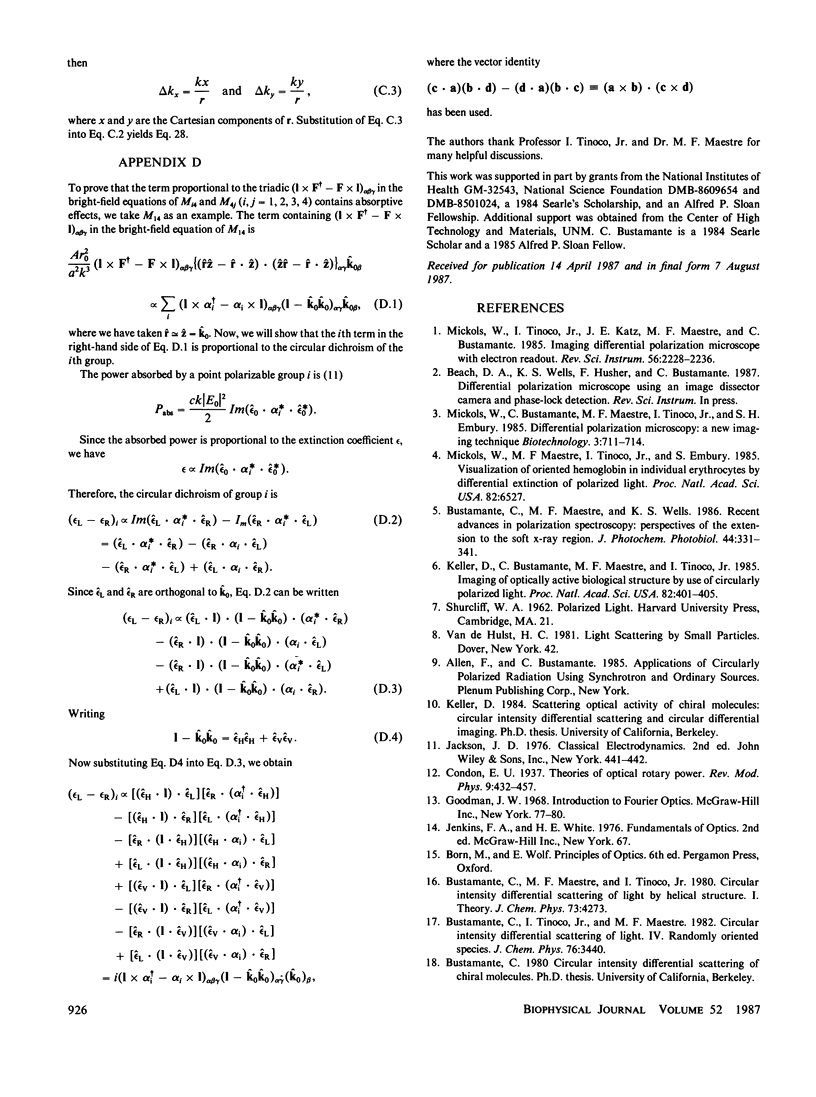
A theory of differential polarization imaging is derived using Mueller calculus. It is shown that, for any arbitrary object, 16 images (in general different) can be obtained by combining different incident polarizations of light and measuring the specific polarization components transmitted or scattered by the object. These are called the Mueller images of the object. Mathematical expressions of these images for an object of arbitrary geometry are derived using classical vector diffraction theory and the paraxial and thin lens approximations. The object is described as a collection of point polarizable groups. The electromagnetic fields are calculated using the first Born-Approximation, but extension of the theory to higher-order approximations is shown to be straightforward. These expressions are obtained for the transmission, or bright-field, geometry, and the scattering, or dark-field, configuration. In both cases, the contributions of scattering, absorption, and background illumination to the Mueller images are characterized. The contributions of linear dichroism, circular dichroism, and linear and circular intensity differential scattering to certain Mueller images are established. It is shown that the Mueller images represent a complete two-dimensional mapping of the molecular anisotropy of the object.
Full text
PDF
Selected References
These references are in PubMed. This may not be the complete list of references from this article.
- Bustamante C., Maestre M. F., Wells K. S. Recent advances in polarization spectroscopy: perspectives of the extension to the soft X-ray region. Photochem Photobiol. 1986 Sep;44(3):331–341. doi: 10.1111/j.1751-1097.1986.tb04672.x. [DOI] [PubMed] [Google Scholar]
- Keller D., Bustamante C., Maestre M. F., Tinoco I., Jr Imaging of optically active biological structures by use of circularly polarized light. Proc Natl Acad Sci U S A. 1985 Jan;82(2):401–405. doi: 10.1073/pnas.82.2.401. [DOI] [PMC free article] [PubMed] [Google Scholar]
- Mickols W., Maestre M. F., Tinoco I., Jr, Embury S. H. Visualization of oriented hemoglobin S in individual erythrocytes by differential extinction of polarized light. Proc Natl Acad Sci U S A. 1985 Oct;82(19):6527–6531. doi: 10.1073/pnas.82.19.6527. [DOI] [PMC free article] [PubMed] [Google Scholar]


