Abstract
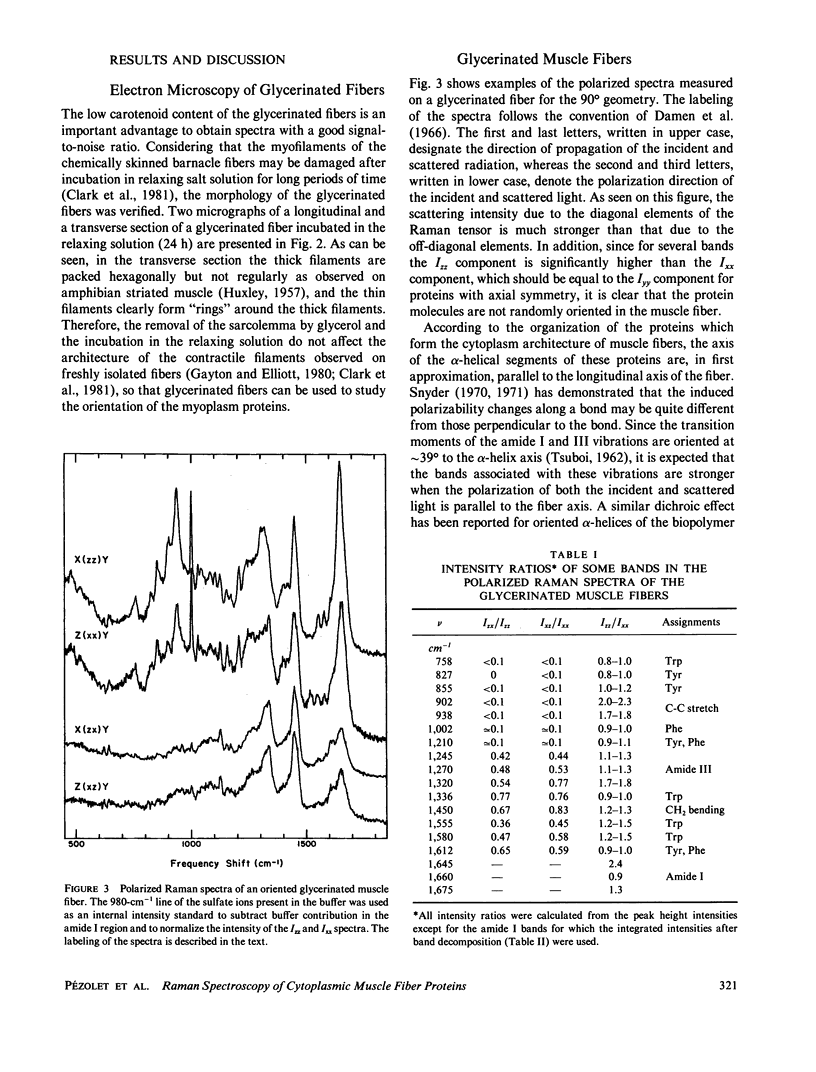
The polarized Raman spectra of glycerinated and intact single muscle fibers of the giant barnacle were obtained. These spectra show that the conformation-sensitive amide I, amide III, and C-C stretching vibrations give Raman bands that are stronger when the electric field of both the incident and scattered radiation is parallel to the fiber axis (Izz). The detailed analysis of the amide I band by curve fitting shows that approximately 50% of the alpha-helical segments of the contractile proteins are oriented along the fiber axis, which is in good agreement with the conformation and composition of muscle fiber proteins. Difference Raman spectroscopy was also used to highlight the Raman bands attributed to the oriented segments of the alpha-helical proteins. The difference spectrum, which is very similar to the spectrum of tropomyosin, displays amide I and amide III bands at 1,645 and 1,310 cm-1, respectively, the bandwidth of the amide I line being characteristic of a highly alpha-helical biopolymer with a small dispersion of dihedral angles. A small dichroic effect was also observed for the band due to the CH2 bending mode at 1,450 cm-1 and on the 1,340 cm-1 band. In the C-C stretching mode region, two bands were detected at 902 and 938 cm-1 and are both assigned to the alpha-helical conformation.
Full text
PDF


Images in this article
Selected References
These references are in PubMed. This may not be the complete list of references from this article.
- Barrett T. W., Peticolas W. L., Robson R. M. Laser Raman light-scattering observations of conformational changes in myosin induced by inorganic salts. Biophys J. 1978 Sep;23(3):349–358. doi: 10.1016/S0006-3495(78)85454-X. [DOI] [PMC free article] [PubMed] [Google Scholar]
- Caillé J. P., Pigeon-Gosselin M., Pézolet M. Effects of organic solutes on the Raman spectra of barnacle muscle fibers. Can J Physiol Pharmacol. 1987 Jul;65(7):1416–1420. doi: 10.1139/y87-222. [DOI] [PubMed] [Google Scholar]
- Caillé J. P., Pigeon-Gosselin M., Pézolet M. Laser Raman study of internally perfused muscle fibers. Effect of Mg2+, ATP and Ca2+. Biochim Biophys Acta. 1983 Jul 29;758(2):121–127. [PubMed] [Google Scholar]
- Carew E. B., Stanley H. E., Seidel J. C., Gergely J. Studies of myosin and its proteolytic fragments by laser Raman spectroscopy. Biophys J. 1983 Nov;44(2):219–224. doi: 10.1016/S0006-3495(83)84294-5. [DOI] [PMC free article] [PubMed] [Google Scholar]
- Carrier D., Pézolet M. Raman spectroscopic study of the interaction of poly-L-lysine with dipalmitoylphosphatidylglycerol bilayers. Biophys J. 1984 Oct;46(4):497–506. doi: 10.1016/S0006-3495(84)84047-3. [DOI] [PMC free article] [PubMed] [Google Scholar]
- Chen M. C., Lord R. C. Laser-excited Raman spectroscopy of biomolecules. VIII. Conformational study of bovine serum albumin. J Am Chem Soc. 1976 Feb 18;98(4):990–992. doi: 10.1021/ja00420a021. [DOI] [PubMed] [Google Scholar]
- Chou P. Y., Fasman G. D. Prediction of protein conformation. Biochemistry. 1974 Jan 15;13(2):222–245. doi: 10.1021/bi00699a002. [DOI] [PubMed] [Google Scholar]
- Fanconi B., Tomlinson B., Nafie L. A., Small W., Peticolas W. L. Polarized laser Raman studies of biological polymers. J Chem Phys. 1969 Nov 1;51(9):3993–4005. doi: 10.1063/1.1672620. [DOI] [PubMed] [Google Scholar]
- HUXLEY H. E. The double array of filaments in cross-striated muscle. J Biophys Biochem Cytol. 1957 Sep 25;3(5):631–648. doi: 10.1083/jcb.3.5.631. [DOI] [PMC free article] [PubMed] [Google Scholar]
- Hinke J. A., Caillé J. P., Gayton D. C. Distribution and state of monovalent ions in skeletal muscle based on ion electrode, isotope, and diffusion analyses. Ann N Y Acad Sci. 1973 Mar 30;204:274–296. doi: 10.1111/j.1749-6632.1973.tb30785.x. [DOI] [PubMed] [Google Scholar]
- Hsu S. L., Moore W. H., Krimm S. Vibrational spectrum of the unordered polypeptide chain: a Raman study of feather keratin. Biopolymers. 1976 Aug;15(8):1513–1528. doi: 10.1002/bip.1976.360150807. [DOI] [PubMed] [Google Scholar]
- Levine R. J., Elfvin M., Dewey M. M., Walcott B. Paramyosin in invertebrate muscles. II. Content in relation to structure and function. J Cell Biol. 1976 Oct;71(1):273–279. doi: 10.1083/jcb.71.1.273. [DOI] [PMC free article] [PubMed] [Google Scholar]
- Lippert J. L., Tyminski D., Desmeules P. J. Determination of the secondary structure of proteins by laser Raman spectroscopy. J Am Chem Soc. 1976 Oct 27;98(22):7075–7080. doi: 10.1021/ja00438a057. [DOI] [PubMed] [Google Scholar]
- Pezolet M., Pigeon-Gosselin M., Caille J. P. Laser Raman investigations of intact single muscle fibers. Protein conformations. Biochim Biophys Acta. 1978 Mar 28;533(1):263–269. doi: 10.1016/0005-2795(78)90570-6. [DOI] [PubMed] [Google Scholar]
- Pezolet M., Pigeon-Gosselin M., Coulombe L. Laser Raman investigation of the conformation of human immunoglobulin G. Biochim Biophys Acta. 1976 Dec 22;453(2):502–512. doi: 10.1016/0005-2795(76)90146-x. [DOI] [PubMed] [Google Scholar]
- Pézolet M., Pigeon-Gosselin M., Nadeau J., Caillé J. P. Laser Raman scattering. A molecular probe of the contractile state of intact single muscle fibers. Biophys J. 1980 Jul;31(1):1–8. doi: 10.1016/S0006-3495(80)85036-3. [DOI] [PMC free article] [PubMed] [Google Scholar]
- Schachar R. A., Solin S. A. The microscopic protein structure of the lens with a theory for cataract formation as determined by Raman spectroscopy of intact bovine lenses. Invest Ophthalmol. 1975 May;14(5):380–396. [PubMed] [Google Scholar]
- Thomas G. J., Jr, Prescott B., Day L. A. Structure similarity, difference and variability in the filamentous viruses fd, If1, IKe, Pf1 and Xf. Investigation by laser Raman spectroscopy. J Mol Biol. 1983 Apr 5;165(2):321–356. doi: 10.1016/s0022-2836(83)80260-5. [DOI] [PubMed] [Google Scholar]
- Williams R. W. Estimation of protein secondary structure from the laser Raman amide I spectrum. J Mol Biol. 1983 Jun 5;166(4):581–603. doi: 10.1016/s0022-2836(83)80285-x. [DOI] [PubMed] [Google Scholar]
- Yates L. D., Greaser M. L. Quantitative determination of myosin and actin in rabbit skeletal muscle. J Mol Biol. 1983 Jul 25;168(1):123–141. doi: 10.1016/s0022-2836(83)80326-x. [DOI] [PubMed] [Google Scholar]
- Yu L. C., Steven A. C., Naylor G. R., Gamble R. C., Podolsky R. J. Distribution of mass in relaxed frog skeletal muscle and its redistribution upon activation. Biophys J. 1985 Mar;47(3):311–321. doi: 10.1016/S0006-3495(85)83921-7. [DOI] [PMC free article] [PubMed] [Google Scholar]
- Yu N. T. Comparison of protein structure in crystals, in lyophilized state, and in solution by laser Raman scattering. 3. Alpha-Lactalbumin. J Am Chem Soc. 1974 Jul 10;96(14):4664–4668. doi: 10.1021/ja00821a049. [DOI] [PubMed] [Google Scholar]
- Yu T. J., Lippert J. L., Peticolas W. L. Laser Raman studies of conformational variations of poly-L-lysine. Biopolymers. 1973;12(9):2161–2175. doi: 10.1002/bip.1973.360120919. [DOI] [PubMed] [Google Scholar]


