

Abstract
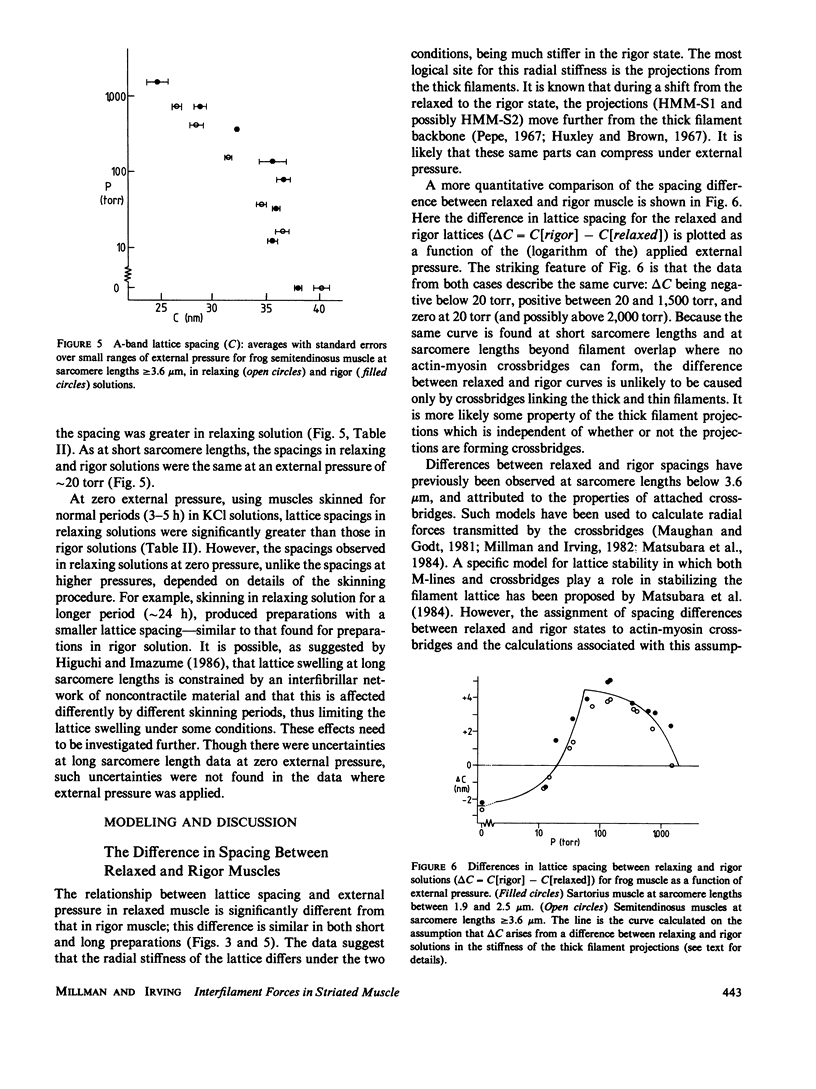
Repulsive pressure in the A-band filament lattice of relaxed frog skeletal muscle has been measured as a function of interfilament spacing using an osmotic shrinking technique. Much improved chemical skinning was obtained when the muscles were equilibrated in the presence of EGTA before skinning. The lattice shrank with increasing external osmotic pressure. At any specific pressure, the lattice spacing in relaxed muscle was smaller than that of muscle in rigor, except at low pressures where the reverse was found. The lattice spacing was the same in the two states at a spacing close to that found in vivo. The data were consistent with an electrostatic repulsion over most of the pressure range. For relaxed muscle, the data lay close to electrostatic pressure curves for a thick filament charge diameter of approximately 26 nm, suggesting that charges stabilizing the lattice are situated about midway along the thick filament projections (HMM-S1). At low pressures, observed spacings were larger than calculated, consistent with the idea that thick filament projections move away from the filament backbone. Under all conditions studied, relaxed and rigor, at short and very long sarcomere lengths, the filament lattice could be modeled by assuming a repulsive electrostatic pressure, a weak attractive pressure, and a radial stiffness of the thick filaments (projections) that differed between relaxed and rigor conditions. Each thick filament projection could be compressed by approximately 5 or 2.6 nm requiring a force of 1.3 or 80 pN for relaxed and rigor conditions respectively.
Full text
PDF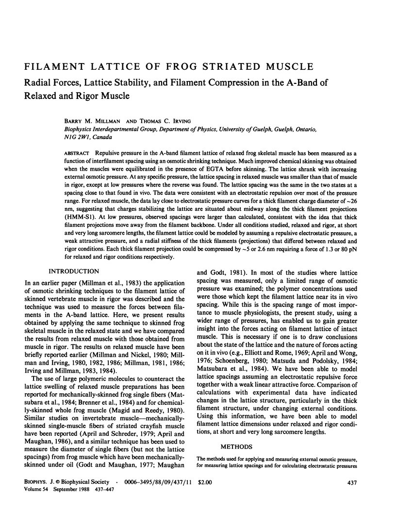


Selected References
These references are in PubMed. This may not be the complete list of references from this article.
- April E. W. Liquid-crystalline characteristics of the thick filament lattice of striated muscle. Nature. 1975 Sep 11;257(5522):139–141. doi: 10.1038/257139a0. [DOI] [PubMed] [Google Scholar]
- April E. W., Maughan D. W. Active force as a function of filament spacing in crayfish skinned muscle fibers. Pflugers Arch. 1986 Oct;407(4):456–460. doi: 10.1007/BF00652634. [DOI] [PubMed] [Google Scholar]
- April E. W., Wong D. Non-isovolumic behavior of the unit cell of skinned striated muscle fibers. J Mol Biol. 1976 Feb 15;101(1):107–114. doi: 10.1016/0022-2836(76)90068-1. [DOI] [PubMed] [Google Scholar]
- Bartels E. M., Elliott G. F. Donnan potentials from the A- and I-bands of glycerinated and chemically skinned muscles, relaxed and in rigor. Biophys J. 1985 Jul;48(1):61–76. doi: 10.1016/S0006-3495(85)83760-7. [DOI] [PMC free article] [PubMed] [Google Scholar]
- Brenner B., Yu L. C., Podolsky R. J. X-ray diffraction evidence for cross-bridge formation in relaxed muscle fibers at various ionic strengths. Biophys J. 1984 Sep;46(3):299–306. doi: 10.1016/S0006-3495(84)84026-6. [DOI] [PMC free article] [PubMed] [Google Scholar]
- Brenner S. L., McQuarrie D. A. Force balances in systems of cylindrical polyelectrolytes. Biophys J. 1973 Apr;13(4):301–331. doi: 10.1016/S0006-3495(73)85987-9. [DOI] [PMC free article] [PubMed] [Google Scholar]
- Cooke P. H., Bartels E. M., Elliott G. F., Hughes R. A. A structural study of gels, in the form of threads, of myosin and myosin rod. Biophys J. 1987 Jun;51(6):947–957. doi: 10.1016/S0006-3495(87)83422-7. [DOI] [PMC free article] [PubMed] [Google Scholar]
- Diamond M. S., Brandt P. W., Kawai M. Comments on "Critical dependence of calcium-activated force on width in highly compressed skinned fibers of the frog". Biophys J. 1986 Dec;50(6):1215–1217. doi: 10.1016/S0006-3495(86)83566-4. [DOI] [PMC free article] [PubMed] [Google Scholar]
- Duncan C. J. Role of calcium in triggering rapid ultrastructural damage in muscle: a study with chemically skinned fibres. J Cell Sci. 1987 May;87(Pt 4):581–594. doi: 10.1242/jcs.87.4.581. [DOI] [PubMed] [Google Scholar]
- Egelman E. H. The structure of F-actin. J Muscle Res Cell Motil. 1985 Apr;6(2):129–151. doi: 10.1007/BF00713056. [DOI] [PubMed] [Google Scholar]
- Elliott G. F., Lowy J., Millman B. M. Low-angle x-ray diffraction studies of living striated muscle during contraction. J Mol Biol. 1967 Apr 14;25(1):31–45. doi: 10.1016/0022-2836(67)90277-x. [DOI] [PubMed] [Google Scholar]
- Elliott G. F. Measurements of the electric charge and ion-binding of the protein filaments in intact muscle and cornea, with implications for filament assembly. Biophys J. 1980 Oct;32(1):95–97. doi: 10.1016/S0006-3495(80)84927-7. [DOI] [PMC free article] [PubMed] [Google Scholar]
- Godt R. E., Maughan D. W. Swelling of skinned muscle fibers of the frog. Experimental observations. Biophys J. 1977 Aug;19(2):103–116. doi: 10.1016/S0006-3495(77)85573-2. [DOI] [PMC free article] [PubMed] [Google Scholar]
- Gulati J., Babu A. Critical dependence of calcium-activated force on width in highly compressed skinned fibers of the frog. Biophys J. 1985 Nov;48(5):781–787. doi: 10.1016/S0006-3495(85)83836-4. [DOI] [PMC free article] [PubMed] [Google Scholar]
- HUXLEY A. F., PEACHEY L. D. The maximum length for contraction in vertebrate straiated muscle. J Physiol. 1961 Apr;156:150–165. doi: 10.1113/jphysiol.1961.sp006665. [DOI] [PMC free article] [PubMed] [Google Scholar]
- Haselgrove J. C. A model of myosin crossbridge structure consistent with the low-angle x-ray diffraction pattern of vertebrate muscle. J Muscle Res Cell Motil. 1980 Jun;1(2):177–191. doi: 10.1007/BF00711798. [DOI] [PubMed] [Google Scholar]
- Haselgrove J. C., Huxley H. E. X-ray evidence for radial cross-bridge movement and for the sliding filament model in actively contracting skeletal muscle. J Mol Biol. 1973 Jul 15;77(4):549–568. doi: 10.1016/0022-2836(73)90222-2. [DOI] [PubMed] [Google Scholar]
- Higuchi H., Umazume Y. Lattice shrinkage with increasing resting tension in stretched, single skinned fibers of frog muscle. Biophys J. 1986 Sep;50(3):385–389. doi: 10.1016/S0006-3495(86)83474-9. [DOI] [PMC free article] [PubMed] [Google Scholar]
- Huxley H. E., Brown W. The low-angle x-ray diagram of vertebrate striated muscle and its behaviour during contraction and rigor. J Mol Biol. 1967 Dec 14;30(2):383–434. doi: 10.1016/s0022-2836(67)80046-9. [DOI] [PubMed] [Google Scholar]
- Huxley H. E. Structural difference between resting and rigor muscle; evidence from intensity changes in the lowangle equatorial x-ray diagram. J Mol Biol. 1968 Nov 14;37(3):507–520. doi: 10.1016/0022-2836(68)90118-6. [DOI] [PubMed] [Google Scholar]
- Kensler R. W., Stewart M. Frog skeletal muscle thick filaments are three-stranded. J Cell Biol. 1983 Jun;96(6):1797–1802. doi: 10.1083/jcb.96.6.1797. [DOI] [PMC free article] [PubMed] [Google Scholar]
- Magid A., Reedy M. K. X-ray diffraction observations of chemically skinned frog skeletal muscle processed by an improved method. Biophys J. 1980 Apr;30(1):27–40. doi: 10.1016/S0006-3495(80)85074-0. [DOI] [PMC free article] [PubMed] [Google Scholar]
- Matsubara I., Elliott G. F. X-ray diffraction studies on skinned single fibres of frog skeletal muscle. J Mol Biol. 1972 Dec 30;72(3):657–669. doi: 10.1016/0022-2836(72)90183-0. [DOI] [PubMed] [Google Scholar]
- Matsubara I., Goldman Y. E., Simmons R. M. Changes in the lateral filament spacing of skinned muscle fibres when cross-bridges attach. J Mol Biol. 1984 Feb 15;173(1):15–33. doi: 10.1016/0022-2836(84)90401-7. [DOI] [PubMed] [Google Scholar]
- Matsuda T., Podolsky R. J. X-ray evidence for two structural states of the actomyosin cross-bridge in muscle fibers. Proc Natl Acad Sci U S A. 1984 Apr;81(8):2364–2368. doi: 10.1073/pnas.81.8.2364. [DOI] [PMC free article] [PubMed] [Google Scholar]
- Maughan D. W., Godt R. E. Radial forces within muscle fibers in rigor. J Gen Physiol. 1981 Jan;77(1):49–64. doi: 10.1085/jgp.77.1.49. [DOI] [PMC free article] [PubMed] [Google Scholar]
- Millman B. M., Irving T. C., Nickel B. G., Loosley-Millman M. E. Interrod forces in aqueous gels of tobacco mosaic virus. Biophys J. 1984 Mar;45(3):551–556. doi: 10.1016/S0006-3495(84)84192-2. [DOI] [PMC free article] [PubMed] [Google Scholar]
- Millman B. M., Nickel B. G. Electrostatic forces in muscle and cylindrical gel systems. Biophys J. 1980 Oct;32(1):49–63. doi: 10.1016/S0006-3495(80)84915-0. [DOI] [PMC free article] [PubMed] [Google Scholar]
- Millman B. M., Racey T. J., Matsubara I. Effects of hyperosmotic solutions on the filament lattice of intact frog skeletal muscle. Biophys J. 1981 Feb;33(2):189–202. doi: 10.1016/S0006-3495(81)84880-1. [DOI] [PMC free article] [PubMed] [Google Scholar]
- Millman B. M., Wakabayashi K., Racey T. J. Lateral forces in the filament lattice of vertebrate striated muscle in the rigor state. Biophys J. 1983 Mar;41(3):259–267. doi: 10.1016/S0006-3495(83)84436-1. [DOI] [PMC free article] [PubMed] [Google Scholar]
- Naylor G. R., Bartels E. M., Bridgman T. D., Elliott G. F. Donnan potentials in rabbit psoas muscle in rigor. Biophys J. 1985 Jul;48(1):47–59. doi: 10.1016/S0006-3495(85)83759-0. [DOI] [PMC free article] [PubMed] [Google Scholar]
- Padrón R., Huxley H. E. The effect of the ATP analogue AMPPNP on the structure of crossbridges in vertebrate skeletal muscles: X-ray diffraction and mechanical studies. J Muscle Res Cell Motil. 1984 Dec;5(6):613–655. doi: 10.1007/BF00713923. [DOI] [PubMed] [Google Scholar]
- Pepe F. A. The myosin filament. II. Interaction between myosin and actin filaments observed using antibody staining in fluorescent and electron microscopy. J Mol Biol. 1967 Jul 28;27(2):227–236. doi: 10.1016/0022-2836(67)90017-4. [DOI] [PubMed] [Google Scholar]
- Schoenberg M. Geometrical factors influencing muscle force development. II. Radial forces. Biophys J. 1980 Apr;30(1):69–77. doi: 10.1016/S0006-3495(80)85077-6. [DOI] [PMC free article] [PubMed] [Google Scholar]
- Stewart M., Kensler R. W. Arrangement of myosin heads in relaxed thick filaments from frog skeletal muscle. J Mol Biol. 1986 Dec 20;192(4):831–851. doi: 10.1016/0022-2836(86)90032-x. [DOI] [PubMed] [Google Scholar]