Abstract
It was previously demonstrated that avirulent Mycoplasma gallisepticum strain Rhigh (passage 164) is lacking three proteins that are expressed in its virulent progenitor, strain Rlow (passage 15). These proteins were identified as the cytadhesin molecule GapA, the putative cytadhesin-related molecule CrmA, and a component of a high-affinity transporter system, HatA. Complementation of Rhigh with wild-type gapA restored expression in the transformant (GT5) but did not restore the cytadherence phenotype and maintained avirulence in chickens. These results suggested that CrmA might play an essential role in the M. gallisepticum cytadherence process. CrmA is encoded by the second gene in the gapA operon and shares significant sequence homology to the ORF6 gene of Mycoplasma pneumoniae, which has been shown to play an accessory role in the cytadherence process. Complementation of Rhigh with wild-type crmA resulted in the transformant (SDCA) that lacked the cytadherence and virulence phenotype comparable to that found in Rhigh and GT5. In contrast, complementation of Rhigh with the entire wild-type gapA operon resulted in the transformant (GCA1) that restored cytadherence to the level found in wild-type Rlow. In vivo pathogenesis trials revealed that GCA1 had regained virulence, causing airsacculitis in chickens. These results demonstrate that both GapA and CrmA are required for M. gallisepticum cytadherence and pathogenesis.
Mycoplasma gallisepticum is one infectious agent initiating the chronic respiratory disease complex in chickens and is the primary agent of infectious sinusitis in turkeys (74). This bacterium has developed a wide array of surface molecules that are involved in cytadherence to host cells (9, 17, 22, 51). GapA is considered the primary cytadhesin. Goh et al. (21) identified its gene in M. gallisepticum based on its nucleotide sequence homology to the Mycoplasma pneumoniae cytadhesin ADP1 gene. Subsequent studies showed that anti-GapA Fab fragments were able to significantly inhibit M. gallisepticum cytadherence (22). Troy (69) reported that GapA was absent in avirulent M. gallisepticum. These data led us to hypothesize that complementation of GapA expression in strain Rhigh via Tn4001 might restore cytadherence and perhaps virulence. Neither cytadherence nor virulence was restored upon the gapA-complemented strain Rhigh, transformant GT5 (56, 57). This indicated that other factors might play an important role in M. gallisepticum cytadherence. Protein profile comparison between virulent and avirulent strains showed that, in addition to GapA, two other proteins are absent in Rhigh (69). One of these proteins was found to be encoded by the second gene of the gapA operon. Its gene product shows significant sequence homology with the precursor of M. pneumoniae ORF6 gene products, which are known to play accessory roles in P1 (ADP1)-mediated cytadherence (35, 36, 40, 66). (MGPC is the new GenBank designation for the M. pneumoniae ORF6 and M. genitalium ORF6 homolog. M. pneumoniae MGPC gene encodes a 130-kDa protein which is posttranslationally cleaved into two products of 40 and 90 kDa by a special proteolytic event [accessory proteins B and C]. M. gallisepticum CrmA does not undergo such posttranslational modification. We will adhere to the new nomenclature in this paper.) This M. gallisepticum protein was designated as cytadherence-related molecule A, or CrmA.
The aim of this study was to evaluate the role of CrmA in M. gallisepticum cytadherence and virulence. We demonstrate that coexpression of gapA and crmA is essential for effective M. gallisepticum cytadherence and virulence.
MATERIALS AND METHODS
Strains and media.
M. gallisepticum strains Rlow and Rhigh (15 and 164 passages, respectively) were provided by Sharon Levisohn (formerly of the Department of Food Animal and Equine Medicine, North Carolina State University, Raleigh, N.C.). Generation of the GT5 transformant has been described previously (57). All strains were cultured at 37°C in Hayflick's medium (10% serum and 5% yeast extract) or on Hayflick's plates with 1% Agar Noble (Difco). Gentamicin was added to the liquid medium (final concentration, 300 μg/ml) for propagating the GT5, SDCA, and GCA1 transformants.
DNA extractions.
Mycoplasma total genomic DNA was extracted according to the method of Hempstead (25). Plasmid DNA was extracted by using QIAprep Spin Miniprep (Qiagen, Valencia, Calif.) or minipreparations according to the protocol described by Engelbrecht at al. (16).
Immunoblot analysis.
Proteins from 2 × 10⁷ CFU of M. gallisepticum Rlow, Rhigh, and transformants were separated by sodium dodecyl sulfate-polyacrylamide gel electrophoresis (38) and then transferred onto a nitrocellulose membrane (Osmonics, Westborough, Mass.) according to the method of Towbin et al. (68). After blocking with 5% bovine serum albumin in phosphate-buffered saline (PBS) for 1 h at room temperature, the membranes were reacted with rabbit anti-GapA or anti-CrmA serum at dilutions of 1:8,000 and 1:50,000, respectively, for 2 h at 4°C with gentle rocking and then washed three times with 0.5% PBS-Tween. Membranes were then incubated with peroxidase-conjugated goat anti-rabbit immunoglobulin G (Sigma, St. Louis, Mo.) at a final concentration of 1:15,000 for 1 h at 4°C with gentle rocking, followed by three consecutive washings with PBS-Tween. Reactions were visualized by the addition of 4-chloro-1-naphthol and hydrogen peroxide.
PCR.
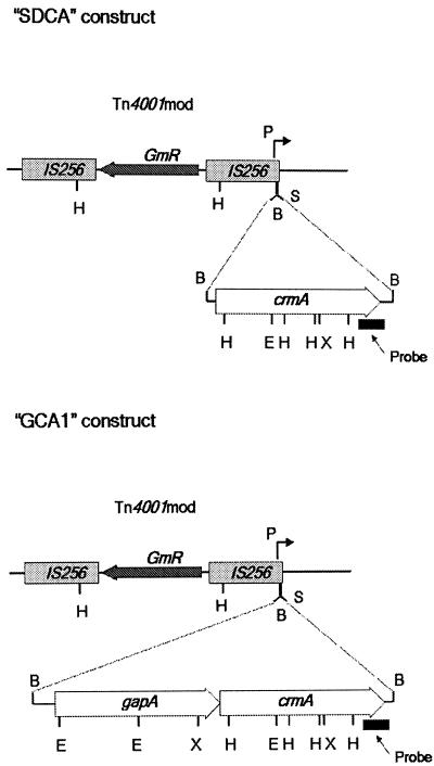
PCRs were performed in a total volume of 50 μl containing 50 ng of template. Primers (Table 1) were synthesized at the University of Connecticut Biotechnology Center. The gapA operon and crmA were amplified with primer pairs SG322-SG721 and SG768-SG721, respectively. The gapA operon was amplified with the same forward primer (SG322), which was used previously to amplify gapA alone to obtain GT5 (57). BamHI sites were incorporated at the 5′ ends of each primer to facilitate cloning into the BamHI site of Tn4001mod in the vector pISM2062 (33) (Fig. 1). The gapA operon and crmA were amplified by using the High-Fidelity Expand PCR kit (Roche Biochemicals, Indianapolis, Ind.) according to the manufacturer's instructions. The AmpliTaq kit (Applied Biosystems, Perkin Elmer, Norwalk, Conn.) was used for other PCRs as previously described (57). Briefly, the amplifications were performed under the following conditions: 30 cycles of denaturation at 94°C for 1 min, annealing at 55°C for 1 min and 72°C for 2 min, followed by 1 cycle at 72°C for 10 min. A total of 2.5 U of AmpliTaq was used in each reaction.
TABLE 1.
Primers used to amplify crmA and the entire gapA operon, the probe for the Southern hybridization, and the gentamicin resistance gene
| Primer | Sequencea (direction) | Gentamicin resistance gene |
|---|---|---|
| SG322 | 5′ GGGGGATCCAGACCAAACTTCCCTAAC 3′ (forward) | gapA |
| SG721 | 5′ GGGGGATCCCCTTATCGTAGAGAAGGGAGGT 3′ (reverse) | crmA |
| SG768 | 5′ AAGGGGATCCGCTCCAGCACCAACTAAGAAAATTGA 3′ (forward) | crmA |
| SG804 | 5′ GGCAATTATGATCATCTTAGGA 3′ (forward) | 3′ crmA |
| SG805 | 5′ TAGAGAAGGGAGGTTATTTT 3′ (reverse) | 3′ crmA |
| SG643 | 5′ ACACAGGAGTCTGGACTTGACTGA 3′ (forward) | GmRb |
| SG665 | 5′ TTACACAGGAGTCTGGACTTGACTCA 3′ (reverse) | GmR |
BamHI restriction endonuclease sites are underlined.
Gentamicin resistance gene from the Tn4001mod transposon.
FIG. 1.
Tn4001mod constructs containing the wild-type gapA and crmA gene from strain Rlow. Schematic representation map of Tn4001mod constructs containing wild-type crmA alone (SDCA) or both gapA and crmA (GCA1, i.e., the whole gapA operon). GmR is the gentamicin resistance gene. P is the outward promoter. B, BamHI; E, EcoRI; H, HindIII; X, XbaI.
Cloning and transformation.
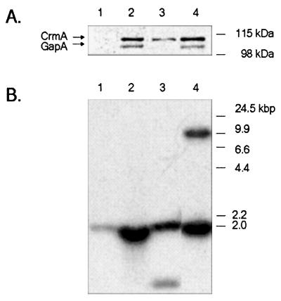
PCR products (gapA operon and crmA) were initially cloned into the pCR 2.1 TOPO vector according to the manufacturer's instructions and maintained in Top 10 Escherichia coli cells (Invitrogen, Carlsbad, Calif.). Extracted plasmids were digested with BamHI (Roche Biochemicals), and the inserts were separated by electrophoresis in a 1% agarose gel. The bands were eluted from the agarose gel and purified with DNA Clean Concentrator 5 (Zymo Research, Orange, Calif.). Fragments were cloned into the BamHI site of the transposon Tn4001mod in the plasmid pISM2062 (33) by using a ligation kit (Epicentre Technologies, Madison, Wis.). The constructs containing the gapA operon and crmA were designated GCA1 and SDCA, respectively (Fig. 1). Rhigh (from the same clonal isolate stock used to generate GT5 [57]) was transformed following the electroporation method of Minion and Kapke (52). Following transformation, single colonies were picked and propagated in Hayflick's medium containing gentamicin. Clones were initially screened by PCR for the gentamicin resistance gene. Positive clones were analyzed by immunoblotting with anti-GapA and anti-CrmA sera (Fig. 2A) and by Southern blot hybridization with a 402-bp 32P-labeled portion of the 5′ end of crmA as a probe (primer pair SG804-SG805) (Table 1 and Fig. 2B). SDCA clone 4 (SDCAc4) and GCA1 clone 5 (GCA1c5) were used for all of the experiments.
FIG. 2.
Characterization of the M. gallisepticum Rhigh transformants SDCA and GCA1 by immunoblotting and DNA hybridization analysis. Lane 1, Rhigh; lane 2, Rlow; lane 3, SDCAc4; lane 4, GCA1c5. (A) Immunoblots developed with mixed anti-GapA and anti-CrmA sera. (B) Southern blot of HindIII-digested total genomic DNAs probed with a 32P-labeled portion of crmA. Lanes in this digitized image have been reordered by using Adobe PhotoShop.
Cytadherence assays.
MRC-5 cell culture cytadherence analysis was performed as described previously (17-19, 57) with tritium-labeled M. gallisepticum Rlow, Rhigh, GT5, SDCA, and GCA1. These assays were performed twice using four replicates per strain or transformant.
Animals.
Four-week-old, specific-pathogen-free White Leghorn chickens (SPAFAS, North Franklin, Conn.) were used for challenge experiments. Upon arrival, the birds were tagged, placed in HEPA filtered isolators (Controlled Isolator Systems, Pittsburgh, Pa.), and allowed to acclimate for 1 week in accordance with the approved Institutional Animal Care and Use Committee protocol. Nonmedicated feed (Blue Seal, Waltham, Mass.) and water were provided ad libitum.
Preparation challenge strains.
Stocks were prepared as follows: 10-ml aliquots of mid-log-phase cultures were centrifuged at 10,000 × g and 4°C for 15 min and then resuspended in 100 μl of fresh medium and stored at −70°C. Four randomly chosen aliquots from each batch were thawed, and serial 10-fold dilution plate counts were performed to determine total CFU. On the day of administration, 100-μl aliquots were thawed, 900 μl of fresh medium was added to each tube, and the cultures were incubated for 5 h at 37°C. The titers of the organisms were established prior to administration to determine the dose administered to ensure consistency among all challenge experiments.
Challenge.
Chickens were divided into five groups of six birds each. Birds were challenged on days 0 and 2 by intratracheal instillation of 100 μl of M. gallisepticum with a micropipette and disposable 200-μl pipette tips (Fisher Scientific, Fairlawn, N.J.). Birds in groups 1, 2, and 4 received 3 × 10⁸ CFU of GT5, SDCAc4, or GCA1c5, respectively. Group 3 and 5 birds received 107 CFU of GCA1c4 or Rlow, respectively. Two weeks postchallenge (day 16), chickens were sacrificed, tracheal swabs were performed, and trachea and air sacs were prepared for histological examination. A previous study (56) demonstrated that 107 CFU of Rlow delivered intratracheally caused significant gross, as well as histopathological, lesions in the lower respiratory tracts of birds, whereas 3 × 10⁸ CFU of GT5 was avirulent. Therefore, groups of birds challenged with 3 × 10⁸ CFU of GT5 and 107 CFU of Rlow were used as negative and positive controls, respectively.
Gross and histopathological examination.
Necropsy was performed as previously described (56). In addition to the collection of samples from tracheas, tissue samples from the cranial and caudal thoracic and abdominal air sacs were collected. Tissue samples were routinely processed, embedded in paraffin blocks, sectioned at 4 μm, stained with hematoxylin and eosin, and evaluated by light microscopy.
All gross necropsy examinations and evaluations of histological sections of trachea and air sacs were performed in a blind fashion. Evaluation of histopathological changes was based on the criteria of Nunoya et al. (54) and modified for the assessment of air sac lesions. Briefly, we implemented the following scoring system similar to that used to evaluate tracheal lesions (54, 56) as follows: 0, no significant findings; 0.5, minimal multifocal lymphocytic infiltrates with one to three discrete, small, widely separated foci; 1, mild multifocal lymphocytic or lymphofollicular infiltrates amounting to four or more discrete foci without stromal edema or heterophils; 2, moderate, multifocal lymphocytic or lymphofollicular infiltrates and loose interstitial infiltrates of lymphocytes, histiocytes, and heterophils with edema, intraepithelial heterophils, and occasional luminal exudates; 3, severe multifocal lymphofollicular aggregates to diffuse infiltrates of lymphocytes, histiocytes, and heterophils with edema, epithelial attenuation and hyperplasia, and luminal exudates.
Reisolation and quantitation of mycoplasmas.
The mucosa of 5- to 7-cm-long segments of trachea was abraded with a cotton swab. Each swab was inoculated into 3 ml of Hayflick's medium in 15-ml sterile culture tubes and then gently vortexed to release attached mycoplasmas into the medium. The samples were filtered through 0.45-μm-pore size filters (Millipore, Bedford, Mass.) and then serially diluted (10-fold). Twenty microliters of each sample dilution was plated onto Hayflick's agar for CFU determination. All cultures were incubated at 37°C for 4 weeks and observed for color change (acid shift) or colony growth on plates.
Statistical analysis.
SAS software version 8.01 (SAS Institute, Cary, N.C.) was used for statistical analysis. Attachment levels (percentages) and lesion scores were initially subjected to the arcsinus of the percentage square root and rank transformation, respectively, because they violated the normal distribution assumption required for parametric statistical analysis (75). Analysis of variance was used to determine the significant differences between groups. When differences were found, a mean separation analysis using Duncan's multiple range test was performed. Cytadherence levels of SDCA and GCA1 were compared to those of Rhigh. Lesion scores of the groups challenged with SDCAc4 and GCA1c5 were compared to those of groups challenged with GT5. Fisher's exact test was used to compare differences regarding the number of isolates between groups.
Sequence analysis.
TMHMM (37, 53), PSIPRED (30), SAPS (10), and SSpro (3) were used to predict protein secondary structure. Sequence homology searches were performed on the BLOCKS, PROSITE, and Pfam databases (4, 11, 26; http://motif.genome.ad.jp/). FastA (58) and Patternmatch (http://workbench.sdsc.edu, University of San Diego Supercomputer Center, San Diego, Calif.) searches were performed with sequences from well-characterized binding domains. Heparin sulfate- and lectin-binding domains were obtained from published reports (27, 48, 49, 64, 65, 67, 70). Amino acid sequence homologies were considered significant if they possessed a ≥30% identity or similarity and the indicated functional motifs. Structural homology searches were performed using three-dimensional predicted protein secondary structure matrices (3D-PSSM) (5), GenTHREADER (29), and 123D+ (1). Vector NTI (Informax, Bethesda, Md.) was used for visualization of the domains in the protein sequence.
RESULTS
Complementation of Rhigh with wild-type crmA and the gapA operon.
Immunoblot analysis indicated that all of the SDCA transformants expressed CrmA (Fig. 2A) and that GCA1 transformants expressed both GapA and CrmA (Fig. 2A). Southern blot hybridization confirmed the presence of the wild-type genes. As shown in Fig. 2B, all of the SDCA and GCA1 transformants possessed two copies of the genes. The common band represents the genomic crmA, and the second band represents the inserted wild-type gene.
Cytadherence assessment of SDCA and GCA1 transformants.
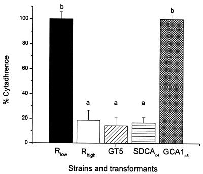
As shown in Fig. 3, SDCAc4 adhered to the MRC-5 cell at the same level as both Rhigh and GT5 (P > 0.05). In contrast, cytadherence of GCA1c5 was significantly high relative to that of Rhigh (P < 0.0001) and comparable to wild-type Rlow levels (P > 0.05).
FIG. 3.
Cytadherence assessment of M. gallisepticum transformants (data presented relative to the Rlow cytadherence level of 100%). Bars labeled with the same letter are not significantly different (P > 0.05). Error bars represent standard deviations.
SDCA and GCA1 challenge experiments.
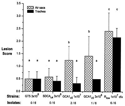
Histopathological evaluations of the respiratory tract lesions are shown in Fig. 4. Birds challenged with 3 × 10⁸ CFU of GT5 had histopathological changes in air sacs that ranged from no significant lesions (score = 0) to mild multifocal lymphocytic infiltrates (score = 1), with most having minimal multifocal stromal expansion by a few, small, widely scattered lymphocytic or lymphofollicular infiltrates (score = 0.5). The same was found in birds challenged with 3 × 10⁸ CFU of SDCAc4. Birds challenged with either 107 CFU or 3 × 10⁸ CFU of GCA1c5 had mild to moderate airsacculitis. Histopathologic lesions in birds challenged with GCA1 ranged from mild multifocal lymphocytic infiltrates (score = 1) to moderate multifocal lymphocytic or lymphofollicular infiltrates and loose interstitial infiltrates of lymphocytes, histiocytes, and heterophils with edema (score = 2). Occasional luminal exudates composed of necrotic and degranulated heterophils and macrophages, along with protein and necrotic epithelial cell debris, were present. In comparison to birds from the GT5 and SDCA challenge groups, GCA1 challenges had increased numbers of lymphofollicular aggregates; interstitial infiltrates of lymphocytes, histiocytes, and heterophils accompanied by edematous separation of stromal collagen and intraepithelial heterophils; and luminal exudates of necrotic cellular debris. Birds challenged with 107 CFU of strain Rlow had the most severe airsacculitis, characterized by marked-to-severe stromal expansion by numerous, densely cellular, follicular lymphocytic aggregates accompanied by multifocal-to-diffuse interstitial infiltrates of lymphocytes, histiocytes, and heterophils. Rlow-challenged birds also had segmental lesions to the epithelium ranging from loss and attenuation to stacking of epithelial cells along short stretches. The most cellular and extensive luminal exudates of necrotic and viable heterophils and macrophages were present in Rlow-challenged birds.
FIG. 4.
Evaluation of the pathogenicities of different M. gallisepticum Rhigh transformants in the lower respiratory tracts of chickens. Bars labeled with the same letter are not significantly different (P > 0.05). Error bars represent standard deviations.
Tracheal lesions in all groups except those challenged with Rlow were absent (score = 0) or consisted of a few, widely separated, minimal, lymphocytic foci (score = 0.5). Birds challenged with Rlow had tracheitis characterized by marked to severe mucosal thickening, diffuse lymphocytic infiltrates with heterophilic stromal and intraepithelial infiltrates, and luminal exudates of necrotic cellular debris.
No significant difference was found between birds challenged with SDCAc4 and those challenged with GT5 (P > 0.05). Birds challenged with GCA1c5 (regardless of the dose) developed airsacculitis (P < 0.05). Three isolates were obtained from birds challenged with GCA1c5 but none from SDCAc4- or GT5-challenged birds. The presence of the transposon carrying the gapA operon was confirmed by PCR in all of the GCA1 isolates (data not shown). None of the groups challenged with GT5, SDCAc4, or GCA1c5 exhibited significant tracheal lesions (P > 0.05).
Sequence analysis.
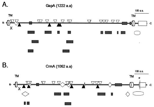
Potential carbohydrate moiety- and heparin sulfate-binding-like domains (confined to regions of 150 to 300 amino acids) were found in the predicted extracellular portion of these cytadhesins (Fig. 5). The predicted secondary structure of these putative domains indicates that they are mostly coils with some partial beta sheets and that their residues are exposed, which emphasizes their potential role in binding (15, 31, 47, 49, 71, 73). Structural homology searches revealed that the GapA and CrmA regions containing predicted binding domains appear similar to the already resolved three-dimensional structures of lectins and other binding proteins in the databases (data not shown). Both the GapA- and CrmA-predicted cytoplasmic tails share significant sequence homology with HMG14/MMG17, MARCKS, histone 2B (H2B), H1, AlgR, DNA polymerase, and CAP protein family motifs (7, 8, 12, 14, 32, 50). Members of these families are well characterized and known to be involved in DNA binding and/or signal transduction.
FIG. 5.
Putative binding and interactive domains of M. gallisepticum GapA (A) and CrmA (B). [ ], transmembrane region (TM); [
 ], putative signal peptidase cleavage site; [
 ], domains shared by carbohydrate binding proteins (determined by performing a BLOCKS database search [26]); [▿], heparin sulfate binding-like domains (determined by performing a FastA search [58] with data from references 27, 48, 64, 65, and 70); [▴], lectin-like recognition domains (determined by performing a Patternmatch search with data from references 49 and 67); [◊], other domains shared by some adhesion molecules (determined by performing a RPS-BLAST search on the Conserved Domain database [2]); [□], interaction domains shared proteins involved in the signal transduction-MARCKS family (determined by performing a BLOCKS database search [26]); [], histone H1, HMG14/HMG17 signature (GapA), H2B and H5 signature, HMG14/HMG17 family, DNA polymerase family X, and AlgR homology (CrmA) (determined by performing a BLOCKS database search [26]). [∗], ATP/GTP-binding site motif A (P-loop) (determined by performing a PROSITE motif search [11]).
DISCUSSION
Levisohn et al. (46) reported that M. gallisepticum strain R lost virulence after 165 successive passages in vitro. Molecular analysis (69) revealed that three proteins that are present in M. gallisepticum Rlow are absent in Rhigh. One of these, GapA, is the ADP1 homologue of M. pneumoniae and Mycoplasma genitalium. Functional characterization of ADP1 proteins in vitro has shown that they mediate cytadherence for these mucosal pathogens (34, 36, 59, 63). Goh at al. (22) demonstrated that GapA not only shares significant sequence homology with these two ADP1 proteins but that incubation of M. gallisepticum with anti-GapA Fab fragments reduced cytadherence by 64%. The fact that GapA was absent in the avirulent Rhigh strain indicated that it might have also affected M. gallisepticum pathogenicity. To test this hypothesis, we reconstituted Rhigh with wild-type gapA and then determined if cytadherence and virulence were restored. Neither cytadherence nor virulence was enhanced upon restoration of GapA expression in Rhigh (57). The fact that GapA alone did not restore M. gallisepticum cytadherence suggested that at least one of the other two proteins absent in Rhigh might play a role in this process. Sequence analysis of the gapA operon revealed that crmA is the second open reading frame following gapA (formerly called ORF3 by Goh et al. [22]). CrmA shares significant sequence homology with the MGPC gene product(s) of M. pneumoniae and M. genitalium. CrmA and the two MGPCs also share sequence homology with the GapA and ADP1 proteins of M. pneumoniae, M. genitalium, and Mycoplasma pirum (57). Together, these proteins constitute a unique mycoplasma adhesin family, the ADP1 family (57, 63). The physical organization of those operons encoding the major cytadhesins and their cytadherence-related molecules in M. pneumoniae, M. genitalium, and M. gallisepticum is conserved.
Layh-Schmitt et al. (6, 39-45, 66) showed that MGPC gene products (accessory proteins B and C) are essential for M. pneumoniae cytadherence. MGPC gene products are found in close proximity to ADP1 as well as to other cytadherence-related molecules, such as HMW1, HMW3, p65, and p30 in M. pneumoniae (42, 45, 66). The M. pneumoniae mutant M5 expresses ADP1 but not the MGPC gene products, and it was shown to be hemadsorption negative and deficient in formation of the tip structure (23, 24, 36, 40). MGPC gene products have not been found in the noncytadsorbing and avirulent M. pneumoniae B176 strain (62, 72), implying that they play a role in virulence as well.
Our data demonstrate that neither GapA nor CrmA alone is sufficient to mediate M. gallisepticum cytadherence and that their coexpression is necessary for efficient cytadherence and virulence. This is consistent with the interactive role of ADP1 and MGPC of M. pneumoniae.
The finding that GCA1c5 caused significant lesions in the air sacs of chickens and that organisms were recovered from challenged birds suggests that GapA-CrmA-mediated cytadherence enabled M. gallisepticum to attach and multiply in the lower respiratory tract of the birds. Surprisingly, GCA1c5 challenge did not result in tracheal lesions, suggesting that GapA and CrmA might effect M. gallisepticum tissue tropism in the host respiratory tract. Taken together, these findings indicate that other factors might be responsible for M. gallisepticum virulence.
Mycoplasma cytadhesins have been shown to bind to sialo- and asialo-glucoconjugates as well as sulfated glycolipids (19, 60, 63). The present belief is that ADP1 molecules are primarily responsible for mycoplasma cytadherence and that MGPC gene product(s) play an accessory role in this process. Dallo et al. (13), Jacobs et al. (28), Gerstenecker and Jacobs (20), and Opitz and Jacobs (55) identified binding domains of M. genitalium and M. pneumoniae ADP1 by immuno-inhibition employing monoclonal antibodies. Binding domains of other mycoplasma ADP1 family adhesins have not been determined, and their three-dimensional structures have not been resolved. Predicted secondary structure of the seven ADP1 family members indicates that they share two transmembrane domains adjacent to their N and C termini and an extracellular portion between the transmembrane domains and a cytoplasmic tail of 68 to 104 amino acids. The extracellular portion of the ADP1 family members represents approximately 90 to 95% of that for each of the proteins, implying that this portion might contain functional cytadherence domains. In addition to GapA and CrmA, a similar modular organization of binding domains has been observed in ADP1 of M. pneumoniae and M. genitalium (13, 20, 28, 55). Lectins have been shown to form different multimeric structures, which accounts for their ligand binding specificity (15, 31, 47, 49, 71, 73). The observed necessity for the coexpression and potential tissue tropism of GapA and CrmA (based on air sac versus tracheal lesions) might be explained by the lectin-like properties of the extracellular portions of mycoplasma cytadherence molecules (61, 62). Sequence analysis of GapA and CrmA cytoplasmic tails revealed features that suggest that they may interact with each other at this intracellular location also. We found that both the GapA and CrmA cytoplasmic tails share significant sequence as well as structural homology with the proteins and protein family motifs involved in DNA binding and protein-protein interactions (Fig. 5.). This suggests that, in addition to cytadherence, these molecules might function as DNA-binding proteins and may play a role in the regulation of gene expression. This hypothesis, if proven, may help to explain mycoplasmal gene regulation in light of the paucity of classic two-component regulatory systems in the genomes of M. pneumoniae, M. genitalium, and M. gallisepticum.
Acknowledgments
We thank Matthew Mikoleit for necropsy technical assistance, Ione Jackman and Sallyann Gemme for tissue processing and histologic preparations, Paul Hudson and Rekha Rangarajan for assistance with reisolation and analysis, and Lawrence Silbart and Timothy S. Gorton for their critical review of the manuscript.
This work was supported by USDA grant 58-1940-0-007 to S.J.G. and a United States-Israel Binational Science Foundation (BSF) grant to S.J.G. and D.Y. This work was supported by the Center of Excellence for Vaccine Research (CEVR #81) and by ag. exp. #2123, Storrs Agricultural Experiment Station, Storrs, Conn.
Editor: J. T. Barbieri
REFERENCES
- 1.Alexandrov, N. N., R. Nussinov, and R. M. Zimmer. 1996. Fast protein fold recognition via sequence to structure alignment and contact capacity potentials, p. 53-72. In L. Hunter and T. E. Klein (ed.), Pacific symposium on biocomputing ’96, World Scientific Publishing Co., Singapore. [PubMed]
- 2.Altschul, S. F., T. L. Madden, A. A. Schäffer, J. Zhang, Z. Zhang, W. Miller, and D. J. Lipman. 1997. Gapped BLAST and PSI-BLAST: a new generation of protein database search programs. Nucleic Acids Res. 25:3389-3402. [DOI] [PMC free article] [PubMed] [Google Scholar]
- 3.Baldi, P., S. Brunak, P. Frasconi, G. Soda, and G. Pollastri. 1999. Exploiting the past and the future in protein secondary structure prediction. Bioinformatics 15:937-946. [DOI] [PubMed] [Google Scholar]
- 4.Bateman, A., E. Birney, R. Durbin, S. R. Eddy, K. L. Howe, and E. L. Sonnhammer. 2000. The Pfam protein families database. Nucleic Acids Res. 28:263-266. [DOI] [PMC free article] [PubMed] [Google Scholar]
- 5.Bates, P. A., L. A. Kelley, R. M. MacCallum, and M. J. Sternberg. 2001. Enhancement of protein modeling by human intervention in applying the automatic programs 3D-JIGSAW and 3D-PSSM. Proteins 45:39-46. [DOI] [PubMed] [Google Scholar]
- 6.Baum, H., A. Strubel, J. Nollert, and G. Layh-Schmitt. 2000. Two cases of fulminant Mycoplasma pneumoniae pneumonia within 4 months. Infection 28:180-183. [DOI] [PubMed] [Google Scholar]
- 7.Baxevanis, A. D., and D. Landsman. 1997. Histone and histone fold sequences and structures: a database. Nucleic Acids Res. 25:272-273. [DOI] [PMC free article] [PubMed] [Google Scholar]
- 8.Blackshear, P. J. 1993. The MARCKS family of cellular protein kinase C substrates. J. Biol. Chem. 268:1501-1504. [PubMed] [Google Scholar]
- 9.Boguslavsky, S., D. Menaker, I. Lysnyansky, T. Liu, S. Levisohn, R. Rosengarten, M. García, and D. Yogev. 2000. Molecular characterization of the Mycoplasma gallisepticum pvpA gene which encodes a putative variable cytadhesin protein. Infect. Immun. 68:3956-3964. [DOI] [PMC free article] [PubMed] [Google Scholar]
- 10.Brendel, V., P. Bucher, I. R. Nourbakhsh, B. E. Blaisdell, and S. Karlin. 1992. Methods and algorithms for statistical analysis of protein sequences. Proc. Natl. Acad. Sci. USA 89:2002-2006. [DOI] [PMC free article] [PubMed] [Google Scholar]
- 11.Bucher, P., and A. Bairoch. 1994. A generalized profile syntax for biomolecular sequences motifs and its function in automatic sequence interpretation, p. 53-61. In R. Altman, D. Brutlag, P. Karp, R. Lathrop, and D. Searls (ed.), Proceedings of the 2nd International Conference on Intelligent Systems for Molecular Biology 1994. AAAI Press, Menlo Park, Calif. [PubMed]
- 12.Bustin, M., and R. Reeves. 1996. High-mobility-group chromosomal proteins: architectural components that facilitate chromatin function. Prog. Nucleic Acid Res. Mol. Biol. 54:35-100. [DOI] [PubMed] [Google Scholar]
- 13.Dallo, S. F., C. J. Su, J. R. Horton, and J. B. Baseman. 1988. Identification of P1 gene domain containing epitope(s) mediating Mycoplasma pneumoniae cytoadherence. J. Exp. Med. 167:718-723. [DOI] [PMC free article] [PubMed] [Google Scholar]
- 14.Deretic, V., and W. M. Konyecsni. 1990. A procaryotic regulatory factor with a histone H1-like carboxy-terminal domain: clonal variation of repeats within algP, a gene involved in regulation of mucoidy in Pseudomonas aeruginosa. J. Bacteriol. 172:5544-5554. [DOI] [PMC free article] [PubMed] [Google Scholar]
- 15.Elgavish, S., and B. Shaanan. 2001. Chemical characteristics of dimer interfaces in the legume lectin family. Protein Sci. 10:753-761. [DOI] [PMC free article] [PubMed] [Google Scholar]
- 16.Engebrecht, J., R. Brent, and M. A. Kaderbhai. 1987. Mini preps of plasmid DNA, p. 1.6.4-1.6.5. In F. M. Ausubel, R. Brent, R. E. Kingston, D. D. Moore, J. G. Seidman, J. A. Smith, and K. StruhI (ed.), Current protocols in molecular biology. John Wiley & Sons, Inc., New York, N.Y.
- 17.Forsyth, M. H., M. E. Tourtellotte, and S. J. Geary. 1992. Localization of an immunodominant 64 kDa lipoprotein (LP 64) in the membrane of Mycoplasma gallisepticum and its role in cytadherence. Mol. Microbiol. 6:2099-2106. [DOI] [PubMed] [Google Scholar]
- 18.Geary, S. J., M. G. Gabridge, and M. F. Gladd. 1988. Utilization of a human lung fibroblast receptor site to identify a 32 kD Mycoplasma pneumoniae antigen involved in attachment. In Proceedings of the 7th Congress of the International Organization for Mycoplasmology. Gustav Fischer Verlag, Stuttgart, Germany.
- 19.Geary, S. J., M. G. Gabridge, R. Intres, D. L. Draper, and M. F. Gladd. 1989. Identification of Mycoplasma binding proteins utilizing a 100 kilodalton lung fibroblast receptor. J. Recept. Res. 9:465-478. [DOI] [PubMed] [Google Scholar]
- 20.Gerstenecker, B., and E. Jacobs. 1990. Topological mapping of the P1-adhesin of Mycoplasma pneumoniae with adherence-inhibiting monoclonal antibodies. J. Gen. Microbiol. 136:471-476. [DOI] [PubMed] [Google Scholar]
- 21.Goh, M. S., M. H. Forsyth, T. S. Gorton, and S. J. Geary. 1994. Cloning and sequence analysis of a Mycoplama gallisepticum cytadhesin gene, p. 669-670. In Proceedings of the 10th International Congress of the International Organization for Mycoplasmology. International Organization for Mycoplasmology, Bordeaux, France.
- 22.Goh, M. S., T. S. Gorton, M. H. Forsyth, K. E. Troy, and S. J. Geary. 1998. Molecular and biochemical analysis of a 105 kDa Mycoplasma gallisepticum cytadhesin (GapA). Microbiology 144:2971-2978. [DOI] [PubMed] [Google Scholar]
- 23.Hansen, E. J., R. M. Wilson, and J. B. Baseman. 1979. Isolation of mutants of Mycoplasma pneumoniae defective in hemadsorption. Infect. Immun. 23:903-906. [DOI] [PMC free article] [PubMed] [Google Scholar]
- 24.Hansen, E. J., R. M. Wilson, W. A. Clyde, Jr., and J. B. Baseman. 1981. Characterization of hemadsorption-negative mutants of Mycoplasma pneumoniae. Infect. Immun. 32:127-136. [DOI] [PMC free article] [PubMed] [Google Scholar]
- 25.Hempstead, P. G. 1990. An improved method for the rapid isolation of chromosomal DNA from Mycoplasma spp. Can. J. Microbiol. 36:59-61. [DOI] [PubMed] [Google Scholar]
- 26.Henikoff, S., J. G. Henikoff, and S. Pietrokovski. 1999. Blocks+: a non-redundant database of protein alignment blocks derived from multiple compilations. Bioinformatics 15:471-479. [DOI] [PubMed] [Google Scholar]
- 27.Herrera, E. M., M. Ming, E. Ortega-Barria, and M. E. Pereira. 1994. Mediation of Trypanosoma cruzi invasion by heparin sulfate receptors on host cells and penetrin counter-receptors on the trypanosomes. Mol. Biochem. Parasitol. 65:73-83. [DOI] [PubMed] [Google Scholar]
- 28.Jacobs, E., B. Gerstenecker, B. Mader, C. H. Huang, P. C. Hu, R. Halter, and W. Bredt. 1989. Binding sites of attachment-inhibiting monoclonal antibodies and antibodies from patients on peptide fragments of the Mycoplasma pneumoniae adhesin. Infect. Immun. 57:685-688. [DOI] [PMC free article] [PubMed] [Google Scholar]
- 29.Jones, D. T. 1999. GenTHREADER: an efficient and reliable protein fold recognition method for genomic sequences. J. Mol. Biol. 287:797-815. [DOI] [PubMed] [Google Scholar]
- 30.Jones, D. T. 1999. Protein secondary structure prediction based on position-specific scoring matrices. J. Mol. Biol. 292:195-202. [DOI] [PubMed] [Google Scholar]
- 31.Karlsson, K. A. 1995. Microbial recognition of target-cell glycoconjugates. Curr. Opin. Struct. Biol. 5:622-635. [DOI] [PubMed] [Google Scholar]
- 32.Kawamukai, M., J. Gerst, J. Field, M. Riggs, L. Rodgers, M. Wigler, and D. Young. 1992. Genetic and biochemical analysis of the adenylyl cyclase-associated protein, cap, in Schizosaccharomyces pombe. Mol. Biol. Cell 3:167-180. [DOI] [PMC free article] [PubMed] [Google Scholar]
- 33.Knudtson, K. L., and F. C. Minion. 1993. Construction of Tn4001lac derivatives to be used as promoter probe vectors in mycoplasmas. Gene 137:217-222. [DOI] [PubMed] [Google Scholar]
- 34.Krause, D. C. 1996. Mycoplasma pneumoniae cytadherence: unravelling the tie that binds. Mol. Microbiol. 20:247-253. [DOI] [PubMed] [Google Scholar]
- 35.Krause, D. C., and M. F. Balish. 2001. Structure, function, and assembly of the terminal organelle of Mycoplasma pneumoniae. FEMS Microbiol. Lett. 198:1-7. [DOI] [PubMed] [Google Scholar]
- 36.Krause, D. C., D. K. Leith, R. M. Wilson, and J. B. Baseman. 1982. Identification of Mycoplasma pneumoniae proteins associated with hemadsorption and virulence. Infect. Immun. 35:809-817. [DOI] [PMC free article] [PubMed] [Google Scholar]
- 37.Krogh, A., B. Larsson, G. von Heijne, and E. L. Sonnhammer. 2001. Predicting transmembrane protein topology with a hidden Markov model: application to complete genomes. J. Mol. Biol. 305:567-580. [DOI] [PubMed] [Google Scholar]
- 38.Laemmli, U. K. 1970. Cleavage of structural proteins during the assembly of the head of bacteriophage T4. Nature 227:680-685. [DOI] [PubMed] [Google Scholar]
- 39.Layh-Schmitt, G. 1993. The ORF6 gene product of the P1 operon of Mycoplasma pneumoniae. Zentbl. Bakteriol. 278:287-295. [DOI] [PubMed] [Google Scholar]
- 40.Layh-Schmitt, G., and M. Harkenthal. 1999. The 40- and 90-kDa membrane proteins (ORF6 gene product) of Mycoplasma pneumoniae are responsible for the tip structure formation and P1 (adhesin) association with the Triton shell. FEMS Microbiol. Lett. 174:143-149. [DOI] [PubMed] [Google Scholar]
- 41.Layh-Schmitt, G., and R. Herrmann. 1992. Localization and biochemical characterization of the ORF6 gene product of the Mycoplasma pneumoniae P1 operon. Infect. Immun. 60:2906-2913. [DOI] [PMC free article] [PubMed] [Google Scholar]
- 42.Layh-Schmitt, G., and R. Herrmann. 1994. Spatial arrangement of gene products of the P1 operon in the membrane of Mycoplasma pneumoniae. Infect. Immun. 62:974-979. [DOI] [PMC free article] [PubMed] [Google Scholar]
- 43.Layh-Schmitt, G., H. Hilbert, and E. Pirkl. 1995. A spontaneous hemadsorption-negative mutant of Mycoplasma pneumoniae exhibits a truncated adhesin-related 30-kilodalton protein and lacks the cytadherence-accessory protein HMW1. J. Bacteriol. 177:843-846. [DOI] [PMC free article] [PubMed] [Google Scholar]
- 44.Layh-Schmitt, G., R. Himmelreich, and U. Leibfried. 1997. The adhesin related 30-kDa protein of Mycoplasma pneumoniae exhibits size and antigen variability. FEMS Microbiol. Lett. 152:101-108. [DOI] [PubMed] [Google Scholar]
- 45.Layh-Schmitt, G., A. Podtelejnikov, and M. Mann. 2000. Proteins complexed to the P1 adhesin of Mycoplasma pneumoniae. Microbiology 146:741-747. [DOI] [PubMed] [Google Scholar]
- 46.Levisohn, S., M. J. Dykstra, M. Y. Lin, and S. H. Kleven. 1986. Comparison of in vivo and in vitro methods for a pathogenicity evaluation for Mycoplasma gallisepticum in respiratory infection. Avian Pathol. 15:233-246. [DOI] [PubMed] [Google Scholar]
- 47.Lis, H., and N. Sharon. 1998. Lectins: carbohydrate-specific proteins that mediate cellular recognition. Chem. Rev. 98:637-674. [DOI] [PubMed] [Google Scholar]
- 48.Locht, C., P. Bertin, F. D. Menozzi, and G. Renauld. 1993. The filamentous haemagglutinin, a multifaceted adhesion produced by virulent Bordetella spp. Mol. Microbiol. 9:653-660. [DOI] [PubMed] [Google Scholar]
- 49.Loris, R., T. W. Hamelryck, J. Bouckaert, and L. Wyns. 1998. Legume lectin structure. Biochim. Biophys. Acta 1383:9-36. [DOI] [PubMed] [Google Scholar]
- 50.Luger, K., A. W. Mader, R. K. Richmond, D. F. Sargent, and T. J. Richmond. 1997. Crystal structure of the nucleosome core particle at 2.8 Å resolution. Nature 389:251-260. [DOI] [PubMed] [Google Scholar]
- 51.Markham, P. F., M. D. Glew, J. E. Sykes, T. R. Bowden, T. D. Pollocks, G. F. Browning, K. G. Whithear, and I. D. Walker. 1994. The organisation of the multigene family which encodes the major cell surface protein, pMGA, of Mycoplasma gallisepticum. FEBS Lett. 352:347-352. [DOI] [PubMed] [Google Scholar]
- 52.Minion, F. C., and P. A. Kapke. 1998. Transformation of Mycoplasmas, p. 227-234. In R. J. Miles and R. A. J. Nicholas (ed.), Mycoplasma protocols, vol. 104. Humana Press Inc., Totowa, N.J. [DOI] [PubMed]
- 53.Moller, S., M. D. Croning, and R. Apweiler. 2001. Evaluation of methods for the prediction of membrane spanning regions. Bioinformatics 17:646-653. [DOI] [PubMed] [Google Scholar]
- 54.Nunoya, T., M. Tajima, T. Yagihashi, and S. Sannai. 1987. Evaluation of respiratory lesions in chickens induced by Mycoplasma gallisepticum. Jpn. J. Vet. Sci. 49:621-629. [DOI] [PubMed] [Google Scholar]
- 55.Opitz, O., and E. Jacobs. 1992. Adherence epitopes of Mycoplasma genitalium adhesin. J. Gen. Microbiol. 138:1785-1790. [DOI] [PubMed] [Google Scholar]
- 56.Papazisi, L., L. K. Silbart, S. Frasca Jr., D. Rood, X. Liao, M. Gladd, M. A. Javed, and S. J. Geary. Modified live Mycoplasma gallisepticum vaccine to protect chickens from respiratory disease, in press. [DOI] [PubMed]
- 57.Papazisi, L., K. E. Troy, T. S. Gorton, X. Liao, and S. J. Geary. 2000. Analysis of cytadherence-deficient, GapA-negative Mycoplasma gallisepticum strain R. Infect. Immun. 68:6643-6649. [DOI] [PMC free article] [PubMed] [Google Scholar]
- 58.Pearson, W. R. 1999. Flexible similarity searching with the FASTA3 program package, p. 185-219. Bioinformatics methods and protocols. Humana Press, Totowa, N.J. [DOI] [PubMed]
- 59.Razin, S. 1999. Adherence of pathogenic mycoplasmas to host cells. Biosci. Rep. 19:367-372. [DOI] [PubMed] [Google Scholar]
- 60.Razin, S. 1985. Mycoplasma adherence, p. 161-202. In S. Razin and M. F. Barile (ed.), The Mycoplasmas, vol. 4. Academic Press, Orlando, Fla.
- 61.Razin, S. 1986. Mycoplasmal adhesins and lectins, p. 217-235. In D. Mirelman (ed.), Microbial lectins and agglutinins: properties and biological activity. Wiley Interscience, New York, N.Y.
- 62.Razin, S., and E. Jacobs. 1992. Mycoplasma adhesion. J. Gen. Microbiol. 138:407-422. [DOI] [PubMed] [Google Scholar]
- 63.Razin, S., D. Yogev, and Y. Naot. 1998. Molecular biology and pathogenicity of mycoplasmas. Microbiol. Mol. Biol. Rev. 62:1094-1156. [DOI] [PMC free article] [PubMed] [Google Scholar]
- 64.Roderiquez, G., T. Oravecz, M. Yanagishita, D. C. Bou-Habib, H. Mostowski, and M. A. Norcross. 1995. Mediation of human immunodeficiency virus type 1 binding by interaction of cell surface heparan sulfate proteoglycans with the V3 region of envelope gp120-gp41. J. Virol. 69:2233-2239. [DOI] [PMC free article] [PubMed] [Google Scholar]
- 65.Rostand, K. S., and J. D. Esko. 1997. Microbial adherence to and invasion through proteoglycans. Infect. Immun. 65:1-8. [DOI] [PMC free article] [PubMed] [Google Scholar]
- 66.Seto, S., G. Layh-Schmitt, T. Kenri, and M. Miyata. 2001. Visualization of the attachment organelle and cytadherence proteins of Mycoplasma pneumoniae by immunofluorescence microscopy. J. Bacteriol. 183:1621-1630. [DOI] [PMC free article] [PubMed] [Google Scholar]
- 67.Sharma, V., and A. Surolia. 1997. Analyses of carbohydrate recognition by legume lectins: size of the combining site loops and their primary specificity. J. Mol. Biol. 267:433-445. [DOI] [PubMed] [Google Scholar]
- 68.Towbin, H., T. Staehelin, and J. Gordon. 1979. Electrophoretic transfer of proteins from polyacrylamide gels to nitrocellulose sheets: procedure and some applications. Proc. Natl. Acad. Sci. USA 76:4350-4354. [DOI] [PMC free article] [PubMed] [Google Scholar]
- 69.Troy, K. E. 1998. M.S. thesis. The University of Connecticut, Storrs, Conn.
- 70.Trybala, E., T. Bergstrom, B. Svennerholm, S. Jeansson, J. C. Glorioso, and S. Olofsson. 1994. Localization of a functional site on herpes simplex virus type 1 glycoprotein C involved in binding to cell surface heparin sulphate. J. Gen. Virol. 75:743-752. [DOI] [PubMed] [Google Scholar]
- 71.Vijayan, M., and N. Chandra. 1999. Lectins. Curr. Opin. Struct. Biol. 9:707-714. [DOI] [PubMed] [Google Scholar]
- 72.Vu, A. C., H. M. Foy, F. D. Cartwright, and G. E. Kenny. 1987. The principal protein antigens of isolates of Mycoplasma pneumoniae as measured by levels of immunoglobulin G in human serum are stable in strains collected over a 10-year period. Infect. Immun. 55:1830-1836. [DOI] [PMC free article] [PubMed] [Google Scholar]
- 73.Weis, W. I. 1997. Cell-surface carbohydrate recognition by animal and viral lectins. Curr. Opin. Struct. Biol. 7:624-630. [DOI] [PubMed] [Google Scholar]
- 74.Yoder, H. W. J. 1978. Mycoplasma gallisepticum infection, p. 236-250. In Diseases of poultry, 7th ed. Iowa State University Press, Ames, Iowa.
- 75.Zar, J. H. 1999. Biostatistical analysis, 4th ed. Prentice Hall, Upper Saddle River, N.J.


