

Abstract
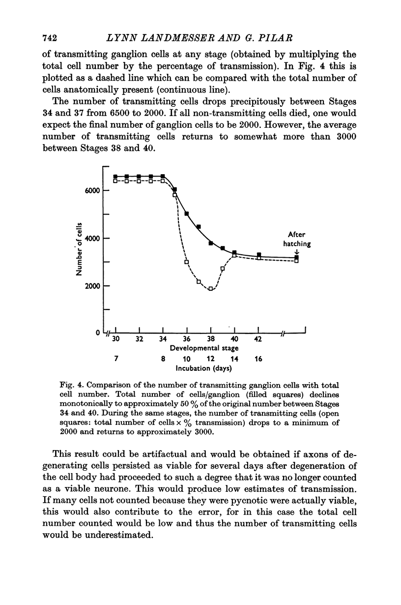
1. During normal embryonic development of the chick ciliary ganglion, cell death over a 4-day period (Stages 35-39) reduces the number of ganglion cells by half, from 6500 to 3200. Both ciliary and choroid populations are affected by approximately the same amount.
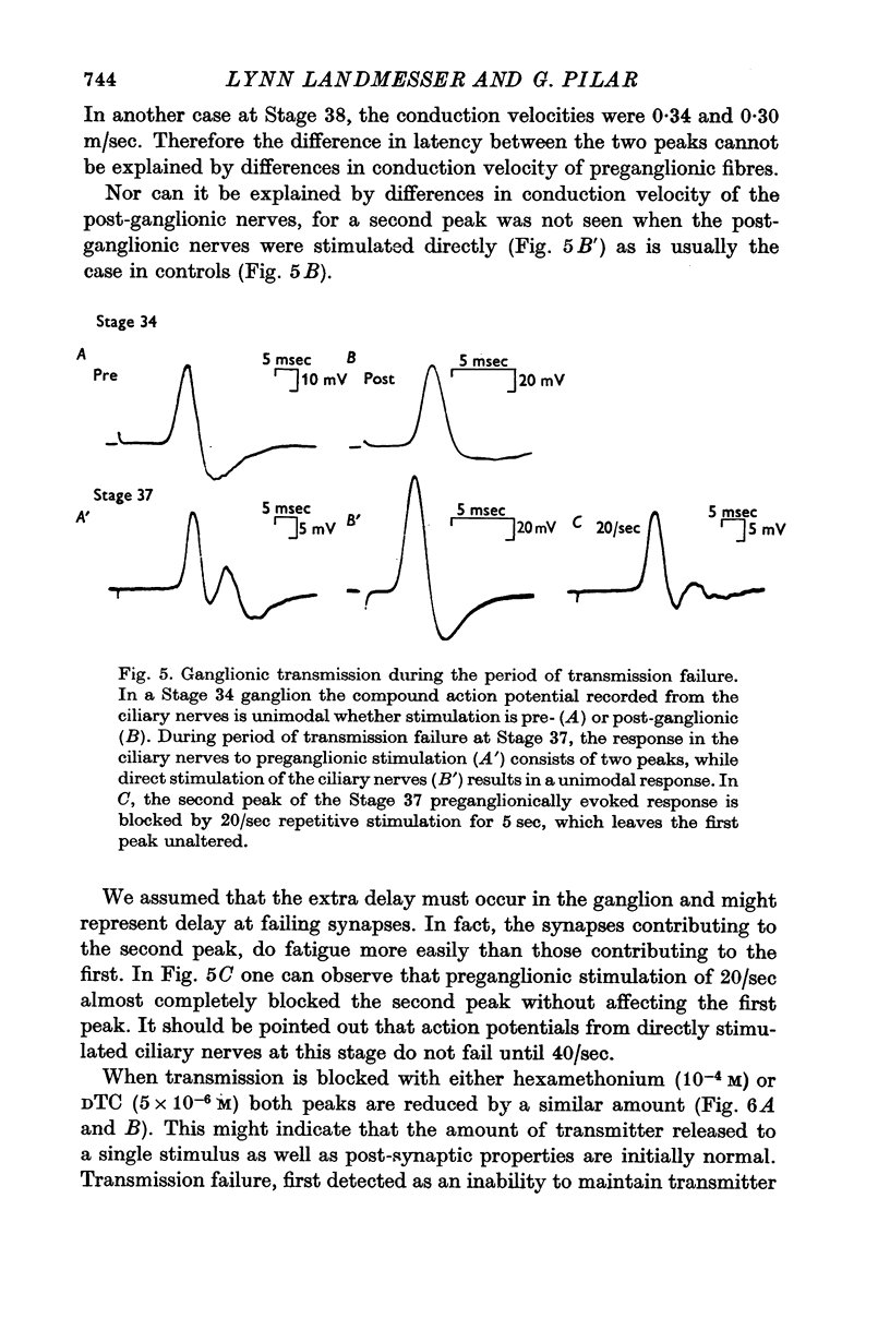
2. Previous to cell death, preganglionic fibres form functional synapses on all ganglion cells, indicating that synapses form on cells which are destined to die.
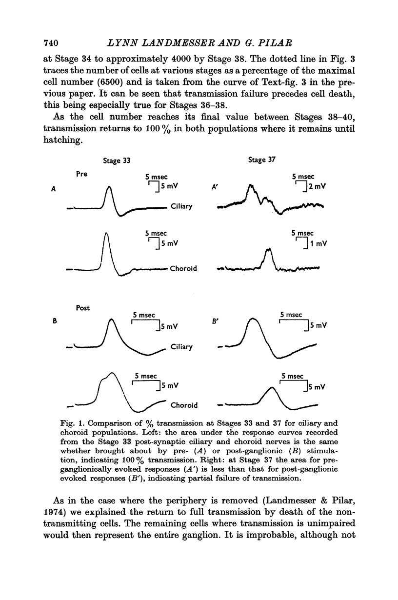
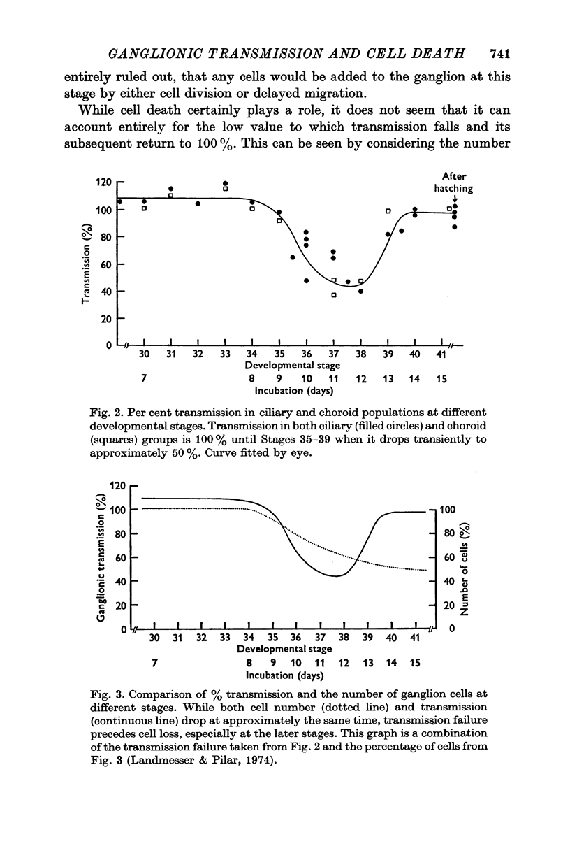
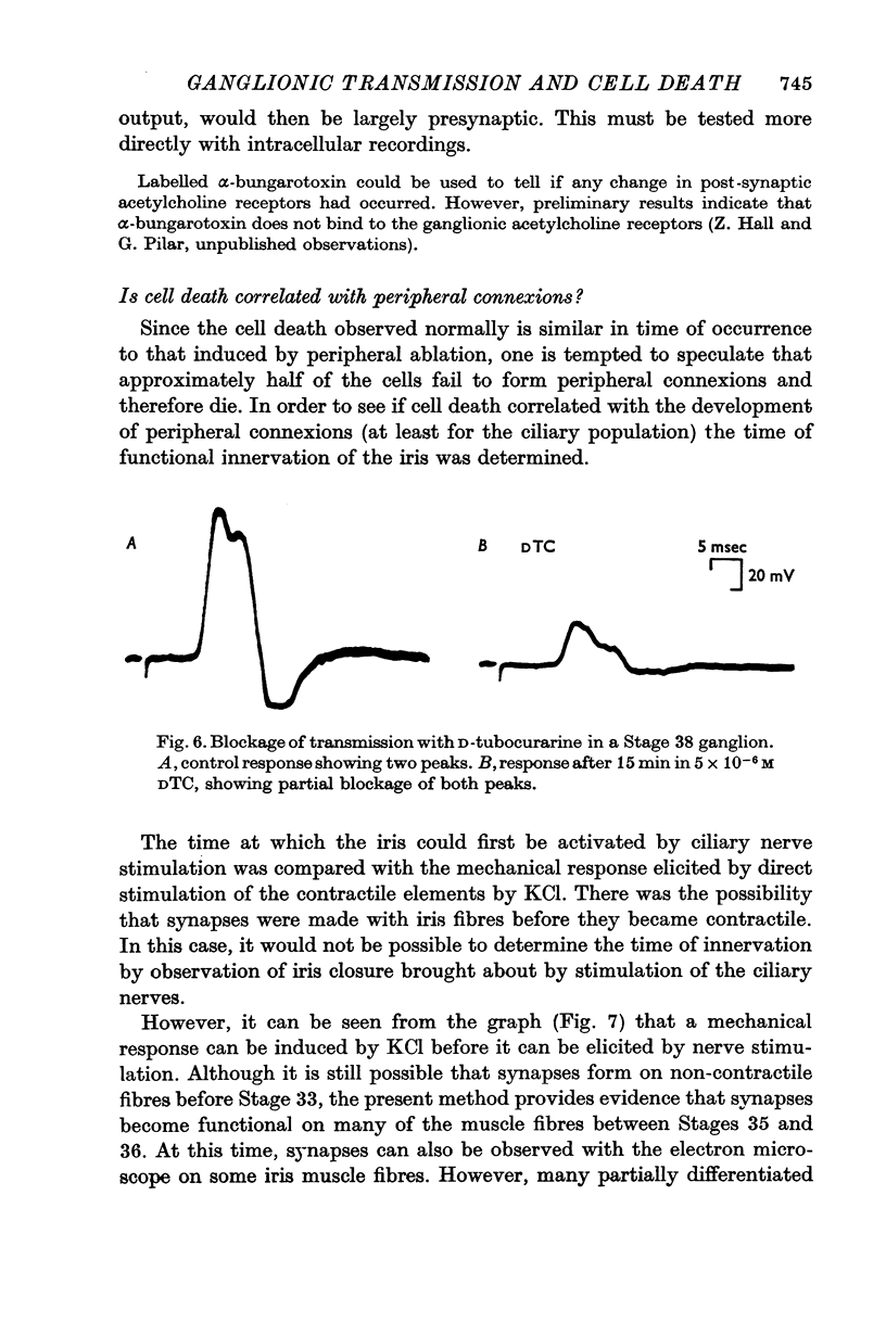
3. Shortly before the period of cell death, there is a failure of transmission in approximately half the cells. Some evidence suggests that transmission failure in at least some of the cells is of preganglionic origin.
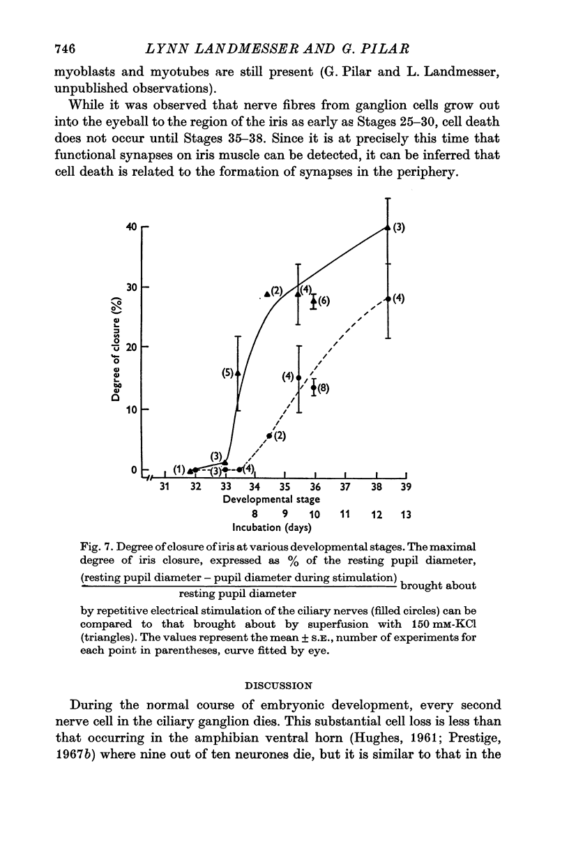
4. Cell death is nearly synchronous with the establishment of peripheral connexions by ganglion cells, at least with respect to the ciliary population which forms functional synapses with iris muscle. This implies that those cells which die do so because they have failed to form adequate peripheral connexions.
5. It is suggested that many of the cells in which transmission has failed die, bringing transmission through the ganglion back to 100%. However, transmission failure appears to be a transitory phenomenon in other cells which survive and probably results from death of their preganglionic elements. Restoration of transmission would then be brought about by the formation of new or more effective synapses by surviving preganglionic fibres.
Full text
PDF

Selected References
These references are in PubMed. This may not be the complete list of references from this article.
- Cowan W. M., Wenger E. Cell loss in the trochlear nucleus of the chick during normal development and after radical extirpation of the optic vesicle. J Exp Zool. 1967 Mar;164(2):267–280. doi: 10.1002/jez.1401640210. [DOI] [PubMed] [Google Scholar]
- Cowan W. M., Wenger E. Degeneration in the nucleus of origin of the preganglionic fibers to the chick ciliary ganglion following early removal of the optic vesicle. J Exp Zool. 1968 May;168(1):105–123. doi: 10.1002/jez.1401680109. [DOI] [PubMed] [Google Scholar]
- Gottlieb D. I., Cowan W. M. Evidence for a temporal factor in the occupation of available synaptic sites during the development of the dentate gyrus. Brain Res. 1972 Jun 22;41(2):452–456. doi: 10.1016/0006-8993(72)90514-8. [DOI] [PubMed] [Google Scholar]
- HAMBURGER V. Regression versus peripheral control of differentiation in motor hypoplasia. Am J Anat. 1958 May;102(3):365–409. doi: 10.1002/aja.1001020303. [DOI] [PubMed] [Google Scholar]
- HUGHES A. Cell degeneration in the larval ventral horn of Xenopus laevis (Daudin). J Embryol Exp Morphol. 1961 Jun;9:269–284. [PubMed] [Google Scholar]
- Hughes A., Egar M. The innervation of the hind limb of Eleutherodactylus martinicensis: further comparison of cell and fiber numbers during development. J Embryol Exp Morphol. 1972 Apr;27(2):389–412. [PubMed] [Google Scholar]
- Landmesser L., Pilar G. Synapse formation during embryogenesis on ganglion cells lacking a periphery. J Physiol. 1974 Sep;241(3):715–736. doi: 10.1113/jphysiol.1974.sp010680. [DOI] [PMC free article] [PubMed] [Google Scholar]
- Landmesser L., Pilar G. The onset and development of transmission in the chick ciliary ganglion. J Physiol. 1972 May;222(3):691–713. doi: 10.1113/jphysiol.1972.sp009822. [DOI] [PMC free article] [PubMed] [Google Scholar]
- Pilar G., Vaughan P. C. Ultrastructure and contractures of the pigeon iris striated muscle. J Physiol. 1971 Dec;219(2):253–266. doi: 10.1113/jphysiol.1971.sp009660. [DOI] [PMC free article] [PubMed] [Google Scholar]
- Prestige M. C. The control of cell number in the lumbar spinal ganglia during the development of Xenopus laevis tadpoles. J Embryol Exp Morphol. 1967 Jun;17(3):453–471. [PubMed] [Google Scholar]