

Abstract
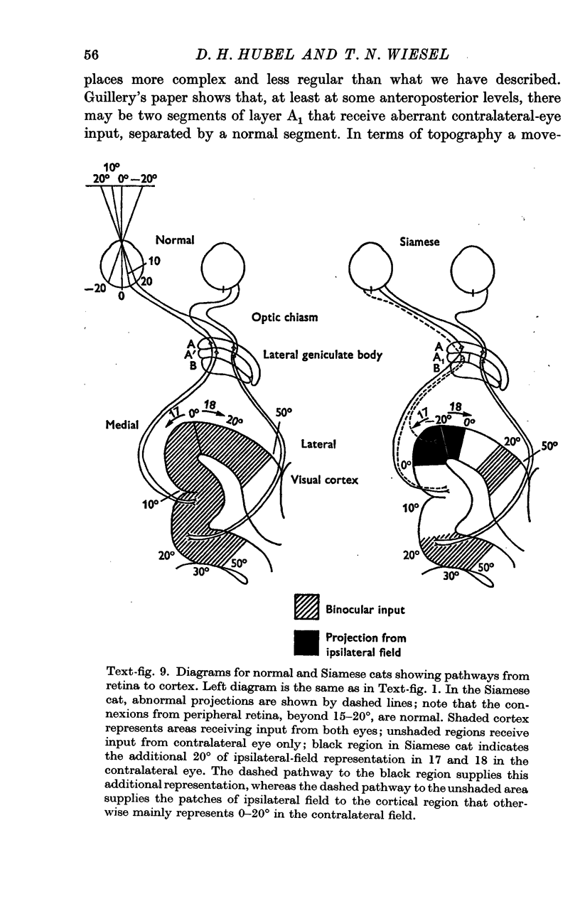
1. Guillery has recently shown that the Siamese cat has a grossly abnormal lateral geniculate body. His anatomical study suggested that certain fibres originating in the temporal retina of each eye cross in the chiasm instead of remaining uncrossed. They thus reach the wrong hemispheres, but in the geniculate they terminate in the regions that the missing fibres from the ipsilateral eye would normally have occupied. The result is that each hemisphere receives an input from parts of the ipsilateral field of vision, this input being entirely from the opposite eye. The purpose of the present work was to study the physiological consequences of this aberrant projection, in the lateral geniculate body and visual cortex.
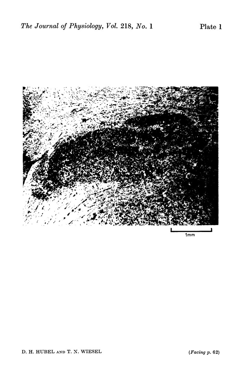
2. Single-cell recordings from the lateral geniculate body confirmed the presence of projections from the ipsilateral visual field of the contralateral eye. The part of layer A1 receiving these projections was arranged so that the receptive fields of the cells were situated at about the same horizontal level and at the same distance from the vertical meridian as the fields of cells in the layers above and below (layers A and B), but were in the ipsilateral visual field instead of the contralateral. They thus occupied a region directly across the mid line from their normal position.
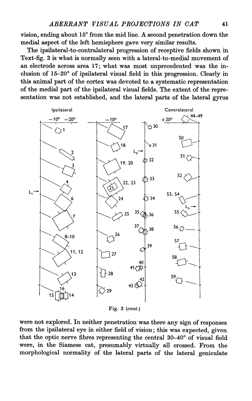
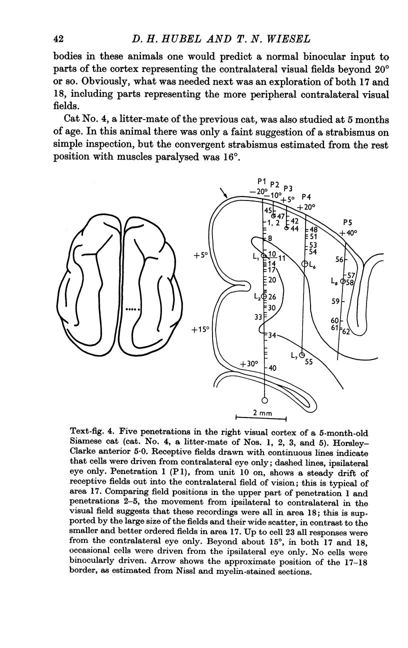
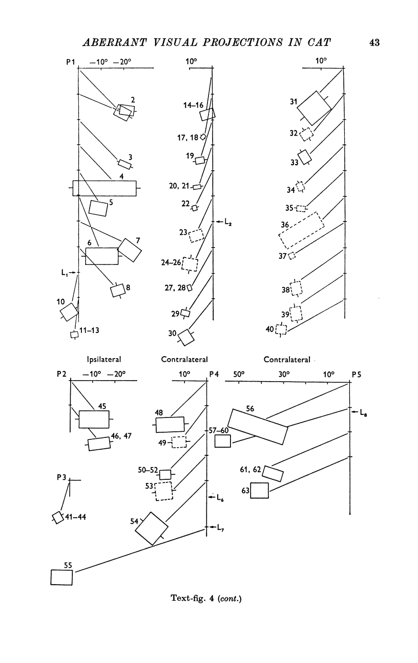
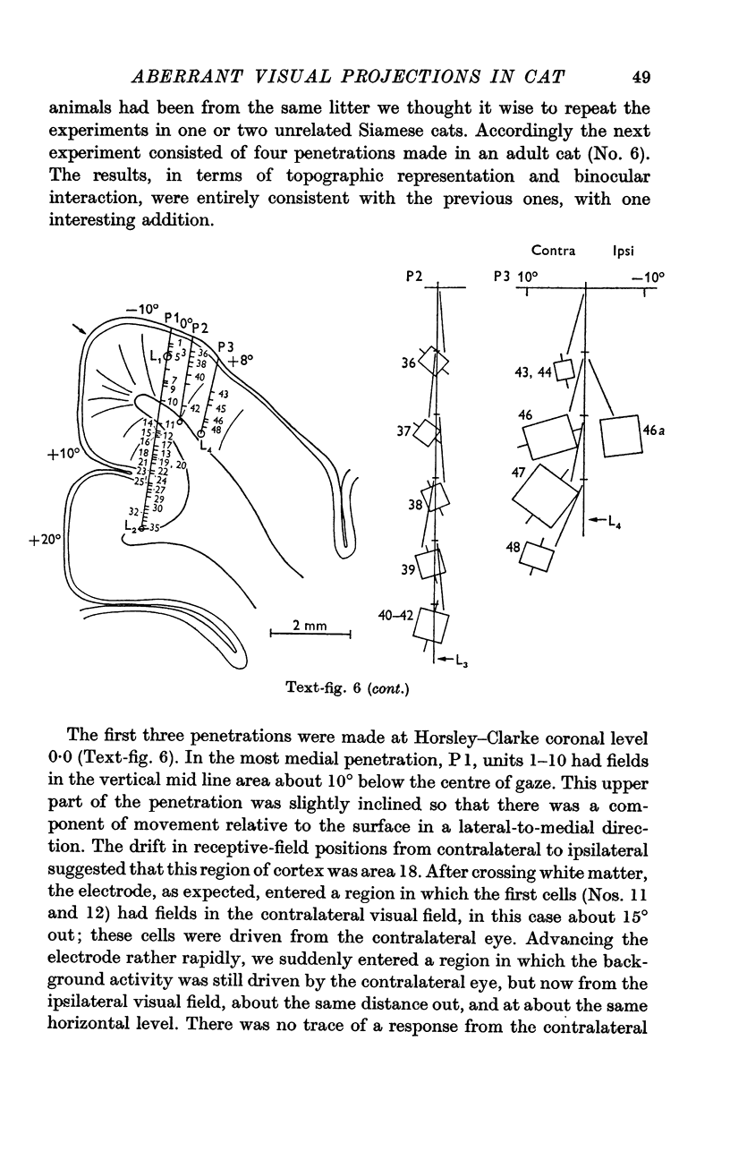
3. In the cortex of all animals studied, we found a systematic representation of part of the ipsilateral visual field, inserted between the usual contralateral representations in areas 17 and 18. When the visual cortex was crossed from medial to lateral the corresponding region of visual field moved from the contralateral periphery to the mid line, and then into the ipsilateral field for 20°. The movement then reversed, with a return to the mid line and a steady progression out into the contralateral field. The entire double representation was, with some possible exceptions, a continuous one. The point of reversal occurred at or near the 17-18 boundary, as judged histologically, and this boundary was in about the same position as in ordinary cats.
4. Cells in the part of the cortex representing the ipsilateral fields had normal receptive fields, simple, complex, or hypercomplex. These fields tended to be larger than those in corresponding parts of the contralateral visual fields. Receptive-field size varied with distance from the area centralis, just as it does in the normal cat, so that cells with the smallest fields, in the area centralis projection, were situated some distance from the 17-18 border.
5. Projections originating from the first 20° from the midvertical in both visual half-fields had their origin entirely in the contralateral eye, as would be expected from the abnormal crossing at the chiasm. Beyond this visual-field region, and out as far as the temporal crescents, there were projections from both eyes, but we found no individual cells with input from the two eyes. The cells were aggregated, with some groups of cells driven by one eye and some by the other.
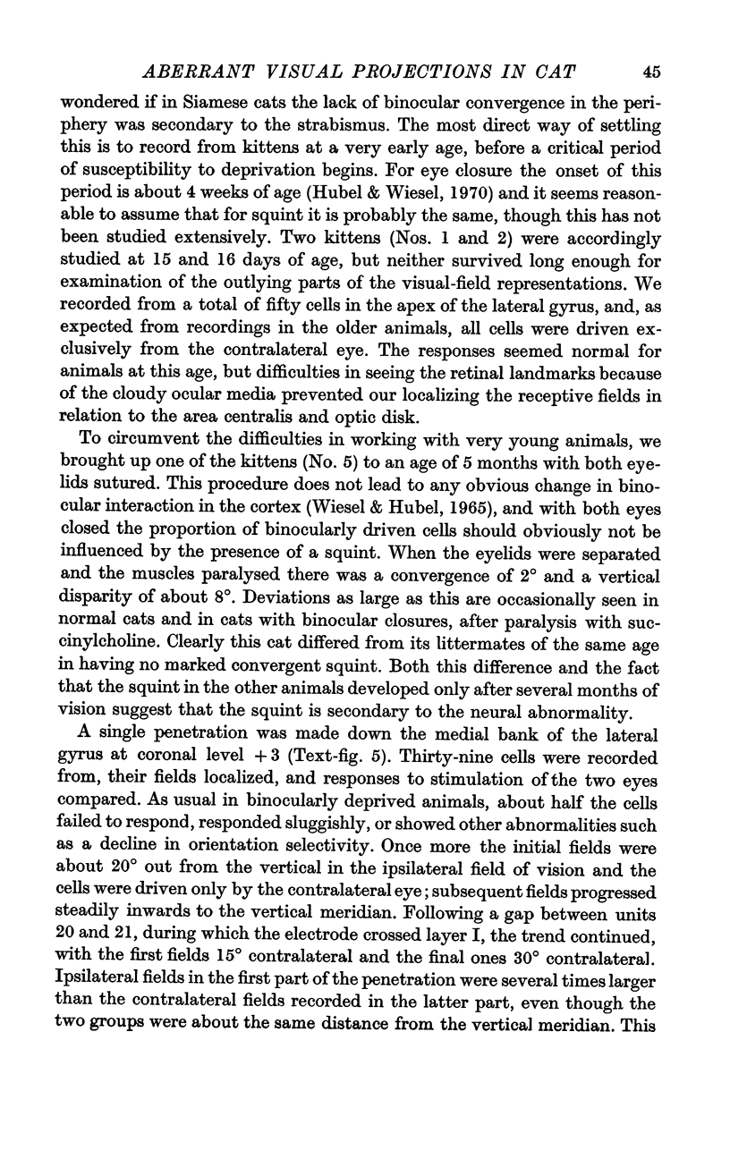
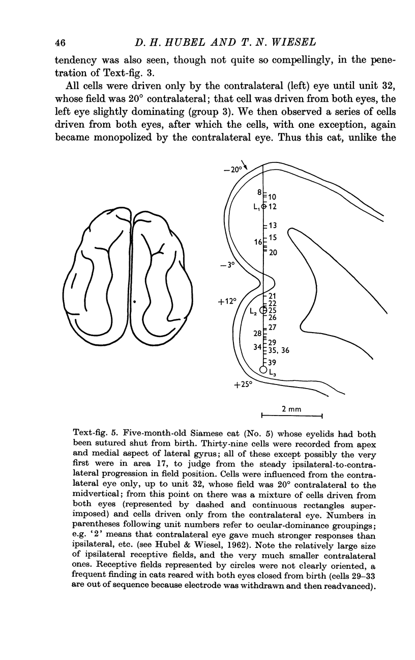
6. From previous work it is known that ordinary cats raised with squint show a decline in the proportion of cells that can be driven binocularly, whereas animals raised with both eyes closed show little or no decline. A Siamese cat raised with both eyes closed had binocular cells in the regions of 17 and 18 subserving the peripheral visual fields, suggesting that the absence of binocular cells seen in the other Siamese cats was indeed secondary to the squint.
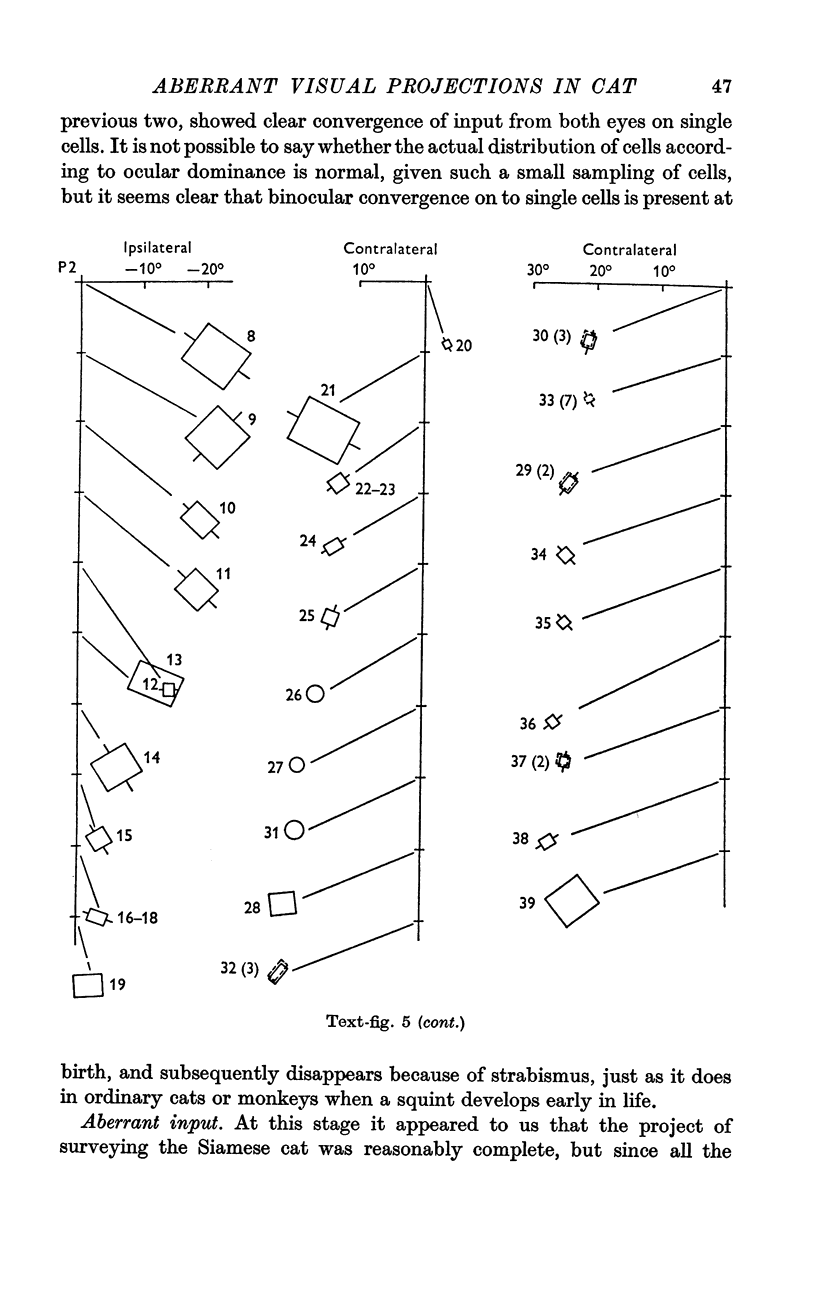
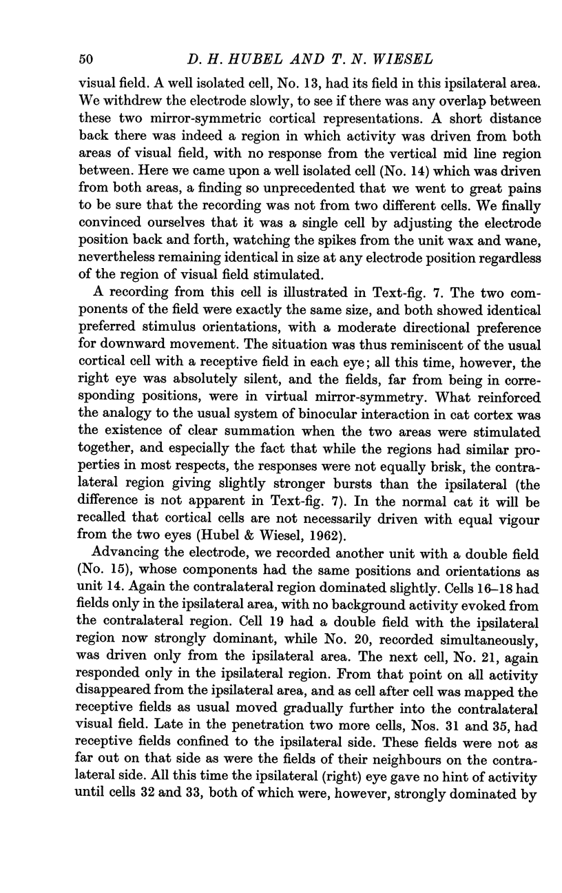
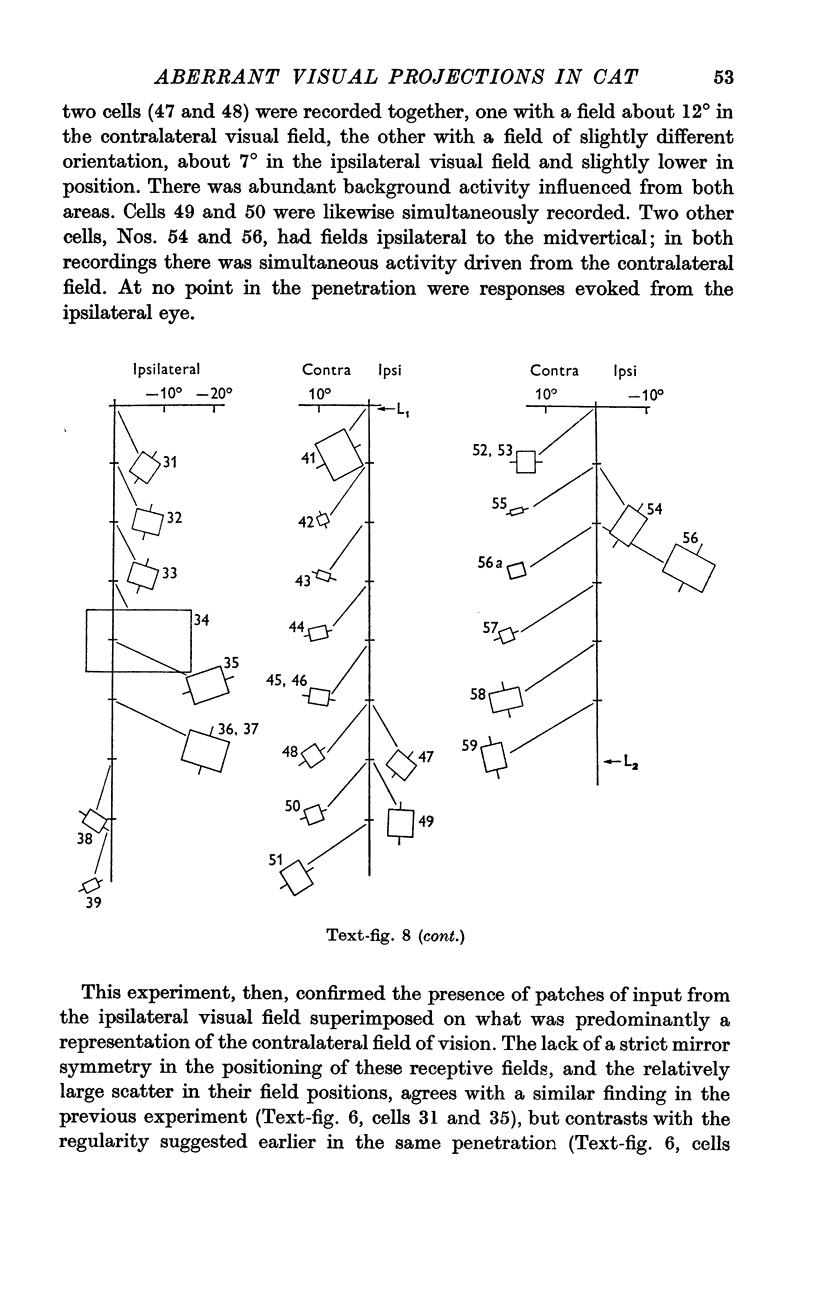
7. In two Siamese cats there were suggestions of an entirely different projection pattern, superimposed upon that described above. In the parts of 17 and 18 otherwise entirely devoted to the contralateral visual field, we observed groups of cells with receptive fields in the ipsilateral field of vision. The electrode would pass from a region where cells were driven from some part of the contralateral visual field, to regions in which they were driven from a part of the ipsilateral field directly opposite, across the vertical mid line. The borders of these groups were not necessarily sharp, for in places there was mixing of the two groups of cells, and a few cells had input from two discrete regions located opposite one another on either side of the vertical mid line. The two receptive-field components of such cells were identical, in terms of orientation, optimum direction of movement, and complexity. Stimulation of the two regions gave a better response than was produced from either one alone, and the relative effectiveness of the two varied from cell to cell. These cells thus behaved in a way strikingly reminiscent of binocular cells in common cats.
8. The apparent existence of two competing mechanisms for determining the projection of visual afferents to the cortex suggests that a number of factors may cooperate in guiding development. There seems, furthermore, not to be a detailed cell-to-cell specificity of geniculocortical connexions, but rather a tendency to topographic order and continuity, with one part of a given area such as 17 able to substitute for another. Whether or not these tentative interpretations are ultimately proved correct, it seems clear that this type of genetic anomaly has potential usefulness for understanding mechanisms of development of the nervous system.
Full text
PDF

Images in this article
Selected References
These references are in PubMed. This may not be the complete list of references from this article.
- BISHOP P. O., KOZAK W., LEVICK W. R., VAKKUR G. J. The determination of the projection of the visual field on to the lateral geniculate nucleus in the cat. J Physiol. 1962 Oct;163:503–539. doi: 10.1113/jphysiol.1962.sp006991. [DOI] [PMC free article] [PubMed] [Google Scholar]
- BISHOP P. O., KOZAK W., VAKKUR G. J. Some quantitative aspects of the cat's eye: axis and plane of reference, visual field co-ordinates and optics. J Physiol. 1962 Oct;163:466–502. doi: 10.1113/jphysiol.1962.sp006990. [DOI] [PMC free article] [PubMed] [Google Scholar]
- GAZE R. M., JACOBSON M., SZEKELY C. The retino-tectal projection in Xenopus with compound eyes. J Physiol. 1963 Mar;165:484–499. doi: 10.1113/jphysiol.1963.sp007072. [DOI] [PMC free article] [PubMed] [Google Scholar]
- Glickstein M., King R. A., Miller J., Berkley M. Cortical projections from the dorsal lateral geniculate nucleus of cats. J Comp Neurol. 1967 May;130(1):55–75. doi: 10.1002/cne.901300104. [DOI] [PubMed] [Google Scholar]
- Guillery RW THE LAMINAR D. a new interpretation. J Comp Neurol. 1970 Mar;138(3):339–366. doi: 10.1002/cne.901380307. [DOI] [PubMed] [Google Scholar]
- Guillery R. W. An abnormal retinogeniculate projection in Siamese cats. Brain Res. 1969 Aug;14(3):739–741. doi: 10.1016/0006-8993(69)90213-3. [DOI] [PubMed] [Google Scholar]
- HUBEL D. H., WIESEL T. N. Integrative action in the cat's lateral geniculate body. J Physiol. 1961 Feb;155:385–398. doi: 10.1113/jphysiol.1961.sp006635. [DOI] [PMC free article] [PubMed] [Google Scholar]
- HUBEL D. H., WIESEL T. N. RECEPTIVE FIELDS AND FUNCTIONAL ARCHITECTURE IN TWO NONSTRIATE VISUAL AREAS (18 AND 19) OF THE CAT. J Neurophysiol. 1965 Mar;28:229–289. doi: 10.1152/jn.1965.28.2.229. [DOI] [PubMed] [Google Scholar]
- HUBEL D. H., WIESEL T. N. RECEPTIVE FIELDS OF CELLS IN STRIATE CORTEX OF VERY YOUNG, VISUALLY INEXPERIENCED KITTENS. J Neurophysiol. 1963 Nov;26:994–1002. doi: 10.1152/jn.1963.26.6.994. [DOI] [PubMed] [Google Scholar]
- HUBEL D. H., WIESEL T. N. Receptive fields, binocular interaction and functional architecture in the cat's visual cortex. J Physiol. 1962 Jan;160:106–154. doi: 10.1113/jphysiol.1962.sp006837. [DOI] [PMC free article] [PubMed] [Google Scholar]
- Hubel D. H., Wiesel T. N. Binocular interaction in striate cortex of kittens reared with artificial squint. J Neurophysiol. 1965 Nov;28(6):1041–1059. doi: 10.1152/jn.1965.28.6.1041. [DOI] [PubMed] [Google Scholar]
- Hubel D. H., Wiesel T. N. The period of susceptibility to the physiological effects of unilateral eye closure in kittens. J Physiol. 1970 Feb;206(2):419–436. doi: 10.1113/jphysiol.1970.sp009022. [DOI] [PMC free article] [PubMed] [Google Scholar]
- OTSUKA R., HASSLER R. [On the structure and segmentation of the cortical center of vision in the cat]. Arch Psychiatr Nervenkr Z Gesamte Neurol Psychiatr. 1962;203:212–234. doi: 10.1007/BF00352744. [DOI] [PubMed] [Google Scholar]
- Wiesel T. N., Hubel D. H. Comparison of the effects of unilateral and bilateral eye closure on cortical unit responses in kittens. J Neurophysiol. 1965 Nov;28(6):1029–1040. doi: 10.1152/jn.1965.28.6.1029. [DOI] [PubMed] [Google Scholar]