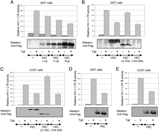
MEDICAL SCIENCES. For the article “Direct interaction of the human I-mfa domain-containing protein, HIC, with HIV-1 Tat results in cytoplasmic sequestration and control of Tat activity,” by Virginie W. Gautier, Noreen Sheehy, Margaret Duffy, Kenichi Hashimoto, and William W. Hall, which appeared in issue 45, November 8, 2005, of Proc. Natl. Acad. Sci. USA (102, 16362–16367; first published October 31, 2005; 10.1073/pnas.0503519102), the authors note that Fig. 3 B and C was mislabeled. “HIC(2–144)” should read “HIC(144–146)” and “HIC(144–146)” should read “HIC(2–144).” The corrected figure and its legend appear below. In addition, the authors note that on page 16364, the fourth sentence of the second full paragraph in the left column, “However, for undetermined reasons HIC(2–144) could not be detected by Western blot,” should read: “However, for undetermined reasons HIC(144–246) could not be detected by Western blot.” These errors do not affect the conclusions of the article.
Fig. 3.
Down-regulation of Tat-mediated transactivation of the HIV-1 LTR by HIC. (A) The 293T cells were transfected with 0.5 μg of reporter pGL3-LTR and 0.05 μg of p-RL-TK in combination with 0.05 μg of pCAGGS-Tat and 0, 2, 4, and 8 μg of pFLAG-HIC. The relative luciferase activity is compared with 100% for Tat transactivation of pGL3-LTR. Error bars indicate the SD of the mean of triplicate samples. (A–E Lower) Western blot shows the corresponding levels of HIC expression. (B) The I-mfa domain is involved in the down-regulation of HIV-1 LTR by HIC. Conditions were as above, but 293T cells were transiently transfected with 0.3 μg of reporter pGL3-LTR and 0.03 μg of p-TK in combination with 0.03 μg of pCAGGS-Tat and 4 μg of pFLAG-HIC, pFLAG-HIC(2–144), or pFLAG-HIC(144–246). (C) As above, but Cos7 cells were transiently transfected with 0.1 μg of reporter pGL3-LTR and 0.03 μgof p-RL-bactin in combination with 0.005 μg of pCAGGS-Tat and 2 μg of pFLAG-HIC, pFLAG-HIC(2–144), or pFLAG-HIC(144–246). (D and E) As described in B and C, but 293T and Cos7 cells were transiently transfected with pGL3-LTRΔ instead of pGL3-LTR.


