

Abstract
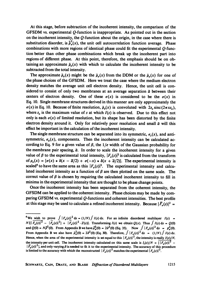
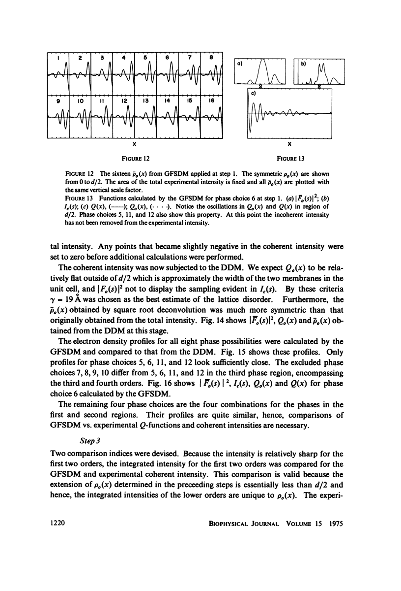
Oriented multilayers containing a membrane pair within the unit cell potentially possess both lattice disorder and substitution disorder. Lattice disorder occurs when there is a lack of long-range order in the lattice spacings produced by a variation in the nearest neighbor distances between unit cells. A simple form of substitution disorder can arise from a variation in the separation of the two membranes within the unit cells in the multilayer. Lattice disorder produces a monotonically increasing width for higher order lamellar "reflections" while simple substitution disorder produces an incoherent intensity underlying the coherent intensity. A generalized Patterson function analysis has been developed for treating lamellar diffraction from lattice disordered multilayers. This analysis allows the identification of the autocorrelation function of the unit cell electron density profile and its subsequent deconvolution to provide the unit cell electron density profile. A recursive procedure has been developed for separating the incoherent intensity from the coherent intensity via a Gaussian probability model of the membrane intra-pair separation. In cases studied so far both disorders can be quantitatively accounted for and eliminated from interfering with the phasing of the coherent intensity or distorting the derived electron density profile. Lamellar X-ray diffraction data from intact retinal rods, using either film or position sensitive detectors, shows severe effects of both forms of disorder which have not been taken into account in past analysis of such data. We have applied our analysis to the data on dark adapted rod outer segments in electrophysiologically intact retinas of Chabre and Cavaggioni (unpublished). An electron density profile is derived at 25 A resolution. The lattice nearest neighbor spacing has a variation of +/- 19 A out of a 295 A repeat. The intra-unit cell membrane pair center to center distance of 88 A varies +/-8 A.
Full text
PDF


Selected References
These references are in PubMed. This may not be the complete list of references from this article.
- Blaurock A. E. Structure of the retinal membrane containing the visual pigments. Adv Exp Med Biol. 1972;24(0):53–63. doi: 10.1007/978-1-4684-8231-7_5. [DOI] [PubMed] [Google Scholar]
- Blaurock A. E., Wilkins M. H. Structure of frog photoreceptor membranes. Nature. 1969 Aug 30;223(5209):906–909. doi: 10.1038/223906a0. [DOI] [PubMed] [Google Scholar]
- Blaurock A. E., Wilkins M. H. Structure of retinal photoreceptor membranes. Nature. 1972 Apr 7;236(5345):313–314. doi: 10.1038/236313a0. [DOI] [PubMed] [Google Scholar]
- Chabre M., Cavaggioni A. Light induced changes in ionic flux in the retinal rod. Nat New Biol. 1973 Jul 25;244(134):118–120. doi: 10.1038/newbio244118a0. [DOI] [PubMed] [Google Scholar]
- Corless J. M. Lamellar structure of bleached and unbleached rod photoreceptor membranes. Nature. 1972 May 26;237(5352):229–231. doi: 10.1038/237229a0. [DOI] [PubMed] [Google Scholar]
- Gras W. J., Worthington C. R. X-ray analysis of retinal photoreceptors. Proc Natl Acad Sci U S A. 1969 Jun;63(2):233–238. doi: 10.1073/pnas.63.2.233. [DOI] [PMC free article] [PubMed] [Google Scholar]
- Lesslauer W., Blasie J. K. Direct determination of the structure of barium stearate multilayers by x-ray diffraction. Biophys J. 1972 Feb;12(2):175–190. doi: 10.1016/S0006-3495(72)86078-8. [DOI] [PMC free article] [PubMed] [Google Scholar]
- Lesslauer W., Cain J., Blasie J. K. On the location of I-anilino-8-naphthalene-sulfonate in lipid model systems. An x-ray diffraction study. Biochim Biophys Acta. 1971 Aug 13;241(2):547–566. doi: 10.1016/0005-2736(71)90054-x. [DOI] [PubMed] [Google Scholar]
- Levine Y. K., Wilkins M. H. Structure of oriented lipid bilayers. Nat New Biol. 1971 Mar 17;230(11):69–72. doi: 10.1038/newbio230069a0. [DOI] [PubMed] [Google Scholar]
- McIntosh T. J., Worthington C. R. Direct determination of the lamellar structure of peripheral nerve myelin at low resolution (17 A). Biophys J. 1974 May;14(5):363–386. doi: 10.1016/S0006-3495(74)85922-9. [DOI] [PMC free article] [PubMed] [Google Scholar]
- Moody M. F. Letter: Structure determination of membranes in swollen lamellar systems. Biophys J. 1974 Sep;14(9):697–702. doi: 10.1016/S0006-3495(74)85945-X. [DOI] [PMC free article] [PubMed] [Google Scholar]
- Pape E. H. X-ray small angle scattering. A new deconvolution method for evaluating electron density distributions from small angle scattering diagrams. Biophys J. 1974 Apr;14(4):284–294. doi: 10.1016/S0006-3495(74)85916-3. [DOI] [PMC free article] [PubMed] [Google Scholar]
- Worthington C. R., King G. I., McIntosh T. J. Direct structure determination of multilayered membrane-type systems which contain fluid layers. Biophys J. 1973 May;13(5):480–494. doi: 10.1016/S0006-3495(73)86001-1. [DOI] [PMC free article] [PubMed] [Google Scholar]
- Worthington C. R., Liu S. C. Structure of sarcoplasmic reticulum membranes at low resolution (17A). Arch Biochem Biophys. 1973 Aug;157(2):573–579. doi: 10.1016/0003-9861(73)90676-0. [DOI] [PubMed] [Google Scholar]
- Worthington C. R. Structure of photoreceptor membranes. Annu Rev Biophys Bioeng. 1974;3(0):53–80. doi: 10.1146/annurev.bb.03.060174.000413. [DOI] [PubMed] [Google Scholar]
- Worthington C. R. X-ray analysis of retinal photoreceptor structure. Exp Eye Res. 1973 Dec 24;17(6):487–501. doi: 10.1016/0014-4835(73)90078-x. [DOI] [PubMed] [Google Scholar]
- Zaccai G., Blasie J. K., Schoenborn B. P. Neutron diffraction studies on the location of water in lecithin bilayer model membranes. Proc Natl Acad Sci U S A. 1975 Jan;72(1):376–380. doi: 10.1073/pnas.72.1.376. [DOI] [PMC free article] [PubMed] [Google Scholar]