Abstract
Targeted gene disruption studies have established that the c-Jun NH2-terminal kinase (JNK) signaling pathway is required for stress-induced release of mitochondrial cytochrome c and apoptosis. Here we demonstrate that activated JNK is sufficient to induce rapid cytochrome c release and apoptosis. However, activated JNK fails to cause death in cells deficient of members of the Bax subfamily of proapoptotic Bcl2-related proteins. Furthermore, exposure to stress fails to activate Bax, cause cytochrome c release, and induce death in JNK-deficient cells. These data demonstrate that proapoptotic members of the Bax protein subfamily are essential for JNK-dependent apoptosis.
The c-Jun NH2-terminal kinase (JNK) group of mitogen-activated protein (MAP) kinases is activated when cells are exposed to environmental stress. Ten members of the JNK family are created by the alternative splicing of transcripts derived from the Jnk1, Jnk2, and Jnk3 genes (5). These isoforms differ in their substrate specificity in vitro (17). Nevertheless, each JNK isoform can phosphorylate members of the AP-1 group of transcription factors (c-Jun, JunB, and JunD) and the AP-1-related transcription factor ATF2 (5). JNK phosphorylates these transcription factors within the NH2-terminal activation domain and increases transcription activity. Indeed, it is established that JNK regulates AP-1 transcription activity in vivo and it is likely that increased AP-1 activity mediates, in part, the effects of the JNK signaling pathway (5).
The physiological function of the JNK signaling pathway is unclear. However, JNK is required for embryonic viability (24) and recent studies have demonstrated roles for JNK in multiple physiological processes (60). Thus, JNK can promote cell survival and JNK is also implicated in some forms of apoptotic cell death (5). JNK is essential for stress-induced apoptosis, including neurotrophic factor withdrawal-induced death (10, 63). Studies of Jnk gene disruption in mice have confirmed that JNK contributes to apoptotic responses. JNK3 is essential for apoptosis of hippocampal neurons following exposure to excitotoxic stress (65). JNK1 and JNK2 are required for thymocyte apoptosis in response to ligation of the T-cell receptor in vivo (41, 44, 46). Furthermore, compound disruptions of both the Jnk1 and Jnk2 genes causes reduced apoptosis in the developing hindbrain neuroepithelium (24, 45) and primary fibroblasts isolated from these embryos are resistant to stress-induced apoptosis (52). Together, these data strongly support a role for JNK in the apoptotic response.
The mechanism that accounts for the proapoptotic actions of JNK has not been elucidated. Recent studies have focused on two different possible mechanisms (5). First, JNK may cause cell death by regulating the expression of death receptor ligands (14). For example, a JNK-dependent element in the Fas ligand promoter that binds c-Jun and ATF2 has been identified (15). It is therefore possible that JNK may induce apoptosis by an autocrine or a juxtacrine mechanism involving the expression of death receptor ligands. A second possible mechanism was identified in biochemical studies of primary Jnk1−/− Jnk2−/− fibroblasts (52). These cells have severe defects in the mitochondrial apoptotic pathway, including the release of cytochrome c. In contrast, these JNK-deficient cells exhibited no defect in the apoptotic response to death receptor signaling. This analysis indicated that JNK may contribute to the apoptotic response by regulating the intrinsic cell death pathway involving the mitochondria (52).
The purpose of the study reported here was to examine the role of JNK in stress-induced apoptosis. Gene knockout studies indicate that JNK is required for stress-induced apoptosis. However, it is unclear whether activated JNK is sufficient for an apoptotic response. Our approach to this question was to examine the effect of expression of constitutively activated JNK in cultured cells. The activated JNK caused rapid apoptosis. Analysis of fibroblasts from knockout mice demonstrated that Bax and the related protein Bak were required for JNK-stimulated apoptosis. Furthermore, Bax was not activated by exposure of JNK-deficient fibroblasts to environmental stress. These data demonstrate that proapoptotic members of the Bcl2 family are essential for JNK-dependent apoptosis.
MATERIALS AND METHODS
Plasmid construction.
Epitope-tagged JNK and MKK7 expression plasmids in pCDNA3 (Invitrogen Inc.) have been described previously (7, 17, 53, 54). The fusion protein expression vectors for MKK7-JNK1 and MKK7-JNK2 were constructed by in-frame ligation of JNK1α1 and JNK2α2 (T4 polymerase blunt-ended NruI-HindIII restriction fragments) into the pCDNA3-Flag-MKKβ2 plasmid at the Bsu36 restriction site (blunt-ended with T4 polymerase). The MKK7-JNK3 vector was constructed by replacing JNK1α1 in the MKK7-JNK1 vector with JNK3α2 (BamHI-SmaI restriction fragments). These fusion proteins encode an NH2-terminal epitope tag (Flag), residues 1 to 443 of MKK7β2 (deletion of 9 COOH-terminal amino acids), a short linker (His-Glu-Ile-Ser-Arg-Arg-Pro-Ser-Tyr-Arg-Lys-Gly-Ser), and the complete sequence of JNK1α1, JNK2α2, or JNK3α2. The fusion protein expression vectors MKK7(K>A)-JNK1 and MKK7-JNK1(APF) were constructed by site-directed mutagenesis to create kinase-negative MKK7 (Lys149 replaced with Ala) and phosphorylation-negative JNK (Thr180-Pro-Tyr182 replaced with Ala-Pro-Phe). The fusion protein expression vector for JNK1-MKK7 was created by in-frame ligation of an MKK7β2 cDNA (T4 polymerase blunt-ended BamHI-HindIII restriction fragment) into the pCDNA3-Flag-JNK1α2 plasmid at the XbaI restriction site (blunt-ended with T4 polymerase). This fusion protein encodes an NH2-terminal epitope tag (Flag), residues 1 to 415 of JNK1α2 (deletion of 11 COOH-terminal amino acids), a short linker (Gly-Ser-Pro-Gly-Ile-Ser-Gly-Gly-Gly-Gly-Gly-Ile-Leu), and the complete sequence of MKK7β2. The plasmid expression vectors for Akt were obtained from Upstate Biotechnologies, Inc. Bcl2 and tBid expression vectors (25) were provided by J. Yuan. The Bax cDNA (EcoRI fragment) and the Bak cDNA (BamHI and EcoRI fragment) were subcloned into the polylinker of pCDNA3 (Invitrogen) to create mammalian expression vectors for Bax and Bak. The activated MEK1 expression vector pCMV-HA-ΔN3-MEK1 (S218E, S222D) (32) was provided by N. Ahn. The activated MKK6 expression vector pCDNA3-Flag-MKK6 (S207E, T211E) has been described previously (40).
Cell culture.
CHO and COS-7 cells were cultured in Ham's F-12 medium with 5% fetal bovine serum (FBS) and Dulbecco's modified Eagle's medium with 5% FBS, respectively. Mouse embryonic fibroblasts were cultured in Dulbecco's modified Eagle's medium with 10% FBS and 0.1 mM mercaptoethanol. Bax−/− Bak−/− and Jnk1−/− Jnk2−/− murine fibroblasts have been described (30, 52). Bid−/− fibroblasts were provided by S. Korsmeyer (67). The culture medium was supplemented with 2 mM l-glutamine, 100 U of penicillin per ml, and 100 μg of streptomycin per ml. Transfection assays were performed using Lipofectamine in Opti-MEM I medium (Invitrogen Inc.).
Cell death assays.
Cell survival was examined by fluorescence microscopy in transfection assays using a green fluorescent protein (GFP) reporter plasmid (pEGFP; Clontech) and a luciferase reporter plasmid (pCDNA3-luciferase [47]) and by crystal violet staining (52). DNA fragmentation was investigated using the Cell Death Detection ELISAPlus kit (Roche).
Immunofluorescence analysis.
Mouse embryonic fibroblasts and CHO cells were cultured on glass coverslips. The cells were washed three times with ice-cold phosphate-buffered saline (PBS), fixed with 4% paraformaldehyde in PBS (10 min), permeabilized with 0.2% Triton X-100 in PBS (5 min), and incubated (15 min) in blocking buffer (PBS with 3% [wt/vol] bovine serum albumin and 0.5% [wt/vol] Tween 20). The coverslips were incubated at room temperature with primary antibody (anti-cytochrome c or anti-Bax; Pharmingen) in blocking buffer (30 min), washed three times, and incubated (30 min) with Texas Red-conjugated secondary antibody (Jackson ImmunoResearch) at room temperature. Nuclei were stained with 4,6-diamidino-2-phenylindole (DAPI; Sigma; 1:10,000 dilution). The coverslips were mounted on slides by use of Vectashield (Vector Labs Inc.) mounting medium. Fluorescence images were examined by conventional microscopy (Zeiss Axioplan).
Microinjection.
CHO cells were cultured on gridded coverslips for 2 days and microinjected with the indicated plasmids together with dog immunoglobulin G (IgG) or fluorescein-conjugated dextran. The number of apoptotic cells, as determined by cell blebbing and detachment from coverslips, was scored every 15 min after injection. Apoptosis in four independent experiments (at least 150 injected cells) was calculated as the mean percentage ± standard deviation (SD) of total injected cells marked by fluorescein-conjugated dextran. Cytochrome c release in cells injected with dog IgG was examined after fixing in 3.7% formaldehyde and staining with primary antibody (mouse anti-cytochrome c; Pharmingen) and two secondary antibodies (rhodamine-conjugated goat anti-mouse IgG and fluorescein-conjugated goat anti-dog IgG; Jackson Immunoresearch).
Biochemical analysis.
Cells were lysed in Triton X-100 lysis buffer (TLB) containing 20 mM Tris (pH 7.4), 1% Triton X-100, 10% glycerol, 137 mM NaCl, 2 mM EDTA, 25 mM β-glycerophosphate, 1 mM sodium orthovanadate, 1 mM phenylmethylsulfonyl fluoride, and 10 μg of aprotinin and leupeptin per ml. Immunoblot analysis was performed by probing using antibodies to Flag (Sigma), hemagglutinin (HA) (Roche), Akt and phospho-Ser473-Akt (Upstate Biotechnologies), p38 MAP kinase and phospho-p38 MAP kinase (Promega), ERK and phospho-ERK (Promega), phospho-JNK (Promega), JNK1/2 (Pharmingen), JNK3 (Upstate Biotechnologies), MKK7 (Zymed), cytochrome c (Pharmingen), Bcl2 (Santa Cruz Biotechnology), Bax (Pharmingen), caspase-3 (Santa Cruz Biotechnology), and activated caspase-3 (48). Immune complexes were detected by chemiluminescence (NEN Life Science Products). Protein kinase assays were performed with TLB cell lysates. The protein kinases were immunoprecipitated using 1 μg of anti-Flag M2 antibody prebound to protein G-Sepharose beads (Sigma). The immunocomplexes were washed three times with TLB and twice with kinase buffer (25 mM HEPES [pH 7.4], 25 mM β-glycerophosphate, 25 mM MgCl2, 2 mM dithiothreitol, 0.1 mM sodium orthovanadate). The kinase reaction was carried out by incubation at 30°C (20 min) in a final volume of 25 μl of kinase buffer containing 1 μg of glutathione S-transferase (GST)-c-Jun and 50 μM [γ-32P]ATP (10 μCi/nmol). The reaction was terminated by adding Laemmli sample buffer. The products were resolved on a sodium dodecyl sulfate-12% polyacrylamide gel and the phosphorylation of GST-c-Jun was detected by autoradiography and quantitated using a PhosphorImager (Molecular Dynamics).
Luciferase reporter gene assays.
Transfection assays using GAL4-ATF2 fusion proteins were performed using CHO cells (18). CHO cells in 35-mm-diameter dishes were transfected with 0.125 μg of pGal4-ATF2(1-109), 0.25 μg of the luciferase reporter plasmid pG5E1bLuc, and 0.125 μg of pRSV β-galactosidase together with 0.4 μg of the indicated plasmids. The cells were lysed at 40 h posttransfection in 1% (vol/vol) Triton X-100, 25 mM glycylglycine (pH 7.8), 15 mM MgSO4, 4 mM EGTA, and 1 mM dithiothreitol. Luciferase and β-galactosidase activities in the cell lysates were measured. Relative luciferase activity was calculated as the luciferase/β-galactosidase activity ratio. Assays of AP-1 activity were performed using the luciferase reporter plasmid 4xTRE (0.5 μg) instead of the pGal4-ATF2 and pG5E1bLuc plasmids.
Statistical analysis.
Mean, SD, and Student's t test calculations were performed using Microsoft Excel 98.
RESULTS
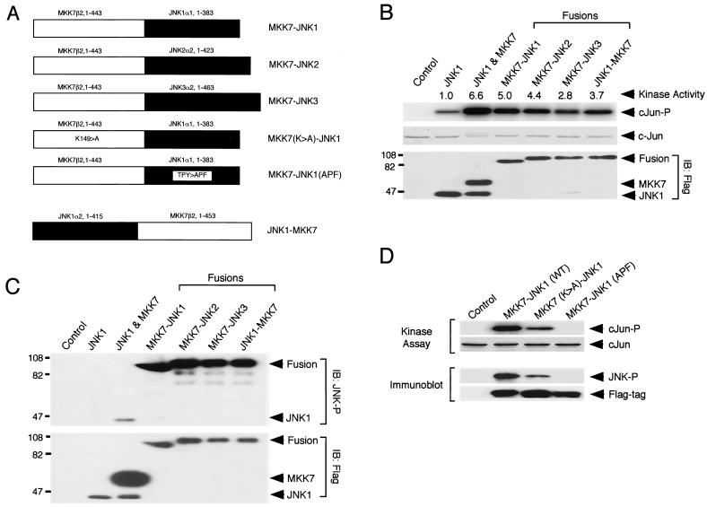
Point mutations that cause JNK activation have not been identified (5). For example, the replacement of the critical phosphorylation sites that cause JNK activation with acidic residues does not cause increased JNK activity. Nevertheless, activated JNK can be constructed using a fusion protein approach that was first reported by Cobb and colleagues (42, 69). This approach involves the fusion of a MAP kinase kinase with a MAP kinase. Thus, fusion of MEK1 to ERK2 causes constitutive ERK2 activity (42). Gene disruption experiments demonstrate that two MAP kinase kinases (MKK4 and MKK7) cause JNK activation (51). MKK4 can activate both JNK and p38 MAP kinase (8, 29). In contrast, MKK7 is a specific activator of JNK (20, 37, 53, 62, 66). This specificity of MKK7 for JNK activation suggests that the fusion of MKK7 to JNK may cause constitutive JNK activation. We therefore prepared a series of proteins in which different JNK isoforms (JNK1α1, JNK2α2, and JNK3α2) were fused to one MKK7 isoform (MKK7β2). We also prepared two mutant MKK7-JNK fusion proteins with point mutations that catalytically inactivate MKK7 (active site Lys149 replaced with Ala) or JNK1 (tripeptide dual-phosphorylation motif Thr-Pro-Tyr replaced with Ala-Pro-Phe). In addition, we constructed a fusion protein with JNK1 and MKK7 in the reverse orientation. These fusion proteins are illustrated schematically in Fig. 1A.
FIG. 1.
Characterization of MKK7-JNK fusion proteins. (A) Plasmid expression vectors were constructed to express fusion proteins with an NH2-terminal epitope tag (Flag). The structure of the fusion proteins is schematically illustrated. The MKK7-JNK fusion proteins contain residues 1 to 443 of MKK7β2 fused to JNK1α1, JNK2α2, and JNK3α2. The JNK1-MKK7 fusion protein contains residues 1 to 415 of JNK1α2 fused to MKK7β2. Point mutations were used to create kinase-negative MKK7 (Lys149 replaced with Ala) and phosphorylation-negative JNK (Thr180-Pro-Tyr182 replaced with Ala-Pro-Phe). (B) Measurement of JNK protein kinase activity. JNK1, JNK1 plus MKK7, and the MKK7-JNK fusion proteins were expressed in CHO cells and were detected by immunoblot (IB) analysis using an antibody to the Flag epitope tag (lower panel). Protein kinase assays were performed using an immune complex assay, with c-Jun and [γ-32P]ATP as the substrates. The reaction products were examined by sodium dodecyl sulfate-polyacrylamide gel electrophoresis. The c-Jun protein was detected by Coomassie staining (middle panel) and phosphorylated c-Jun was detected by autoradiography (upper panel). The relative kinase activity was quantitated by phosphorimager analysis. The numbers on the left correspond to the migration of molecular mass standards (in kilodaltons). (C) The activation state of JNK was examined by immunoblot analysis using an antibody to phospho-JNK. The expression of JNK1, MKK7, and the MKK7-JNK fusion proteins in CHO cell lysates was examined by immunoblot analysis using an antibody to the Flag epitope tag (lower panel) and to phospho-JNK (upper panel). (D) Comparison of the wild-type MKK7-JNK1 fusion protein with the catalytically inactive mutant proteins MKK7(K>A)-JNK1 and MKK7-JNK1(APF). Upper panel, the phosphorylation of c-Jun was examined in an in vitro Flag-immune complex protein kinase assay. c-Jun was detected by staining with Coomassie blue. Phosphorylated c-Jun was detected by autoradiography. Lower panel, the activation state of JNK was examined by immunoblot analysis by probing with antibodies to the epitope tag (Flag) and phospho-JNK.
To test whether the MKK7-JNK fusion proteins exhibited constitutive JNK activity, we expressed the fusion proteins in CHO cells (Fig. 1B). Immunoblot analysis demonstrated that the amount of JNK, MKK7, and MKK7-JNK fusion proteins was similar. Immune complex protein kinase assays using the substrate c-Jun indicated that cotransfection of JNK1 together with MKK7 caused markedly increased JNK activity compared with assays with JNK1 alone. Increased kinase activity was also observed in assays using each of the fusion proteins, including MKK7-JNK1 and JNK1-MKK7 (Fig. 1B). These data indicate that the MKK7-JNK fusion proteins exhibit constitutive JNK activity. However, we found no evidence that these fusion proteins caused activation of endogenous JNK in transfection assays (data not shown). To confirm whether JNK in the fusion proteins was activated, we performed immunoblot analysis with an antibody that detects JNK phosphorylated on the tripeptide dual-phosphorylation motif Thr-Pro-Tyr. Each of the fusion proteins was found to contain activated JNK (Fig. 1C). In contrast, the mutated fusion protein MKK7-JNK1(APF) was found to lack JNK activity, while the mutated fusion protein MKK7(K>A)-JNK1 exhibited low basal JNK activity (Fig. 1D).
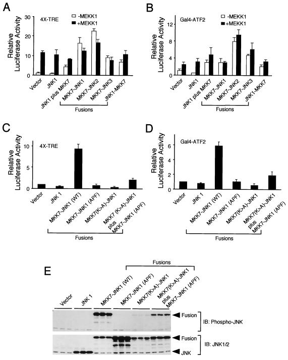
The JNK signaling pathway can regulate AP-1 transcription activity. We therefore tested the effect of the MKK7-JNK fusion proteins in cotransfection assays using an AP-1 reporter plasmid (Fig. 2A). Expression of JNK1 caused no significant change in AP-1 activity, as expected. In contrast, expression of MEKK1, a strong activator of the JNK pathway, caused markedly increased AP-1 transcription activity. Similarly, expression of the MKK7-JNK fusion proteins caused increased AP-1 activity. Increased activity was detected in experiments using JNK1, JNK2, and JNK3 fusion proteins and in experiments using both MKK7-JNK1 and JNK1-MKK7 fusion proteins. Interestingly, MEKK1 was unable to increase AP-1 activity in cells expressing the MKK7-JNK fusion proteins. Together, these data demonstrate that the constitutively activated JNK fusion proteins are functional in vivo and cause increased AP-1 activity (Fig. 2A). Studies of the transcription factor ATF2, which is phosphorylated and activated by JNK (18, 31), confirmed this conclusion (Fig. 2B).
FIG. 2.
The MKK7-JNK fusion proteins increase AP-1 transcription activity. (A, C) AP-1 transcription activity was examined in a transfection assay using a reporter plasmid containing four AP-1 binding sites. CHO cells were cotransfected with the reporter plasmid p4XTRE-luciferase, a β-galactosidase expression vector, and the indicated plasmid expression vectors. The normalized luciferase activity detected in the cell lysates is presented. The data shown are the means ± SD for three independent experiments. (B, D) ATF2 transcription activity was monitored in transfection assays using a GAL4-ATF2 fusion protein. CHO cells were cotransfected with Gal4-ATF2(1-109), reporter plasmid pG5E1bLuc, a β-galactosidase expression vector, and the indicated plasmids. The normalized luciferase activity detected in the cell lysates is presented. The data shown are the means ± SD for three independent experiments. (E) Immunoblot analysis of transfected CHO cells. Triplicate samples from one experiment (panel C) were probed with antibody to JNK1/2 (lower panel) and phospho-JNK (upper panel).
To examine whether the effect of the MKK7-JNK fusion proteins on transcription activity depends upon increased JNK activity, we examined the effect of catalytically inactive fusion proteins on AP-1 activity in luciferase reporter assays (Fig. 2C) and ATF2 luciferase reporter assays (Fig. 2D). These data demonstrated that no increased transcription activity was detected in experiments using inactivated JNK (replacement of the tripeptide dual-phosphorylation motif Thr-Pro-Tyr with Ala-Pro-Phe) in the fusion protein MKK7-JNK1(APF). Similarly, no increased transcription activity was detected in experiments using inactive MKK7 (replacement of active site Lys149 with Ala) in the fusion protein MKK7(K>A)-JNK1. Thus, catalytically active MKK7 and JNK are required for the fusion proteins to increase AP-1 activity.
A small increase in AP-1 and ATF2 transcription activity was observed when the two inactive fusion proteins were coexpressed [MKK7-JNK1(APF) plus MKK7(K>A)-JNK1]. This may be caused by functional complementation between the two mutated fusion proteins. To test this hypothesis, we examined JNK activation by immunoblot analysis using an antibody that binds dually phosphorylated JNK. As expected, the MKK7-JNK1(APF) fusion proteins did not contain activated JNK (Fig. 2E), but a low level of activated JNK was detected in experiments using the MKK7(K>A)-JNK1 fusion protein. In contrast, activated JNK was detected when the two inactive fusion proteins were coexpressed. These data indicate partial complementation between the defective functions of these inactive fusion proteins. The simplest hypothesis is that MKK7 in the MKK7-JNK1(APF) fusion protein can phosphorylate and activate JNK in the MKK7(K>A)-JNK1 fusion protein when these two proteins are coexpressed.
Activated JNK causes cell death.
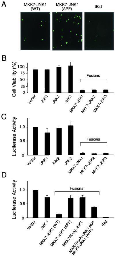
The JNK signal transduction pathway can contribute to stress-induced apoptosis (5). However, it is unclear whether activated JNK is sufficient to induce an apoptotic response. We therefore tested the effect of activated JNK on the survival of cultured cells (Fig. 3A). In initial experiments, we cotransfected GFP together with the inactive fusion protein MKK7-JNK1(APF). Fluorescence microscopy demonstrated numerous transfected cells expressing GFP. In contrast, a marked reduction in GFP-positive cells was detected in cultures transfected with the activated fusion protein MKK7-JNK1 (Fig. 3A). As a positive control, we demonstrated that a strong inducer of apoptosis, truncated Bid (tBid), also caused a marked reduction in the number of GFP-positive cells. These data indicate that activated JNK can induce cell death. To confirm this observation, we measured viable cells by crystal violet staining of the adherent population (Fig. 3B). A marked reduction in cell viability was detected in cultures expressing activated JNK1, JNK2, or JNK3 fusion proteins, but not when these cultures were transfected with wild-type JNK protein kinases (Fig. 3B). Similar results were obtained in experiments using an alternative assay of cell viability that employs a luciferase reporter plasmid (Fig. 3C). Control studies demonstrated that the inactive fusion proteins MKK7-JNK1(APF) and MKK7(K>A)-JNK1 did not cause marked cell death (Fig. 3D). However, coexpression of these inactive fusion proteins did cause some cell death (Fig. 3D), consistent with the observed partial functional complementation between these inactive fusion proteins (Fig. 2E).
FIG. 3.
Activated JNK causes cell death. (A) CHO cells were cotransfected with a GFP expression vector together with expression vectors for the MKK7-JNK1 fusion protein (wild type and phosphorylation defective) or an active fragment of the BH3-only protein Bid (tBid). The cells were examined at 40 h posttransfection by fluorescence microscopy. The data presented are representative of three independent experiments. (B) CHO cells were transfected with empty vector or with the indicated expression plasmids. The cells were harvested after 40 h and stained with crystal violet. The data shown are the means ± SD for three independent experiments. (C, D) CHO cells were cotransfected with the indicated expression vectors and a luciferase expression vector (pCDNA3-Luc) to monitor cell viability. The cells were harvested at 40 h posttransfection and luciferase activity was measured. The data shown are the means ± SD for three independent experiments.
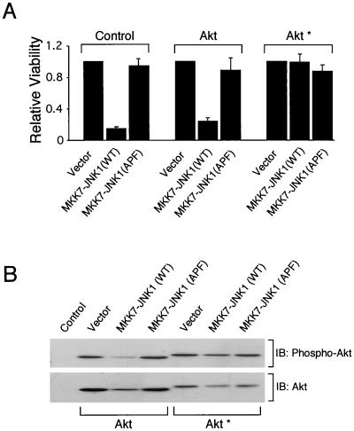
The cell death caused by activated JNK could be the result of nonspecific toxicity. To exclude this possibility, we examined whether the cell death caused by activated JNK could be rescued by increased survival signaling. The Akt signaling pathway is established as an important mediator of cell survival (4). We therefore examined the effect of overexpression of Akt on activated JNK-induced cell death. Expression of wild-type Akt caused no significant reduction in JNK-induced death (Fig. 4A). In contrast, activated Akt (modified by NH2-terminal myristoylation) caused a marked reduction in activated JNK-induced cell death (Fig. 4A). These data suggest that the cell death caused by activated JNK is not the result of nonspecific toxicity but is the result of increased signal transduction by JNK.
FIG. 4.
JNK-stimulated apoptosis is inhibited by Akt. (A) CHO cells were transfected with vector control, Akt, or activated Akt∗ (myristoylated Akt) together with the indicated plasmid expression vectors. Decreased cell survival caused by activated JNK (MKK7-JNK) was prevented by activated Akt. (B) The cell lysates were examined by immunoblot analysis using antibodies to Akt and phospho-Ser473-Akt.
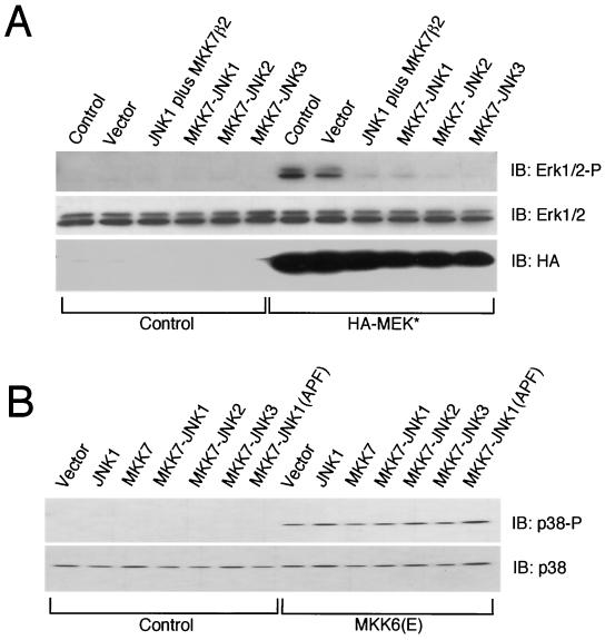
The observation that activated Akt, but not wild-type Akt, blocked cell death mediated by activated JNK is interesting because we had not anticipated that overexpressed wild-type Akt would not suppress JNK-induced cell death. One possible explanation is that activated myristoylated Akt has different signaling properties than wild-type Akt (35). Indeed, we found that activated JNK caused decreased amounts of activated wild-type Akt, but not membrane-bound myristoylated and activated Akt (Fig. 4B). This decrease in the amount of activated Akt was mediated, in part, by decreased expression of Akt (Fig. 4B). This observation prompted us to examine other signaling pathways in cells expressing activated JNK. These studies demonstrated that the expression of activated JNK did not cause changes in the activity of endogenous JNK (data not shown). However, activated JNK did markedly inhibit activation of the ERK1/2 MAP kinases caused by phorbol ester (data not shown) and MEK1 (Fig. 5A). In contrast, activated JNK caused no change in the p38 MAP kinase signaling pathway (Fig. 5B). The selective effect of activated JNK to suppress ERK1/2 activation could be mediated by increased expression of a MAP kinase phosphatase (2). Together, these data demonstrate that activated JNK alters the regulation of Akt and the ERK-1/2 MAP kinases. Since these signaling pathways have been implicated in survival signaling by previous studies (4, 63), it is possible that these changes may contribute to cell death caused by activated JNK.
FIG. 5.
Activated JNK causes decreased ERK activity. (A) CHO cells were transfected with an empty expression vector (control) or with an expression vector for activated MEK1 (HA-ΔN3-MEK1 [S218E, S222D]) together with the indicated expression vectors. The cells were harvested at 40 h posttransfection. Endogenous ERK1 and ERK2 were examined by immunoblot analysis using an antibody to ERK and phospho-ERK. The expression of transfected MEK1 was examined by immunoblot analysis using an antibody to the HA epitope tag. (B) Activated JNK does not cause decreased p38 MAP kinase activity. CHO cells were transfected with an empty expression vector (control) or with an expression vector for activated MKK6 (Flag-MKK6 [S207E, T211E]) together with the indicated expression vectors. The cells were harvested at 40 h posttransfection. Endogenous p38 MAP kinase was examined by immunoblot analysis using an antibody to p38 and phospho-p38.
Activated JNK causes apoptosis.
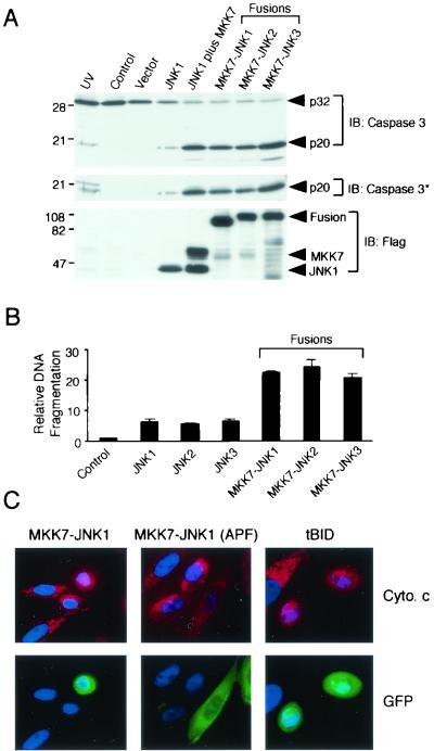
The cell death caused by activated JNK could be mediated by several different mechanisms. To test if the cell death was caused by apoptosis, we examined cells expressing activated JNK for biochemical characteristics of the apoptotic response. The activation of effector caspases, including caspase-3, is a key step in the apoptotic program. Expression of activated JNK caused a reduction in the amount of 32-kDa pro-caspase-3 and a corresponding increase in the amount of processed and activated caspase-3 (Fig. 6A). The activation of caspase-3 was confirmed by probing immunoblots with an antibody that binds selectively to a 20-kDa processed fragment of caspase-3 (Fig. 6A). These data indicate that JNK causes caspase activation. One caspase-dependent process in apoptotic cells is nucleosomal fragmentation of genomic DNA. Indeed, increased nucleosomal DNA fragmentation was caused by activated JNK1, JNK2, and JNK3 (Fig. 6B). The effects of activated JNK on DNA fragmentation were blocked by incubation of the cells with the caspase inhibitor zVAD-fmk (data not shown).
FIG. 6.
Activated JNK causes apoptosis. (A) Cell lysates prepared at 40 h posttransfection were examined by immunoblot analysis using antibodies to the Flag epitope tag (lower panel), activated caspase-3 (middle panel), and caspase-3 (upper panel). Equal amounts of total cell lysate (50 μg) were examined in each lane. The effect of exposure of the cells to UV radiation (80 J/m2) 12 h prior to harvesting was examined. The numbers on the left correspond to the migration of molecular mass standards (in kilodaltons). (B) CHO cells were transfected with empty vector (control) or the indicated expression vectors. Nucleosomal DNA fragmentation was examined at 40 h posttransfection. The data presented are the means ± SD of the results obtained for three independent experiments. (C) Activated JNK causes cytochrome c release from the mitochondria. CHO cells were cotransfected with GFP and the indicated expression vectors. The cells were fixed after 24 h, processed for immunofluorescence analysis, and stained with DAPI. GFP (green), DNA (blue), and cytochrome c (red) are illustrated. The cells were incubated with 100 μM zVAD-fmk to inhibit apoptosis. Transfected cells (green) show punctate mitochondrial cytochrome c (red) in experiments using the kinase-negative fusion protein MKK7-JNK1(APF). Expression of tBid or MKK7-JNK1 caused the release of cytochrome c from the mitochondria and the redistribution of cytochrome c throughout the cytoplasm and nucleus.
JNK causes release of mitochondrial cytochrome c by a caspase-independent mechanism.
JNK could activate apoptosis by stimulating death receptor signal transduction or by inducing the intrinsic cell death pathway involving mitochondria. To test the involvement of the intrinsic pathway, we examined cytochrome c release in cells expressing activated JNK. The active fusion protein MKK7-JNK1 was sufficient to induce cytochrome c release and was not inhibited by the caspase inhibitor zVAD-fmk (Fig. 6C). In contrast, the inactive fusion protein MKK7-JNK1 (APF) failed to release cytochrome c. These data indicate that JNK causes cytochrome c release by a caspase-independent mechanism.
JNK causes a rapid apoptotic response.
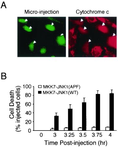
The observation that activated JNK causes apoptosis in transfection assays does not provide reliable information concerning the time course of this effect of JNK. It is possible that JNK causes apoptosis by an indirect mechanism that requires long periods of JNK activation. In contrast, activated JNK may lead to rapid apoptosis. To distinguish between these possibilities, we performed microinjection experiments. The wild-type fusion protein MKK7-JNK1 caused cytochrome c release (Fig. 7A). Time course analysis demonstrated that 33% of the microinjected cells were dead within 3 h postinjection and that 84% of the microinjected cells were dead within 4 h (Fig. 7B). No marked changes in the viability of cells microinjected with the inactive fusion protein MKK7-JNK1(APF) were detected during this time period (Fig. 7B). Control studies using a tBid expression vector demonstrated that this protein caused apoptosis (39% of cells within 1 h and 88% of cells within 2 h postinjection) that was more rapid than that caused by activated JNK (data not shown). The kinetics of tBid-stimulated cytochrome c release are consistent with the known function of tBid to bind, induce conformational changes in, and cause mitochondrial redistribution of Bax and Bak (9, 58). The slightly slower kinetics of JNK-stimulated apoptosis may either reflect a lower expression threshold for the function of tBid or indicate that several biochemical steps separate JNK activation from the mechanism of cytochrome c release. Nevertheless, these data demonstrate that activated JNK does cause rapid apoptosis and that the observed cell death is not a long-term consequence of JNK activity.
FIG. 7.
JNK causes rapid release of cytochrome c and apoptosis. (A) Activated JNK causes cytochrome c release from the mitochondria. CHO cells were microinjected with the MKK7-JNK1 expression plasmid together with dog IgG. The injected cells were visualized by staining with fluorescein-conjugated anti-dog IgG (green, indicated by arrowheads) and cytochrome c was stained with mouse anti-cytochrome c and rhodamine-conjugated goat anti-mouse antibodies. Cells releasing cytochrome c showed diffuse cytoplasmic and nuclear staining (red). Punctate mitochondrial staining was observed in the noninjected cells. (B) Time course of apoptosis induced by microinjection of MKK7-JNK1. CHO cells were microinjected with the indicated plasmids and apoptosis was evaluated by membrane blebbing and cell detachment. The data presented represent the means ± SD of data obtained for four independent experiments.
Activated JNK may regulate the function of Bcl2 family proteins.
It has been established in previous studies that JNK can phosphorylate the antiapoptotic proteins Bcl2 and Bcl-XL in vitro (6, 13, 23, 34, 64). This phosphorylation has been reported to inhibit the prosurvival function of Bcl2 and Bcl-XL (13, 23, 34, 64), although this conclusion is controversial since some studies indicate that phosphorylation may enhance the antiapoptotic actions of Bcl2 (6, 22, 43) and may stabilize Bcl2 (and thus increase prosurvival signaling) by preventing ubiquitin-mediated degradation of Bcl2 (3, 11). Taken together, these data indicate that Bcl2 and Bcl-XL represent potential sites of regulation by JNK, although the exact role of JNK-regulated phosphorylation of these proteins is unclear.
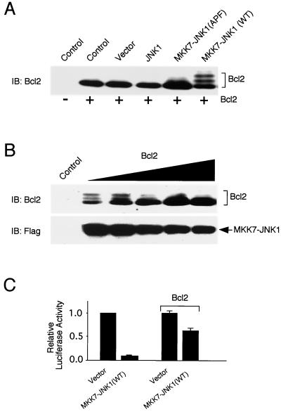
We examined Bcl2 phosphorylation in cells expressing the activated JNK fusion proteins. Immunoblot analysis demonstrated that the MKK7-JNK1 fusion protein caused a marked decrease in the electrophoretic mobility of Bcl2, consistent with phosphorylation (Fig. 8A). However, only a fraction of the Bcl2 was found to become phosphorylated in cells overexpressing MKK7-JNK1 (Fig. 8A). This partial phosphorylation of Bcl2 was unexpected because previous studies have suggested that Bcl2 is a JNK substrate and we had expected that the overexpression of activated JNK would lead to stoichiometric phosphorylation of Bcl2. To examine further the extent of Bcl2 phosphorylation caused by activated JNK, we investigated the effect of changes in the expression of Bcl2. These studies demonstrated that Bcl2 phosphorylation was only observed when Bcl2 was expressed at a low level (Fig. 8B). Interestingly, expression of Bcl2 partially inhibited apoptosis caused by the MKK7-JNK1 fusion protein (Fig. 8C).
FIG. 8.
Regulation of Bcl2 by activated JNK. (A) Activated JNK causes Bcl2 phosphorylation in vivo. CHO cells were transfected without and with a plasmid expression vector for human Bcl2 (20 ng). The cells were cotransfected with the empty expression vector or expression vectors for JNK1 and MKK7-JNK1 fusion proteins. The cells were harvested at 40 h posttransfection and the expression of Bcl2 was examined by immunoblot analysis. (B) Bcl2 phosphorylation is suppressed by increased expression of Bcl2. CHO cells were cotransfected with an expression vector for the Flag-MKK7-JNK1 fusion protein together with increasing amounts of an expression vector for human Bcl2 (0, 10, 20, 40, 60, and 80 ng). The cells were harvested at 40 h posttransfection and the expression of Bcl2 and Flag-MKK7-JNK1 was examined by immunoblot analysis. (C) CHO cells were transfected with an empty vector (control) or an expression vector for the MKK7-JNK1 fusion protein. The effect of expression of Bcl2 was examined (20 ng of plasmid). Cell viability at 40 h posttransfection was examined using a luciferase reporter plasmid. The data presented represent the means ± SD of data obtained for three independent experiments.
The observation that only a fraction of the total population of Bcl2 molecules can be phosphorylated in cells with activated JNK is interesting. Whether this partial phosphorylation of Bcl2 is sufficient to account for the ability of activated JNK to cause apoptosis is unclear. If this phosphorylation does cause reduced antiapoptotic signaling (13, 23, 34, 64), it is possible that the partial phosphorylation decreases Bcl2 function below a threshold that is required to maintain survival. In contrast, if Bcl2 phosphorylation causes increased antiapoptotic signaling (3, 6, 11), it is not obvious how the partial Bcl2 phosphorylation might contribute to JNK-stimulated apoptosis.
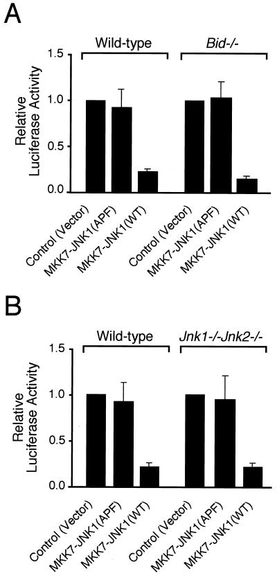
In addition to the antiapoptotic protein Bcl2, previous studies have also implicated the proapoptotic BH3-only protein Bid in JNK-stimulated apoptosis (52). It was observed that UV radiation caused Bid cleavage by a caspase-independent mechanism and that this processing of Bid was absent in Jnk-null cells (52). These data suggested that Bid might contribute to JNK-stimulated apoptosis. To test this hypothesis, we examined the effect of activated JNK on wild-type and Bid−/− fibroblasts (Fig. 9A). The MKK7-JNK1 fusion protein efficiently killed the Bid−/− cells (Fig. 9A). Control studies demonstrated that Jnk-null cells were also efficiently killed by the MKK7-JNK1 fusion protein (Fig. 9B). In contrast, the kinase-inactive mutant fusion protein MKK7-JNK1(APF) did not cause apoptosis of wild-type, Jnk-null, or Bid−/− cells. These data demonstrate that Bid does not play an essential role in JNK-dependent apoptosis.
FIG. 9.
The proapoptotic Bcl2 family protein Bid is not required for JNK-dependent apoptosis. (A) Bid is not required for JNK-dependent apoptosis. Wild-type and Bid−/− mouse fibroblasts were transfected with empty vector (control) or expression vectors for MKK7-JNK fusion proteins. Cell viability at 40 h posttransfection was examined using a luciferase reporter plasmid. The data presented represent the means ± SD of data obtained for three independent experiments. (B) Endogenous JNK is not required for apoptosis caused by ectopic expression of activated JNK. Wild-type and Jnk1−/− Jnk2−/− mouse fibroblasts were transfected with empty vector (control) or expression vectors for MKK7-JNK fusion proteins. Cell viability at 40 h posttransfection was examined using a luciferase reporter plasmid. The data presented represent the means ± SD of data obtained for three independent experiments.
The observation that JNK activation does regulate Bcl2 phosphorylation (Fig. 8A) and Bid processing (52) suggests that Bcl2 family proteins may contribute to JNK-stimulated apoptosis. However, Bid is not required for JNK-dependent apoptosis and the importance of Bcl2 phosphorylation is unclear (Fig. 8 and 9). These considerations suggest that other members of the Bcl2 family may contribute to JNK-dependent apoptosis. Other members of the Bcl2 family that are potentially relevant include the proapoptotic proteins Bax and Bak.
Bax and Bak are required for JNK-dependent apoptosis.
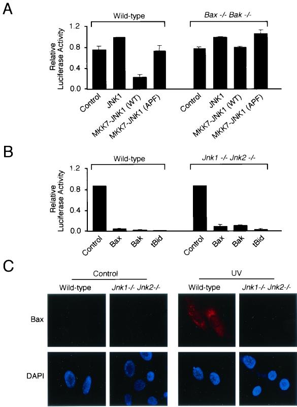
Members of the proapoptotic Bax subfamily of Bcl2-like proteins represent potential targets of the JNK-dependent apoptosis signaling pathway (36). Recent studies indicate redundant functions of Bax and the related protein Bak (30). Fibroblasts with compound mutations in both the Bax and Bak genes are remarkably resistant to stress-induced cytochrome c release and apoptosis (59, 70). This phenotype is similar to JNK-deficient primary fibroblasts (52). Thus, Bax and Bak may be required for JNK-dependent apoptosis. To test this hypothesis, we examined fibroblasts derived from wild-type and Bax−/− Bak−/− mice (Fig. 10A). Expression of activated JNK caused apoptosis of wild-type fibroblasts. In contrast, the Bax−/− Bak−/− fibroblasts were resistant to the apoptotic effects of activated JNK (Fig. 10A). Together, these data demonstrate that the proapoptotic proteins Bax and Bak are essential for JNK-dependent apoptosis.
FIG. 10.
The proapoptotic Bcl2 family proteins Bax and Bak are required for JNK-dependent apoptosis. (A) Bax and Bak are required for JNK-dependent apoptosis. Wild-type and Bax−/− Bak−/− mouse fibroblasts were transfected with empty vector (control) or expression vectors for JNK1 or MKK7-JNK fusion proteins. Cell viability at 40 h posttransfection was examined using a luciferase reporter plasmid. The data presented represent the means ± SD of data obtained for three independent experiments. (B) Endogenous JNK is not required for apoptosis caused by Bax and Bak. Wild-type and Jnk1−/− Jnk2−/− mouse fibroblasts were transfected with empty vector (control) or expression vectors for Bax, Bak, or tBid. Cell viability at 40 h posttransfection was examined using a luciferase reporter plasmid. The data presented represent the means ± SD of data obtained for three independent experiments. (C) Bax is not activated by UV radiation in JNK-deficient murine fibroblasts. Wild-type and Jnk1−/− Jnk2−/− fibroblasts were exposed to UV radiation (80 J/m2) and incubated (24 h). The cells were fixed and stained with a conformation-specific antibody to Bax and Texas Red-conjugated goat anti-mouse antibody. The Bax antibody stains activated and membrane-targeted Bax but not cytoplasmic Bax. DNA was stained with DAPI (blue).
The defect in JNK-stimulated apoptosis observed in Bax−/− Bak−/− fibroblasts (Fig. 10A) suggests that Bax and Bak may act as downstream components of a JNK-stimulated apoptotic signaling pathway. If this mechanism is correct, it would be predicted that Jnk-null cells would not be resistant to the proapoptotic effects of Bax and Bak. Control studies demonstrated that the BH3-only protein tBid caused apoptosis of wild-type and Jnk-null cells (Fig. 10B). Similarly, the expression of Bax or Bak also caused apoptosis of wild-type and Jnk-null cells (Fig. 10B). These data are consistent with the hypothesis that Bax and Bak function as downstream components of a JNK-stimulated apoptosis signaling pathway.
JNK is required for the stress-induced activation of Bax.
The requirement of Bax and Bak for JNK-dependent apoptosis (Fig. 10A) suggests that JNK deficiency may cause a defect in the normal function of Bax or Bak. To test this hypothesis, we examined the effect of UV radiation on Bax activation using a conformation-specific monoclonal antibody that binds activated Bax (21). Immunofluorescence microscopy demonstrated that UV radiation caused Bax activation in wild-type fibroblasts but not in JNK-deficient (Jnk1−/− Jnk2−/−) fibroblasts (Fig. 10C). Control studies using immunoblot analysis demonstrated that the wild-type and JNK-deficient fibroblasts expressed similar amounts of Bax (52). Together, these data demonstrate that Bax activation is JNK dependent in cells exposed to stress.
The defective Bax activation in JNK-deficient cells exposed to stress (Fig. 10C) might reflect a specific role of JNK in the response of cells to particular stimuli, or it might reflect a general requirement of JNK for Bax function. To distinguish between these possibilities, we examined the effect of the BH3-only protein tBid, which is known to induce Bax and Bak oligomerization and insertion into the mitochondrial outer membrane (9, 58, 59, 70). The effects of tBid on cytochrome c release and apoptosis require Bax and Bak (58, 59, 70). Microinjection experiments demonstrated that tBid caused efficient cytochrome c release and apoptosis in both wild-type and JNK-deficient (Jnk1−/− Jnk2−/−) fibroblasts (data not shown). Furthermore, transfection assays confirmed that tBid can kill Jnk-null cells (Fig. 10B). Thus, JNK is required for Bax and Bak activation caused by exposure of cells to stress (Fig. 10C) but is not required for cell death initiated by BH3-only proteins (Fig. 10B). Together, these data indicate that BH3-only members of the Bcl2 family may function downstream of JNK and upstream of Bax and Bak in a stress-induced apoptotic signaling pathway.
DISCUSSION
Activated JNK causes cell death.
Expression of activated JNK in cultured cells leads to rapid cell death (Fig. 7). Time course measurements in cells microinjected with a plasmid expression vector demonstrated that most of the cells died within 4 h. The cell death observed was inhibited by the broad-spectrum caspase inhibitor zVAD-fmk, indicating that the JNK-stimulated cell death was apoptosis. Indeed, several characteristics of apoptosis were detected in cells expressing activated JNK, including membrane blebbing, chromatin condensation, DNA fragmentation, and activation of caspase-3 (Fig. 6 and 7). Together, these data demonstrate that activated JNK can be sufficient for apoptosis. This conclusion complements the results of previous studies that indicate a requirement of JNK for some forms of stress-induced apoptosis (52, 63).
Interestingly, the expression of activated Akt, a kinase that is known to promote cell survival (4), inhibited apoptosis caused by activated JNK (Fig. 4A). Furthermore, the JNK-stimulated apoptosis was suppressed by coexpression of the antiapoptotic protein Bcl2 (Fig. 8C). Since JNK-stimulated apoptosis can be suppressed by increased survival signaling, the decision to die in response to JNK activation in vivo will depend upon multiple factors, including the cytokine environment, and may be cell-type specific.
Activated JNK regulates the mitochondrial apoptotic pathway.
It is established that JNK is essential for some forms of stress-induced apoptosis (5). Studies of primary embryo fibroblasts isolated from mice with compound disruptions of the Jnk genes indicate that JNK is required for apoptosis in response to environmental stress but not in response to death receptor ligands (52). Biochemical studies demonstrate that these JNK-deficient cells exhibit defects in the mitochondrial apoptotic pathway. Several apoptotic molecules have been shown to be released from mitochondria. These include the flavoprotein AIF, which causes chromosomal DNA condensation and fragmentation (49); endonuclease G, which degrades chromosomal DNA (27, 38); Smac/Diablo, which antagonizes the actions of IAP molecules (12, 56); the protease Omi/HtrA2, which also antagonizes IAP function (19, 33, 50, 55, 57); and cytochrome c, which activates the Apaf-1/caspase-9 complex (26, 28). JNK-deficient primary embryo fibroblasts fail to release mitochondrial proapoptotic molecules in response to environmental stress (52). In this study, we demonstrate that the expression of activated JNK in cultured cells causes the release of mitochondrial cytochrome c (Fig. 6 and 7). This effect of activated JNK was not reduced by the broad-spectrum caspase inhibitor zVAD-fmk. Thus, JNK-induced activation of the mitochondrial apoptosis pathway is not a secondary consequence of caspase activation. Indeed, biochemical studies have demonstrated that JNK can cause the release of cytochrome c in vitro (1). Together, these data establish that JNK is both necessary and sufficient for activation of the intrinsic cell death pathway involving the mitochondria in response to environmental stresses, including UV radiation.
Members of the Bcl2 group of apoptotic regulatory proteins may contribute to signaling by JNK.
Three subfamilies of Bcl2 proteins have been identified to play important roles in the apoptotic response. The Bcl2 subfamily (e.g., Bcl2 and Bcl-XL) functions to inhibit apoptosis, while the Bax subfamily (e.g., Bax, Bak, Bok, and Bcl-XS) and the BH3-only subfamily (Bid, Bad, Bik, Bim, Bmf, Noxa, Puma, and a number of others) promote apoptosis (16). Recently, these Bcl2 family proteins have been implicated in JNK-dependent apoptosis (5).
The antiapoptotic proteins Bcl2 and Bcl-XL have been reported to be phosphorylated and inactivated by JNK (6, 13, 23, 34, 64), although this conclusion is controversial since some studies indicate that phosphorylation may enhance the antiapoptotic actions of Bcl2 (6, 22, 43) and may stabilize Bcl2 (and thus increase prosurvival signaling) by preventing ubiquitin-mediated degradation of Bcl2 (3, 11). Taken together, these data indicate that Bcl2 and Bcl-XL represent potential sites of regulation by JNK. Consistent with this hypothesis, we found that the expression of activated JNK in cultured cells caused increased Bcl2 phosphorylation (Fig. 8A). It is possible that Bcl2 phosphorylation mediates, in part, the effects of JNK on apoptosis. If Bcl2 phosphorylation inhibits prosurvival signaling (6, 13, 23, 34, 64), the phosphorylation of Bcl2 may decrease the level of functional Bcl2 below a threshold that is required for cell survival. Alternatively, if phosphorylation of Bcl2 increases cell survival signaling (3, 6, 11, 22, 43), the role of Bcl2 phosphorylation in JNK-stimulated apoptosis is unclear.
It is interesting that the overexpression of activated JNK leads only to partial phosphorylation of Bcl2 (Fig. 8A). Furthermore, it is intriguing that this phosphorylation of Bcl2 is only observed at low levels of Bcl2 expression (Fig. 8B). At higher levels of Bcl2 expression, JNK-stimulated apoptosis is inhibited (Fig. 8C) and Bcl2 is not phosphorylated (Fig. 8B). These data suggest that Bcl2 phosphorylation correlates with apoptosis rather than with JNK activation. Consistent with this hypothesis are previous studies that demonstrate that JNK activation caused by UV radiation does not result in Bcl2 phosphorylation (51). Furthermore, Bcl2 phosphorylation caused by paclitaxel (Taxol) occurs in the absence of JNK activation (51). Together, these data indicate that JNK may not be the physiologically relevant kinase that causes Bcl2 phosphorylation in vivo. In contrast, it appears that activated JNK may regulate another kinase (or a phosphatase) that causes increased Bcl2 phosphorylation.
Several studies have implicated BH3-only members of the Bcl2-related family in JNK-dependent apoptosis. JNK-deficient fibroblasts were reported to exhibit defects in the UV-stimulated, caspase-independent processing and activation of Bid (52). However, studies of Bid−/− fibroblasts do not support a (nonredundant) role for Bid in JNK-dependent apoptosis (Fig. 9A). Furthermore, microinjection studies demonstrated that tBid, which is myristoylated and targeted to the mitochondria (68), efficiently killed both wild-type and JNK-deficient fibroblasts (Fig. 10B). These data demonstrate that JNK is not required for Bid-stimulated apoptosis and that Bid is not essential for JNK-stimulated apoptosis. However, other members of the BH3-only subfamily may be required for JNK-dependent apoptosis. Indeed, a recent study indicates that the expression of the proapoptotic BH3-only protein Bim may be JNK dependent (61), although an independent study indicates that JNK is not required for Bim expression but may be required for Bim function (39).
While the role of Bcl2 and Bcl-XL phosphorylation and BH3-only proteins in JNK-dependent apoptosis remains unclear, the results of the present study establish that the proapoptotic Bax subfamily of Bcl2-related proteins is essential for JNK-dependent apoptosis.
The Bax subfamily of Bcl2-related proteins is essential for JNK-induced apoptosis.
Members of the proapoptotic Bax subfamily of Bcl2-related proteins, including Bax and Bak, are cytoplasmic proteins that undergo a conformational change in response to environmental stress and redistribute to the mitochondria (16). It is likely that Bax (and related members of this subfamily) mediates the release of proapoptotic molecules from the mitochondria. Interestingly, the mitochondrial redistribution of, and conformational changes in, Bax are not observed in JNK-deficient cells exposed to stress (Fig. 10C). This observation suggests that members of the Bax subfamily may mediate the effects of JNK on apoptosis. The functions of Bax are partially redundant with those of the related molecule Bak (30). Consequently, studies of Bax deficiency require the analysis of compound mutations of Bax and Bak. Interestingly, Bax−/− Bak−/− embryo fibroblasts exhibit defects in stress-induced apoptosis (59) that are similar to those in JNK-deficient fibroblasts (52). Furthermore, Bax−/− Bak−/− fibroblasts are resistant to the apoptotic effects of activated JNK (Fig. 10A).
The results of the present study demonstrate that JNK is required for stress-induced activation of Bax (Fig. 10C). In addition, Bax is essential for JNK-stimulated apoptosis (Fig. 10A). These data are consistent with the hypothesis that members of the proapoptotic Bax subfamily of Bcl2-related proteins function downstream of JNK in an apoptotic signal transduction pathway that is initiated by stress.
A goal for future studies will be to define the mechanism by which JNK regulates Bax and Bak. It is established that BH3-only members of the Bcl2 family can engage Bax and Bak to release mitochondrial cytochrome c (9, 58, 59). Gene knockout studies have confirmed this conclusion (70). These considerations suggest that the effects of JNK to activate the mitochondrial apoptotic signaling pathway via Bax and Bak may be mediated by increased expression or function of one or more BH3-only proteins.
Acknowledgments
We thank S. Korsmeyer for providing Bid−/− fibroblasts; N. Ahn for providing the activated MEK1 expression vector; T. Barrett, S. Stone, and S. A. Quail for technical assistance; and K. Gemme for administrative assistance.
This study was supported in part by grant CA65861 from the National Cancer Institute. R.A.F. and R.J.D. are investigators of the Howard Hughes Medical Institute.
REFERENCES
- 1.Aoki, H., P. M. Kang, J. Hampe, K. Yoshimura, T. Noma, M. Matsuzaki, and S. Izumo. 2002. Direct activation of mitochondrial apoptosis machinery by c-Jun N-terminal kinase in adult cardiac myocytes. J. Biol. Chem. 277:10244-10250. [DOI] [PubMed] [Google Scholar]
- 2.Bokemeyer, D., A. Sorokin, M. Yan, N. G. Ahn, D. J. Templeton, and M. J. Dunn. 1996. Induction of mitogen-activated protein kinase phosphatase 1 by the stress-activated protein kinase signaling pathway but not by extracellular signal-regulated kinase in fibroblasts. J. Biol. Chem. 271:639-642. [DOI] [PubMed] [Google Scholar]
- 3.Breitschopf, K., J. Haendeler, P. Malchow, A. M. Zeiher, and S. Dimmeler. 2000. Posttranslational modification of Bcl-2 facilitates its proteasome-dependent degradation: molecular characterization of the involved signaling pathway. Mol. Cell. Biol. 20:1886-1896. [DOI] [PMC free article] [PubMed] [Google Scholar]
- 4.Datta, S. R., A. Brunet, and M. E. Greenberg. 1999. Cellular survival: a play in three Akts. Genes Dev. 13:2905-2927. [DOI] [PubMed] [Google Scholar]
- 5.Davis, R. J. 2000. Signal transduction by the JNK group of MAP kinases. Cell 103:239-252. [DOI] [PubMed] [Google Scholar]
- 6.Deng, X., L. Xiao, W. Lang, F. Gao, P. Ruvolo, and W. S. May, Jr. 2001. Novel role for JNK as a stress-activated Bcl2 kinase. J. Biol. Chem. 276:23681-23688. [DOI] [PubMed] [Google Scholar]
- 7.Derijard, B., M. Hibi, I. H. Wu, T. Barrett, B. Su, T. Deng, M. Karin, and R. J. Davis. 1994. JNK1: a protein kinase stimulated by UV light and Ha-Ras that binds and phosphorylates the c-Jun activation domain. Cell 76:1025-1037. [DOI] [PubMed] [Google Scholar]
- 8.Derijard, B., J. Raingeaud, T. Barrett, I. H. Wu, J. Han, R. J. Ulevitch, and R. J. Davis. 1995. Independent human MAP-kinase signal transduction pathways defined by MEK and MKK isoforms. Science 267:682-685. [DOI] [PubMed] [Google Scholar]
- 9.Desagher, S., A. Osen-Sand, A. Nichols, R. Eskes, S. Montessuit, S. Lauper, K. Maundrell, B. Antonsson, and J. C. Martinou. 1999. Bid-induced conformational change of Bax is responsible for mitochondrial cytochrome c release during apoptosis. J. Cell Biol. 144:891-901. [DOI] [PMC free article] [PubMed] [Google Scholar]
- 10.Dickens, M., J. S. Rogers, J. Cavanagh, A. Raitano, Z. Xia, J. R. Halpern, M. E. Greenberg, C. L. Sawyers, and R. J. Davis. 1997. A cytoplasmic inhibitor of the JNK signal transduction pathway. Science 277:693-696. [DOI] [PubMed] [Google Scholar]
- 11.Dimmeler, S., K. Breitschopf, J. Haendeler, and A. M. Zeiher. 1999. Dephosphorylation targets Bcl-2 for ubiquitin-dependent degradation: a link between the apoptosome and the proteasome pathway. J. Exp. Med. 189:1815-1822. [DOI] [PMC free article] [PubMed] [Google Scholar]
- 12.Du, C., M. Fang, Y. Li, L. Li, and X. Wang. 2000. Smac, a mitochondrial protein that promotes cytochrome c-dependent caspase activation by eliminating IAP inhibition. Cell 102:33-42. [DOI] [PubMed] [Google Scholar]
- 13.Fan, M., M. Goodwin, T. Vu, C. Brantley-Finley, W. A. Gaarde, and T. C. Chambers. 2000. Vinblastine-induced phosphorylation of Bcl-2 and Bcl-XL is mediated by JNK and occurs in parallel with inactivation of the Raf-1/MEK/ERK cascade. J. Biol. Chem. 275:29980-29985. [DOI] [PubMed] [Google Scholar]
- 14.Faris, M., N. Kokot, K. Latinis, S. Kasibhatla, D. R. Green, G. A. Koretzky, and A. Nel. 1998. The c-Jun N-terminal kinase cascade plays a role in stress-induced apoptosis in Jurkat cells by up-regulating Fas ligand expression. J. Immunol. 160:134-144. [PubMed] [Google Scholar]
- 15.Faris, M., K. M. Latinis, S. J. Kempiak, G. A. Koretzky, and A. Nel. 1998. Stress-induced Fas ligand expression in T cells is mediated through a MEK kinase 1-regulated response element in the Fas ligand promoter. Mol. Cell. Biol. 18:5414-5424. [DOI] [PMC free article] [PubMed] [Google Scholar]
- 16.Gross, A., J. M. McDonnell, and S. J. Korsmeyer. 1999. Bcl-2 family members and the mitochondria in apoptosis. Genes Dev. 13:1899-1911. [DOI] [PubMed] [Google Scholar]
- 17.Gupta, S., T. Barrett, A. J. Whitmarsh, J. Cavanagh, H. K. Sluss, B. Derijard, and R. J. Davis. 1996. Selective interaction of JNK protein kinase isoforms with transcription factors. EMBO J. 15:2760-2770. [PMC free article] [PubMed] [Google Scholar]
- 18.Gupta, S., D. Campbell, B. Derijard, and R. J. Davis. 1995. Transcription factor ATF2 regulation by the JNK signal transduction pathway. Science 267:389-393. [DOI] [PubMed] [Google Scholar]
- 19.Hegde, R., S. M. Srinivasula, Z. Zhang, R. Wassell, R. Mukattash, L. Cilenti, G. DuBois, Y. Lazebnik, A. S. Zervos, T. Fernandes-Alnemri, and E. S. Alnemri. 2002. Identification of Omi/HtrA2 as a mitochondrial apoptotic serine protease that disrupts inhibitor of apoptosis protein-caspase interaction. J. Biol. Chem. 277:432-438. [DOI] [PubMed] [Google Scholar]
- 20.Holland, P. M., M. Suzanne, J. S. Campbell, S. Noselli, and J. A. Cooper. 1997. MKK7 is a stress-activated mitogen-activated protein kinase kinase functionally related to hemipterous. J. Biol. Chem. 272:24994-24998. [DOI] [PubMed] [Google Scholar]
- 21.Hsu, Y. T., and R. J. Youle. 1997. Nonionic detergents induce dimerization among members of the Bcl-2 family. J. Biol. Chem. 272:13829-13834. [DOI] [PubMed] [Google Scholar]
- 22.Ito, T., X. Deng, B. Carr, and W. S. May. 1997. Bcl-2 phosphorylation required for anti-apoptosis function. J. Biol. Chem. 272:11671-11673. [DOI] [PubMed] [Google Scholar]
- 23.Kharbanda, S., S. Saxena, K. Yoshida, P. Pandey, M. Kaneki, Q. Wang, K. Cheng, Y. N. Chen, A. Campbell, T. Sudha, Z. M. Yuan, J. Narula, R. Weichselbaum, C. Nalin, and D. Kufe. 2000. Translocation of SAPK/JNK to mitochondria and interaction with Bcl-x(L) in response to DNA damage. J. Biol. Chem. 275:322-327. [DOI] [PubMed] [Google Scholar]
- 24.Kuan, C. Y., D. D. Yang, D. R. Samanta Roy, R. J. Davis, P. Rakic, and R. A. Flavell. 1999. The Jnk1 and Jnk2 protein kinases are required for regional specific apoptosis during early brain development. Neuron 22:667-676. [DOI] [PubMed] [Google Scholar]
- 25.Li, H., H. Zhu, C. J. Xu, and J. Yuan. 1998. Cleavage of BID by caspase 8 mediates the mitochondrial damage in the Fas pathway of apoptosis. Cell 94:491-501. [DOI] [PubMed] [Google Scholar]
- 26.Li, K., Y. Li, J. M. Shelton, J. A. Richardson, E. Spencer, Z. J. Chen, X. Wang, and R. S. Williams. 2000. Cytochrome c deficiency causes embryonic lethality and attenuates stress-induced apoptosis. Cell 101:389-399. [DOI] [PubMed] [Google Scholar]
- 27.Li, L. Y., X. Luo, and X. Wang. 2001. Endonuclease G is an apoptotic DNase when released from mitochondria. Nature 412:95-99. [DOI] [PubMed] [Google Scholar]
- 28.Li, P., D. Nijhawan, I. Budihardjo, S. M. Srinivasula, M. Ahmad, E. S. Alnemri, and X. Wang. 1997. Cytochrome c and dATP-dependent formation of Apaf-1/caspase-9 complex initiates an apoptotic protease cascade. Cell 91:479-489. [DOI] [PubMed] [Google Scholar]
- 29.Lin, A., A. Minden, H. Martinetto, F. X. Claret, C. Lange-Carter, F. Mercurio, G. L. Johnson, and M. Karin. 1995. Identification of a dual specificity kinase that activates the Jun kinases and p38-Mpk2. Science 268:286-290. [DOI] [PubMed] [Google Scholar]
- 30.Lindsten, T., A. J. Ross, A. King, W. X. Zong, J. C. Rathmell, H. A. Shiels, E. Ulrich, K. G. Waymire, P. Mahar, K. Frauwirth, Y. Chen, M. Wei, V. M. Eng, D. M. Adelman, M. C. Simon, A. Ma, J. A. Golden, G. Evan, S. J. Korsmeyer, G. R. MacGregor, and C. B. Thompson. 2000. The combined functions of proapoptotic Bcl-2 family members Bak and Bax are essential for normal development of multiple tissues. Mol. Cell 6:1389-1399. [DOI] [PMC free article] [PubMed] [Google Scholar]
- 31.Livingstone, C., G. Patel, and N. Jones. 1995. ATF-2 contains a phosphorylation-dependent transcriptional activation domain. EMBO J. 14:1785-1797. [DOI] [PMC free article] [PubMed] [Google Scholar]
- 32.Mansour, S. J., W. T. Matten, A. S. Hermann, J. M. Candia, S. Rong, K. Fukasawa, G. F. Vande Woude, and N. G. Ahn. 1994. Transformation of mammalian cells by constitutively active MAP kinase kinase. Science 265:966-970. [DOI] [PubMed] [Google Scholar]
- 33.Martins, L. M., I. Iaccarino, T. Tenev, S. Gschmeissner, N. F. Totty, N. R. Lemoine, J. Savopoulos, C. W. Gray, C. L. Creasy, C. Dingwall, and J. Downward. 2002. The serine protease Omi/HtrA2 regulates apoptosis by binding XIAP through a reaper-like motif. J. Biol. Chem. 277:439-444. [DOI] [PubMed] [Google Scholar]
- 34.Maundrell, K., B. Antonsson, E. Magnenat, M. Camps, M. Muda, C. Chabert, C. Gillieron, U. Boschert, E. Vial-Knecht, J. C. Martinou, and S. Arkinstall. 1997. Bcl-2 undergoes phosphorylation by c-Jun N-terminal kinase/stress-activated protein kinases in the presence of the constitutively active GTP-binding protein Rac1. J. Biol. Chem. 272:25238-25242. [DOI] [PubMed] [Google Scholar]
- 35.Meier, R., and B. A. Hemmings. 1999. Regulation of protein kinase B. J. Recept Signal Transduct Res. 19:121-128. [DOI] [PubMed] [Google Scholar]
- 36.Miller, T. M., K. L. Moulder, C. M. Knudson, D. J. Creedon, M. Deshmukh, S. J. Korsmeyer, and E. M. Johnson, Jr. 1997. Bax deletion further orders the cell death pathway in cerebellar granule cells and suggests a caspase-independent pathway to cell death. J. Cell Biol. 139:205-217. [DOI] [PMC free article] [PubMed] [Google Scholar]
- 37.Moriguchi, T., F. Toyoshima, N. Masuyama, H. Hanafusa, Y. Gotoh, and E. Nishida. 1997. A novel SAPK/JNK kinase, MKK7, stimulated by TNF alpha and cellular stresses. EMBO J. 16:7045-7053. [DOI] [PMC free article] [PubMed] [Google Scholar]
- 38.Parrish, J., L. Li, K. Klotz, D. Ledwich, X. Wang, and D. Xue. 2001. Mitochondrial endonuclease G is important for apoptosis in C.elegans. Nature 412:90-94. [DOI] [PubMed] [Google Scholar]
- 39.Putcha, G. V., K. L. Moulder, J. P. Golden, P. Bouillet, J. A. Adams, A. Strasser, and E. M. Johnson. 2001. Induction of BIM, a proapoptotic BH3-only BCL-2 family member, is critical for neuronal apoptosis. Neuron 29:615-628. [DOI] [PubMed] [Google Scholar]
- 40.Raingeaud, J., A. J. Whitmarsh, T. Barrett, B. Derijard, and R. J. Davis. 1996. MKK3- and MKK6-regulated gene expression is mediated by the p38 mitogen-activated protein kinase signal transduction pathway. Mol. Cell. Biol. 16:1247-1255. [DOI] [PMC free article] [PubMed] [Google Scholar]
- 41.Rincon, M., R. A. Flavell, and R. J. Davis. 2001. Signal transduction by MAP kinases in T lymphocytes. Oncogene 20:2490-2497. [DOI] [PubMed] [Google Scholar]
- 42.Robinson, M. J., S. A. Stippec, E. Goldsmith, M. A. White, and M. H. Cobb. 1998. A constitutively active and nuclear form of the MAP kinase ERK2 is sufficient for neurite outgrowth and cell transformation. Curr. Biol. 8:1141-1150. [DOI] [PubMed] [Google Scholar]
- 43.Ruvolo, P. P., X. Deng, and W. S. May. 2001. Phosphorylation of Bcl2 and regulation of apoptosis. Leukemia 15:515-522. [DOI] [PubMed] [Google Scholar]
- 44.Sabapathy, K., Y. Hu, T. Kallunki, M. Schreiber, J. P. David, W. Jochum, E. F. Wagner, and M. Karin. 1999. JNK2 is required for efficient T-cell activation and apoptosis but not for normal lymphocyte development. Curr. Biol. 9:116-125. [DOI] [PubMed] [Google Scholar]
- 45.Sabapathy, K., W. Jochum, K. Hochedlinger, L. Chang, M. Karin, and E. F. Wagner. 1999. Defective neural tube morphogenesis and altered apoptosis in the absence of both JNK1 and JNK2. Mech. Dev. 89:115-124. [DOI] [PubMed] [Google Scholar]
- 46.Sabapathy, K., T. Kallunki, J. P. David, I. Graef, M. Karin, and E. F. Wagner. 2001. c-Jun NH2-terminal kinase (JNK)1 and JNK2 have similar and stage-dependent roles in regulating T cell apoptosis and proliferation. J. Exp. Med. 193:317-328. [DOI] [PMC free article] [PubMed] [Google Scholar]
- 47.Seth, A., S. Gupta, and R. J. Davis. 1993. Cell cycle regulation of the c-Myc transcriptional activation domain. Mol. Cell. Biol. 13:4125-4136. [DOI] [PMC free article] [PubMed] [Google Scholar]
- 48.Srinivasan, A., F. Li, A. Wong, L. Kodandapani, R. Smidt, Jr., J. F. Krebs, L. C. Fritz, J. C. Wu, and K. J. Tomaselli. 1998. Bcl-xL functions downstream of caspase-8 to inhibit Fas- and tumor necrosis factor receptor 1-induced apoptosis of MCF7 breast carcinoma cells. J. Biol. Chem. 273:4523-4529. [DOI] [PubMed] [Google Scholar]
- 49.Susin, S. A., H. K. Lorenzo, N. Zamzami, I. Marzo, B. E. Snow, G. M. Brothers, J. Mangion, E. Jacotot, P. Costantini, M. Loeffler, N. Larochette, D. R. Goodlett, R. Aebersold, D. P. Siderovski, J. M. Penninger, and G. Kroemer. 1999. Molecular characterization of mitochondrial apoptosis-inducing factor. Nature 397:441-446. [DOI] [PubMed] [Google Scholar]
- 50.Suzuki, Y., Y. Imai, H. Nakayama, K. Takahashi, K. Takio, and R. Takahashi. 2001. A serine protease, HtrA2, is released from the mitochondria and interacts with XIAP, inducing cell death. Mol. Cell 8:613-621. [DOI] [PubMed] [Google Scholar]
- 51.Tournier, C., C. Dong, T. K. Turner, S. N. Jones, R. A. Flavell, and R. J. Davis. 2001. MKK7 is an essential component of the JNK signal transduction pathway activated by pro-inflammatory cytokines. Genes Dev. 15:1419-1426. [DOI] [PMC free article] [PubMed] [Google Scholar]
- 52.Tournier, C., P. Hess, D. D. Yang, J. Xu, T. K. Turner, A. Nimnual, D. Bar-Sagi, S. N. Jones, R. A. Flavell, and R. J. Davis. 2000. Requirement of JNK for stress-induced activation of the cytochrome c-mediated death pathway. Science 288:870-874. [DOI] [PubMed] [Google Scholar]
- 53.Tournier, C., A. J. Whitmarsh, J. Cavanagh, T. Barrett, and R. J. Davis. 1997. Mitogen-activated protein kinase kinase 7 is an activator of the c-Jun NH2-terminal kinase. Proc. Natl. Acad. Sci. USA 94:7337-7342. [DOI] [PMC free article] [PubMed] [Google Scholar]
- 54.Tournier, C., A. J. Whitmarsh, J. Cavanagh, T. Barrett, and R. J. Davis. 1999. The MKK7 gene encodes a group of c-Jun NH2-terminal kinase kinases. Mol. Cell. Biol. 19:1569-1581. [DOI] [PMC free article] [PubMed] [Google Scholar]
- 55.van Loo, G., M. van Gurp, B. Depuydt, S. M. Srinivasula, I. Rodriguez, E. S. Alnemri, K. Gevaert, J. Vandekerckhove, W. Declercq, and P. Vandenabeele. 2002. The serine protease Omi/HtrA2 is released from mitochondria during apoptosis. Omi interacts with caspase-inhibitor XIAP and induces enhanced caspase activity. Cell Death Differ. 9:20-26. [DOI] [PubMed] [Google Scholar]
- 56.Verhagen, A. M., P. G. Ekert, M. Pakusch, J. Silke, L. M. Connolly, G. E. Reid, R. L. Moritz, R. J. Simpson, and D. L. Vaux. 2000. Identification of DIABLO, a mammalian protein that promotes apoptosis by binding to and antagonizing IAP proteins. Cell 102:43-53. [DOI] [PubMed] [Google Scholar]
- 57.Verhagen, A. M., J. Silke, P. G. Ekert, M. Pakusch, H. Kaufmann, L. M. Connolly, C. L. Day, A. Tikoo, R. Burke, C. Wrobel, R. L. Moritz, R. J. Simpson, and D. L. Vaux. 2002. HtrA2 promotes cell death through its serine protease activity and its ability to antagonize inhibitor of apoptosis proteins. J. Biol. Chem. 277:445-454. [DOI] [PubMed] [Google Scholar]
- 58.Wei, M. C., T. Lindsten, V. K. Mootha, S. Weiler, A. Gross, M. Ashiya, C. B. Thompson, and S. J. Korsmeyer. 2000. tBID, a membrane-targeted death ligand, oligomerizes BAK to release cytochrome c. Genes Dev. 14:2060-2071. [PMC free article] [PubMed] [Google Scholar]
- 59.Wei, M. C., W. X. Zong, E. H. Cheng, T. Lindsten, V. Panoutsakopoulou, A. J. Ross, K. A. Roth, G. R. MacGregor, C. B. Thompson, and S. J. Korsmeyer. 2001. Proapoptotic BAX and BAK: a requisite gateway to mitochondrial dysfunction and death. Science 292:727-730. [DOI] [PMC free article] [PubMed] [Google Scholar]
- 60.Weston, C. R., and R. J. Davis. 2002. The JNK signal transduction pathway. Curr. Opin. Genet. Dev. 12:14-21. [DOI] [PubMed] [Google Scholar]
- 61.Whitfield, J., S. J. Neame, L. Paquet, O. Bernard, and J. Ham. 2001. Dominant-negative c-Jun promotes neuronal survival by reducing BIM expression and inhibiting mitochondrial cytochrome c release. Neuron 29:629-643. [DOI] [PubMed] [Google Scholar]
- 62.Wu, Z., J. Wu, E. Jacinto, and M. Karin. 1997. Molecular cloning and characterization of human JNKK2, a novel Jun NH2-terminal kinase-specific kinase. Mol. Cell. Biol. 17:7407-7416. [DOI] [PMC free article] [PubMed] [Google Scholar]
- 63.Xia, Z., M. Dickens, J. Raingeaud, R. J. Davis, and M. E. Greenberg. 1995. Opposing effects of ERK and JNK-p38 MAP kinases on apoptosis. Science 270:1326-1331. [DOI] [PubMed] [Google Scholar]
- 64.Yamamoto, K., H. Ichijo, and S. J. Korsmeyer. 1999. Bcl-2 is phosphorylated and inactivated by an ASK1/Jun N-terminal protein kinase pathway normally activated at G(2)/M. Mol. Cell. Biol. 19:8469-8478. [DOI] [PMC free article] [PubMed] [Google Scholar]
- 65.Yang, D. D., C. Y. Kuan, A. J. Whitmarsh, M. Rincon, T. S. Zheng, R. J. Davis, P. Rakic, and R. A. Flavell. 1997. Absence of excitotoxicity-induced apoptosis in the hippocampus of mice lacking the Jnk3 gene. Nature 389:865-870. [DOI] [PubMed] [Google Scholar]
- 66.Yao, Z., K. Diener, X. S. Wang, M. Zukowski, G. Matsumoto, G. Zhou, R. Mo, T. Sasaki, H. Nishina, C. C. Hui, T. H. Tan, J. P. Woodgett, and J. M. Penninger. 1997. Activation of stress-activated protein kinases/c-Jun N-terminal protein kinases (SAPKs/JNKs) by a novel mitogen-activated protein kinase kinase. J. Biol. Chem. 272:32378-32383. [DOI] [PubMed] [Google Scholar]
- 67.Yin, X. M., K. Wang, A. Gross, Y. Zhao, S. Zinkel, B. Klocke, K. A. Roth, and S. J. Korsmeyer. 1999. Bid-deficient mice are resistant to Fas-induced hepatocellular apoptosis. Nature 400:886-891. [DOI] [PubMed] [Google Scholar]
- 68.Zha, J., S. Weiler, K. J. Oh, M. C. Wei, and S. J. Korsmeyer. 2000. Posttranslational N-myristoylation of BID as a molecular switch for targeting mitochondria and apoptosis. Science 290:1761-1765. [DOI] [PubMed] [Google Scholar]
- 69.Zheng, C., J. Xiang, T. Hunter, and A. Lin. 1999. The JNKK2-JNK1 fusion protein acts as a constitutively active c-Jun kinase that stimulates c-Jun transcription activity. J. Biol. Chem. 274:28966-28971. [DOI] [PubMed] [Google Scholar]
- 70.Zong, W. X., T. Lindsten, A. J. Ross, G. R. MacGregor, and C. B. Thompson. 2001. BH3-only proteins that bind pro-survival Bcl-2 family members fail to induce apoptosis in the absence of Bax and Bak. Genes Dev. 15:1481-1486. [DOI] [PMC free article] [PubMed] [Google Scholar]


