

Abstract
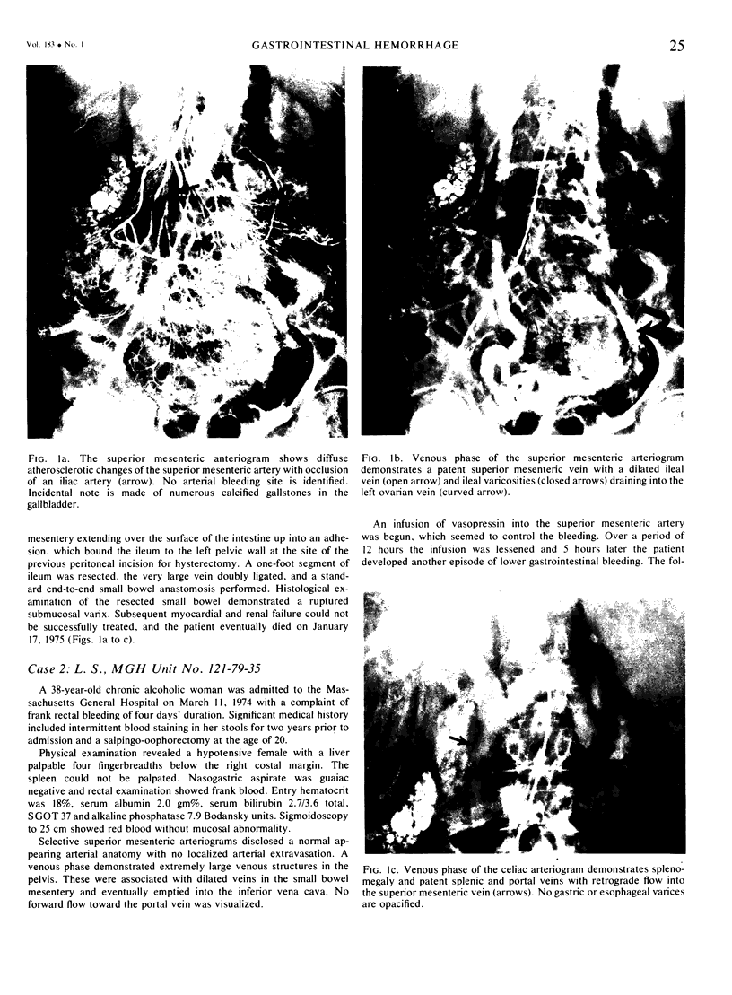
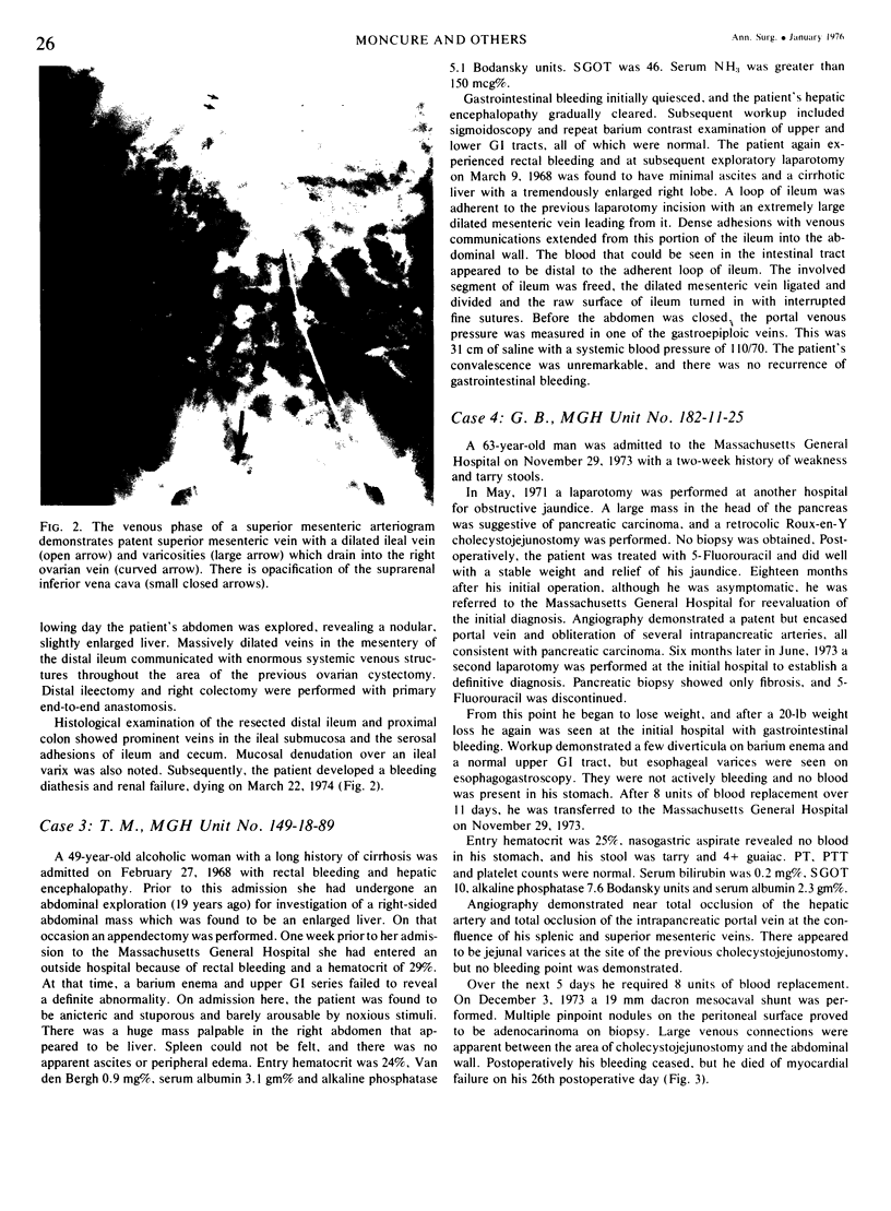
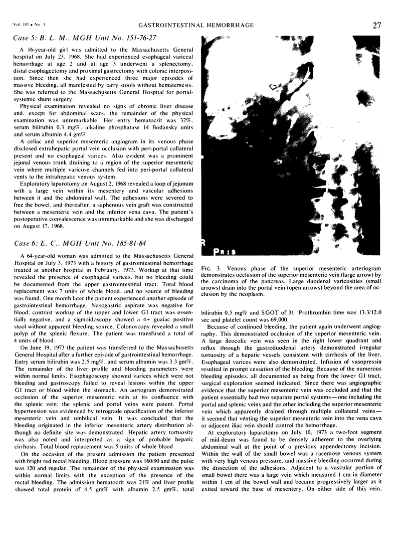
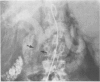
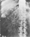
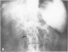
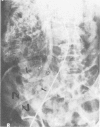
As a result of this retrospective analysis of hemorrhage from a porta-systemic venous shunt occurring within the small intestine, we believe that the early diagnosis of the syndrome is strongly suggested by the presence of varices in unusual locations demonstrated by the venous phase of mesenteric arteriography. In all patients portal hypertension was present, and in all the affected bowel was adherent to postoperative adhesions on old suture lines. The syndrome was treated variously with lysis of adhesions, bowel resection, or portal-systemic shunt. Those patients with excellent hepatic reserve survived and had no further gastrointestinal bleeding.
Full text
PDF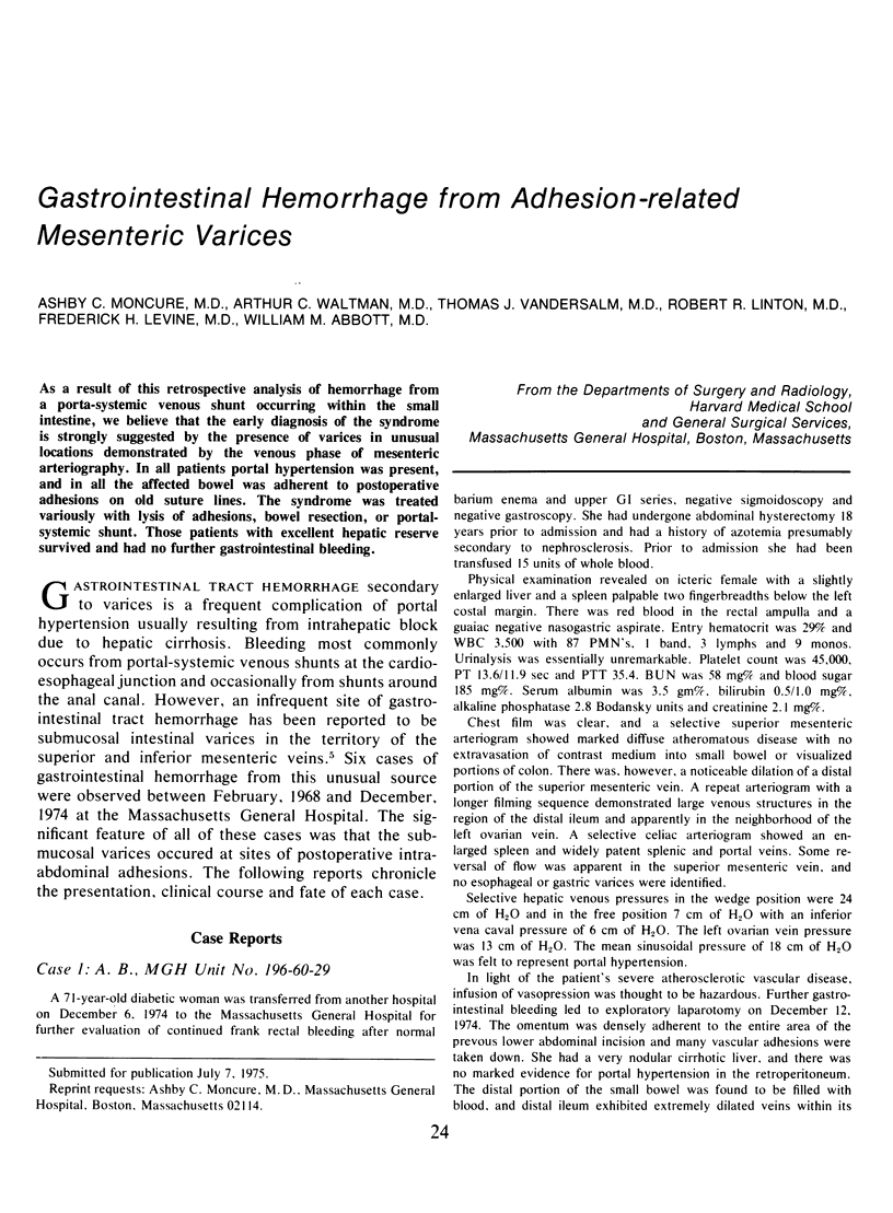
Images in this article

Selected References
These references are in PubMed. This may not be the complete list of references from this article.
- Börjesson B., Olsson A. M., Vang J. O. Hemorrhage from a portal-systemic venous shunt with unusual localization in a case of portal hypertension. Scand J Gastroenterol. 1974;9(6):571–573. [PubMed] [Google Scholar]
- EDWARDS E. A. Functional anatomy of the porta-systemic communications. AMA Arch Intern Med. 1951 Aug;88(2):137–154. doi: 10.1001/archinte.1951.03810080005002. [DOI] [PubMed] [Google Scholar]
- Gray R. K., Grollman J. H., Jr Acute lower gastrointestinal bleeding secondary to varices of the superior mesenteric venous system. Radiology. 1974 Jun;111(3):559–561. doi: 10.1148/111.3.559. [DOI] [PubMed] [Google Scholar]
- Hamlyn A. N., Morris J. S., Lunzer M. R., Puritz H., Dick R. Portal hypertension with varices in unusual sites. Lancet. 1974 Dec 28;2(7896):1531–1534. doi: 10.1016/s0140-6736(74)90283-9. [DOI] [PubMed] [Google Scholar]