

Abstract
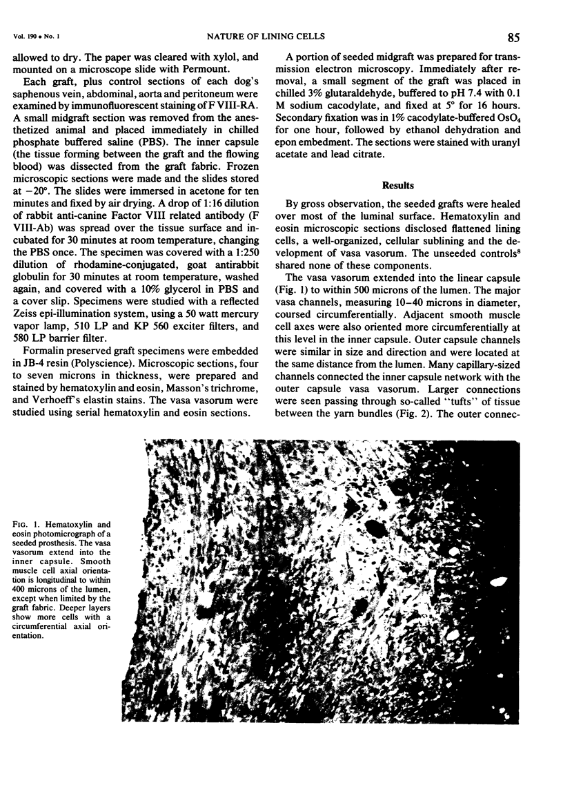
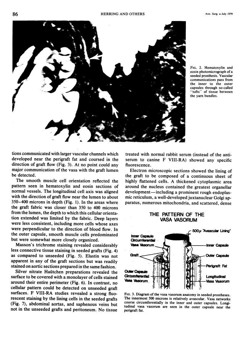
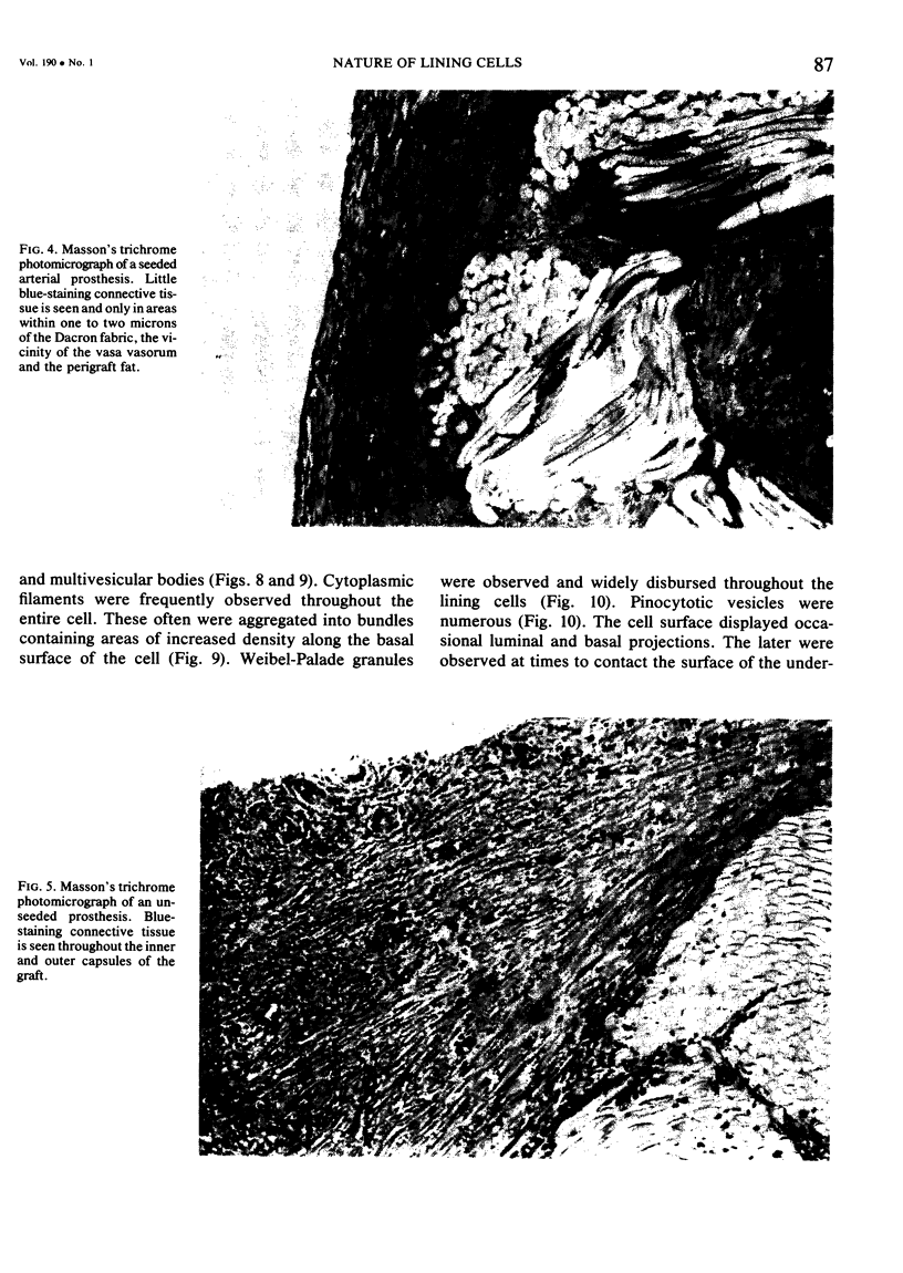
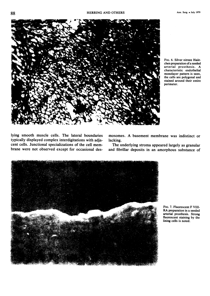
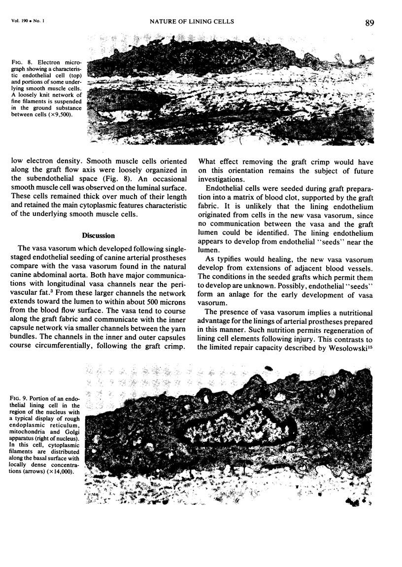
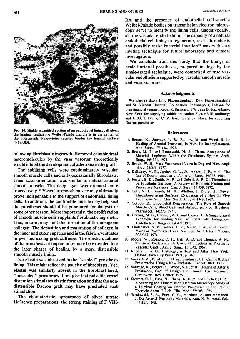
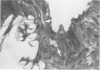
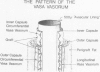
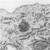
Arterial prostheses seeded with autogenous vascular endothelium demonstrate a well-organized, cellular, inner lining. To determine the nature of the lining cells, six animals underwent replacement of the infrarenal aorta with Dacron prostheses. During the preparation of three such grafts, endothelium was scraped from the saphenous vein with a steel wool pledget, suspended in chilled Sack's solution, and mixed with blood used to preclot the graft. This suspension was omitted from the three control grafts. After six weeks, the grafts were removed, rinsed and examined. Fluorescent Factor VIII related antigen (F VIII-RA) strongly stained the lining cells. Silver nitrate Haütchen and electron microscopy preparations revealed a lining pattern characteristic of vascular endothelium. Endothelial cell-specific Weibel-Palade bodies were identified in the lining cell cytoplasm. Masson's trichrome staining revealed a relatively collagen-poor connective tissue within the seeded fabric. Transmission electron microscopy disclosed vascular smooth muscle cells between the seeded graft fabric and the lining cells. Vasa vasorum, arising from the outer capsule, penetrated the fabric to supply the inner capsules of the seeded grafts. It is concluded that the cells lining seeded canine arterial prostheses are true vascular endothelium supported by vascular smooth muscle cells, that the lining contains minimal connective tissue, and that vasa vasorum develop. Unseeded control grafts lacked these features.
Full text
PDF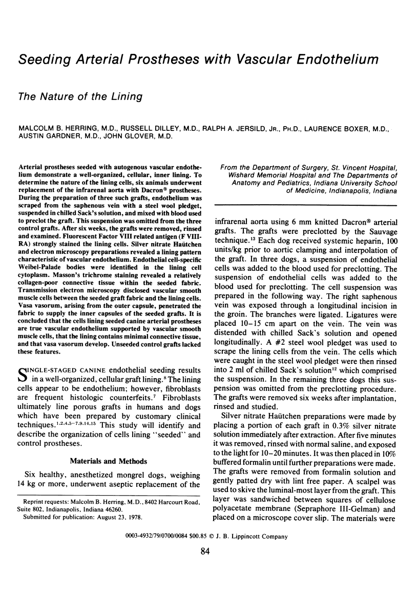
Images in this article

Selected References
These references are in PubMed. This may not be the complete list of references from this article.
- Berger K., Sauvage L. R., Rao A. M., Wood S. J. Healing of arterial prostheses in man: its incompleteness. Ann Surg. 1972 Jan;175(1):118–127. doi: 10.1097/00000658-197201000-00018. [DOI] [PMC free article] [PubMed] [Google Scholar]
- Brais M. P., Braunwald N. S. Tissue acceptance of materials implanted within the circulatory system. Arch Surg. 1974 Sep;109(3):351–358. doi: 10.1001/archsurg.1974.01360030003002. [DOI] [PubMed] [Google Scholar]
- Brook W. H. Vasa vasorum of veins in dog and man. Angiology. 1977 May;28(5):351–360. doi: 10.1177/000331977702800507. [DOI] [PubMed] [Google Scholar]
- DEBAKEY M. E., JORDAN G. L., Jr, ABBOTT J. P., HALPERT B., O'NEAL R. M. THE FATE OF DACRON VASCULAR GRAFTS. Arch Surg. 1964 Nov;89:757–782. [PubMed] [Google Scholar]
- Foster E. D., Smith M. R., Dobell A. R. Thrombosis on prosthetic surfaces: a review of etiologic factors and preventive measures. Can J Surg. 1972 Nov;15(6):339–349. [PubMed] [Google Scholar]
- Gott V. L., Ameli M. M., Whiffen J. D., Leininger R. I., Falb R. D. Newer thromboresistant surfaces: evaluation with a new in-vivo technique. Surg Clin North Am. 1967 Dec;47(6):1443–1452. doi: 10.1016/s0039-6109(16)38394-3. [DOI] [PubMed] [Google Scholar]
- Gottlob R. Endothelial regeneration: the role of smooth muscle cells, blood cells and histiocytes. Prog Biochem Pharmacol. 1977;13:276–282. [PubMed] [Google Scholar]
- Herring M., Gardner A., Glover J. A single-staged technique for seeding vascular grafts with autogenous endothelium. Surgery. 1978 Oct;84(4):498–504. [PubMed] [Google Scholar]
- Moore W. S., Rosson C. T., Hall A. D., Thomas A. N. Transient bacteremia. A cause of infection in prosthetic vascular grafts. Am J Surg. 1969 Mar;117(3):342–343. doi: 10.1016/0002-9610(69)90368-7. [DOI] [PubMed] [Google Scholar]
- Stewart G. J., Essa N., Chang K. H., Reichle F. A. A scanning and transmission electron microscope study of the luminal coating on Dacron prostheses in the canine thoracic aorta. J Lab Clin Med. 1975 Feb;85(2):208–226. [PubMed] [Google Scholar]
- Wesolowski S. A., Fries C. C., Martinez A., McMahon J. D. Arterial prosthetic materials. Ann N Y Acad Sci. 1968 Jan;146(1):325–344. doi: 10.1111/j.1749-6632.1968.tb20291.x. [DOI] [PubMed] [Google Scholar]